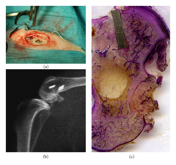
Figure 2.

Clinical view of the device placement in rabbit proximal tibial epiphysis (a). X-ray micrograph of the surgical site (b). Histological ground section of an implant device. The implant is medially and laterally surrounded by cortical and trabecular host bone. LM, toluidine blue, and acid fuchsin staining, 1x.
