Abstract
Oxidative stress during fetal development, delivery, or early postnatal life is a major cause of neuropathology, as both hypoxic and hyperoxic insults can significantly damage the developing brain. Despite the obvious need for reliable cerebral oxygenation monitoring, no technology currently exists to monitor cerebral oxygen metabolism continuously and noninvasively in infants at high risk for developing brain injury. Consequently, a rational approach to titrating oxygen supply to cerebral oxygen demand – and thus avoiding hyperoxic or hypoxic insults – is currently lacking. We present a promising method to close this crucial technology gap in the important case of neonates on conventional ventilators. By using cerebral near-infrared spectroscopy (NIRS) and signals from conventional ventilators, along with arterial oxygen saturation, we derive continuous (breath-by-breath) estimates of cerebral venous oxygen saturation, cerebral oxygen extraction fraction, cerebral blood flow, and cerebral metabolic rate of oxygen. The resultant estimates compare very favorably to previously reported data obtained by non-continuous and invasive means from preterm infants in neonatal critical care.
Keywords: cerebral venous oxygen saturation, cerebral blood flow, cerebral metabolic rate of oxygen, cerebral oxygen metabolism, preterm infant, brain injury, near-infrared spectroscopy, continuous monitoring
Introduction
Brain injury in the preterm infant comes with tremendous personal hardship and immense societal cost over the lifetime of the affected individual. The primary mode of damage to the developing brain is hypoxic-ischemic injury sustained during antenatal, intrapartum, and early neonatal life. [1]. Likewise, hyperoxic insults to the developing brain are also injurious due to a particular susceptibility of developing neural tissue to oxygen free-radicals. Unfortunately, no technology currently exists to monitor cerebral oxygen utilization at the bedside. Clinicians therefore find themselves in the situation of having to titrate oxygen therapy without reliable and sensitive tools for continuous monitoring of cerebral oxygen metabolism.
Cerebral near-infrared spectroscopy (NIRS) has the potential to fill this crucial technology gap, as it provides for continuous and noninvasive assessment of changes in the oxy- and deoxyhemoglobin concentrations in cerebral tissue (comprising arteries, veins, and cerebral microcirculation). In other words, NIRS measures cerebral hemoglobin saturation in the intravascular compartment as a whole, without distinguishing between the arterial or venous compartments. To relate these signals to contributions of individual vascular compartments, and thus to identify important variables of cerebral oxygen metabolism, changes in hemoglobin content need to be induced selectively in one compartment or another. Jugular venous occlusion, for example, induces changes primarily on the venous side of the cerebral vasculature, and therefore provides important insights into cerebral venous oxygen saturation [2]. Nevertheless, repeated temporary occlusion of a preterm infant’s cerebral venous outflow is neither a practical nor a safe option. However, positive-pressure ventilation commonly required during the early period after premature birth might induce such fluctuations in cerebral venous drainage on a breath-by-breath basis. This forms the basis of the work presented here. Specifically, we examined NIRS signals during the brief interval of impeded venous return imposed by every inspiratory breath by conventional positive-pressure ventilation, to estimate important variables of cerebral oxygen metabolism continuously and noninvasively.
Materials and Methods
Estimation algorithm
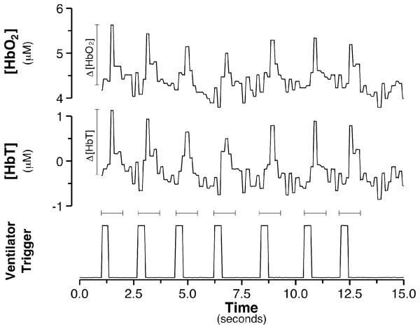
Our algorithm for estimating cerebral venous oxygen saturation (SvO2), cerebral oxygen extraction fraction (OEF), cerebral blood flow (CBF), and cerebral metabolic rate of oxygen (CMRO2) is based on the principle that during conventional ventilation, the positive-pressure inspiratory phase increases intra-thoracic pressure, transiently impeding venous return to the heart. Figure 1 shows an example of the ventilator-associated changes in cerebral oxyhemoglobin and total hemoglobin concentrations in one of our subjects.
Figure 1.
Oxyhemoglobin concentration (top), total hemoglobin concentration (middle), and ventilator trigger signal (bottom). Also indicated are the windows over which the [HbO2] and [HbT] signals are analyzed following the start of each inspiratory cycle.
It is commonly assumed that cerebral arterial blood volume remains unaffected over the immediate brief inspiratory period, so the changes in cerebral oxy-hemoglobin concentration, Δ[HbO2], and total hemoglobin concentration, Δ[HbT] (=Δ[HbO2]+ Δ[Hb]) imprinted in the NIRS signals reflect changes in the cerebral venous compartment [2, 3]. The ratio Δ[HbO2]/Δ[HbT] gated to the inspiratory phase of each positive-pressure breath thus provides an estimate of cerebral SvO2 on a breath-by-breath basis. By averaging in a least-square-error manner over adjacent breaths, SvO2 can be estimated on a continuous (i.e., breath-by-breath) basis and in a robust manner. From a continuous measurement of systemic arterial oxygen saturation, SaO2 (gated to arterial pulsations) and estimates of cerebral SvO2, we can derive continuous estimates of cerebral oxygen extraction (OE = SaO2 − SvO2) and cerebral oxygen extraction fraction (OEF = [SaO2−SvO2]/ SaO2).
Using a similar approach, we can also estimate tissue-specific CBF (CBF per 100 g of brain tissue), by further analyzing the NIRS-derived [HbT] signal around the inspiratory phase of the respiratory cycle. Assuming that mean arterial blood pressure, systemic oxygen saturation, and cerebral oxygen metabolism remain constant over the brief period of ventilator-induced inspiration, the change in total cerebral hemoglobin concentration, Δ[HbT], induced by the transient retardation of venous outflow, is directly related to changes in cerebral venous blood volume in the sample volume interrogated by NIRS. The maximum slope of the Δ[HbT] signal is therefore a measure of regional cerebral blood flow (per 100g of brain tissue) [2]:
where k is a constant that incorporates the patient’s hemoglobin concentration and serves to convert the units of the time derivative of [HbT] to cerebral blood flow (per 100 g tissue). This amounts to a patient-specific calibration of hemoglobin flow. We approximate the derivative above by the ratio of the maximum increase Δ[HbT] during the analysis window for each breath (as labeled in Figure 1) to the time required to reach this value after the start of the inspiratory cycle.
Finally, the cerebral metabolic rate of oxygen (CMRO2 per 100g of tissue) is the product of cerebral oxygen extraction and cerebral blood flow (CMRO2 = OE · CBF).
Study population
We tested our algorithm on data archived from eight preterm infants born before 32 weeks of gestation and requiring neonatal intensive care with ventilatory support. The research protocol was approved by the institutional review board at Brigham and Women’s Hospital, Boston, USA, and we obtained parental written informed consent in all cases.
Instrumentation
To track the systemic cardio-respiratory states of the neonates, we continuously recorded arterial blood pressure (through an umbilical artery catheter), systemic arterial oxygen saturation (by pulse oximetry), the electrocardiogram, and ventilator signals (peak airway pressure, ventilator trigger, and airway flow). We assessed cerebral tissue oxygenation by bihemispheric near-infrared spectroscopy (NIRS; Hamamatsu NIRO-200) and electrocortical activity through continuous recording of a single-channel EEG. The NIRO-200 provides estimates of changes in tissue oxy- and deoxy-hemoglobin concentrations from some unknown baseline. The systemic cardiovascular and NIRS signals were streamed to an analog-to-digital converter, sampled at 4kHz, and archived in a time-locked manner. A trained bedside observer was present continuously to log details of any manipulation or changes in management.
Data pre-processing
To apply our estimation algorithms, we down-sampled the ventilator trigger, NIRS signals, and systemic oxygen saturation recording to 20 Hz. We used custom-made algorithms to detect the onset of each trigger pulse, which marks the onset of the inspiratory phase of the positive-pressure ventilator. We then defined a window of one-second duration starting with the beginning of the inspiratory period, over which the [HbT] and [HbO2] signals are analyzed as outlined above.
As shown in Figure 1, the ventilator-induced changes induced in the NIRS signals can vary from one breath to the next. To mitigate the effect of such fluctuations, and to arrive at robust estimates of SvO2, CBF, and CMRO2, we estimate these quantities in a least-square-error manner over a rolling window containing 20 breaths.
Results
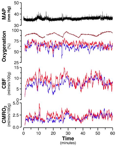
Figure 2 shows measured mean arterial blood pressure (MAP; black) along with the measured SaO2 (maroon) and continuous estimates of cerebral SvO2, cerebral CBF, and CMRO2 as derived from the right (red tracing) and the left (blue tracing) hemisphere, over the course of one hour in a preterm neonate with a gestational age of 27 weeks. Although the estimates of SvO2, CBF, and CMRO2 were derived independently for the right and left hemisphere, the resultant bilateral estimates bear a notable degree of correspondence at various timescales, though also some differences and offsets.
Fig. 2.
From top to bottom: Mean arterial blood pressure; systemic arterial and cerebral venous oxygen saturation; cerebral blood flow; and cerebral metabolic rate of oxygen. Red: right hemispheric estimates; blue: left hemispheric estimates.
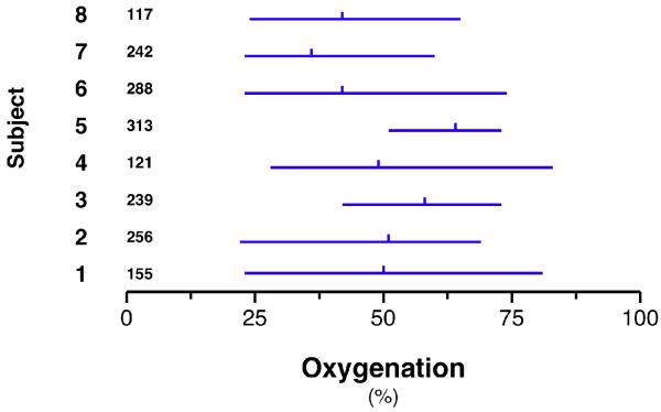
Figure 3 summarizes the range and median values of SvO2 in eight premature infants from our database. (The results shown in Figure 2 represent a snapshot of the data of Subject 5 in Figure 3.)
Fig. 3.
Range and median (tick mark) of cerebral venous oxygen saturation in eight premature infants. Numbers to the right of the subject identifier signify the duration (in minutes) over which the ranges were computed.
Discussion
Accurate titration of cerebral oxygen therapy to cerebral oxygen demand remains an important and unresolved problem in neonatal care. The importance of this issue relates to the fact that both excessive and insufficient cerebral oxygenation may be injurious to the immature brain. Furthermore, normal intrinsic autoregulatory systems for regulation of oxygen delivery may be disrupted in these infants. Rational clinical decision making regarding brain-tissue oxygenation is currently impaired by our inability to monitor continuously and noninvasively important variables of cerebral oxygen metabolism (such as oxygen extraction and cerebral metabolic rate of oxygen) that reflect oxygen delivery to, and oxygen demand by, the brain. Here, we present an algorithmic approach to estimating these critical variables of cerebral oxygen metabolism from changes in near-infrared spectroscopy signals imprinted by cyclic variations in intrathoracic pressure associated with positive-pressure ventilation. The same strategy has been leveraged by Wolf et al. [3] to estimate cerebral venous oxygen saturation, though they only report intermittent estimates and the details of our respective approaches differ.
Like others [2, 3], we assume that the ventilator-induced changes in the NIRS signal reflect changes in the cerebral venous vascular compartment. Neglecting possible changes in cerebral arterial volume is a common assumption, though it remains to be properly verified.
Our patient-specific estimates of SvO2, CBF, and CMRO2 compare very favorably to what has been reported in the medical literature for these variables in neonates, using other, highly invasive measurements. For example, Volpe [4] summarizes tissue-specific cerebral blood flow measurements for preterm infants from different studies that used intravenous Xenon-133 clearance (see Table 6-28, p. 301 in [4]). For mechanically ventilated infants, CBF measurements pooled from different studies seem to cluster around 10 ml/min/100g. When averaged over the course of a four-hour period, the specific cerebral blood flow for the patient whose data are shown in Figure 2 is (6.1±3.4) ml/min/100g and (7.5± 4.1) ml/min/100g for the left and the right hemisphere, respectively1. Our estimates of CBF might underestimate true tissue-specific CBF if the ventilator-induced changes in intrathoracic pressure fail to completely halt cerebral venous outflow temporarily. Furthermore, the computation of the derivative to obtain estimated CBF tends to be susceptible to noise in the data, though computing the slope in a least-squares manner over a window of adjacent breaths mitigates this effect. Finally, we have used a representative value for the hemoglobin concentration in nonates to calculate CBF in this retrospective study. Future application of this method should rely on actual hemoglobin measurements from each patient to convert the time derivative of [HbT] to CBF.
Using positron emission tomography, Altman [5] and co-workers determined CMRO2 in a group of sixteen preterm infants (with mean gestational age of 27 weeks and mean birth weight of 925 g). They report a value of 0.06 ml/min/100g, which is significantly lower than the blood flow values for the left and right hemisphere in the patient presented in Figure 2 [(1.5±1.4) ml/min/100g and (1.8±1.3) ml/min/100g]. Inspection of Altman’s data suggests that this difference is largely due to the comparatively large oxygen extraction fraction of our subject (0.30±0.09), which resembles that of a term newborn (in the neighborhood of 0.21) rather than the value reported in [4] for preterm neonates (in the neighborhood of 0.06).
Estimates for the right and left hemisphere show a remarkable concordance. The origins of fluctuations seen in the estimated signals remain to be elucidated, but might be reflective of changing regional metabolic demand in response to changing electrocortical activity. This question is currently under investigation with concurrent EEG measures.
Estimates in cerebral SvO2 can vary substantially over the course of two to four hours, as indicated by the wide ranges shown in Figure 3. Some of these variations correlate with variations in SaO2 (not shown). In other cases, the reasons for the excursions of estimated cerebral SvO2 are less obvious.
Future work will focus on evaluating these estimates against gold-standard measurements in animal studies, as well as investigating the effects of changing peak-inspiratory pressure on the robustness and accuracy of our estimates.
Acknowledgments
This work was supported in part by the United States National Institutes of Health through grants R01EB001659, K24NS057568, and R21HD056009 by the National Institute of Biomedical Imaging and Bioengineering, the National Institute of Neurological Disorders and Stroke, and the National Institute of Child Health and Human Development, respectively. Further support was provided by the Lifebridge Fund.
Footnotes
Note that over the course of the four-hour period, significant trends in cerebral blood flow exist, which contribute to the relatively large standard deviation.
Declaration of conflict of interest The authors declare that they have no conflict of interest.
References
- 1.Volpe JJ. Brain injury in premature infants: a complex amalgam of destructive and developmental disturbances. Lancet Neurology. 2009;8:110–124. doi: 10.1016/S1474-4422(08)70294-1. [DOI] [PMC free article] [PubMed] [Google Scholar]
- 2.Victor S, Weindling M. Near-infrared spectroscopy and its use for the assessment of tissue perfusion in the neonate. In: Kleinman CS, Seri I, editors. Hemodynamics and Cardiology -- Neonatology Questions and Controversies. Saunders; 2008. Chapter 6. [Google Scholar]
- 3.Wolf M, Duc G, Keel M, Niederer P, von Siebenthal K, Bucher HU. Continuous noninvasive measurements of cerebral arterial and venous oxygen saturation at the bedside in mechanically ventilated neonates. Critical Care Medicine. 1997;25(9):1579–82. doi: 10.1097/00003246-199709000-00028. [DOI] [PubMed] [Google Scholar]
- 4.Volpe JJ. Neurology of the Newborn. 5th edition Saunders Elsevier; 2008. [Google Scholar]
- 5.Altman DI, Powers WJ, Perlman JM, Herscovitch P, Volpe SL, Volpe JJ. Cerebral blood flow requirement for brain viability in newborn infants is lower than in adults. Annals of Neurology. 1988;24(2):218–226. doi: 10.1002/ana.410240208. [DOI] [PubMed] [Google Scholar]


