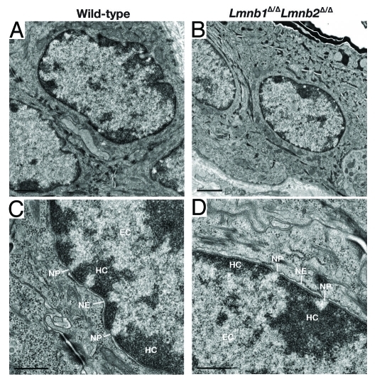
Figure 4.
Electron micrographs of keratinocyte nuclei in the skin from wild-type and Lmnb1Δ/ΔLmnb2Δ/Δ mice, showing heterochromatin (HC), euchromatin (EC), nuclear pores (NP), and nuclear envelope (NE). Magnification in A–B, × 22700. Scale bar, 1.0 μm. Magnification in C–D, × 74200. Scale bar, 0.5 μm. Reproduced with permission from Yang et al.23

