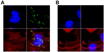
FIG 4 .
Confocal analysis of FdeC expression in contact with cultured epithelial cells. Monolayers of UM-UC-3 cells were infected with IHE3034 (A) and the respective fdeC knockout mutant strain (B). At the end of the infection, the samples were fixed and stained for confocal immunofluorescence microscopy. FdeC was detected using specific antibodies and visualized using a fluorescent secondary antibody (green). DAPI was used to stain the host cell nuclei and visualize bacteria (blue). Cellular actin was stained with fluorescent phalloidin (red).

