Abstract
DEC205/CD205 is a C-type multilectin receptor, expressed highly in dendritic cells (DCs). Previous efforts to generate anti-human DEC205 (anti-hDEC205) monoclonal antibodies (mAbs) from mice immunized with subdomain proteins of hDEC205 resulted in a few mAbs. Recently, we expressed and utilized a full-length extracellular domain protein of hDEC205 to successfully generate 5 strong anti-hDEC205 mAbs from mice. In this study, DEC205 knockout (KO) mice were immunized with this full-length extracellular domain protein of hDEC205. One of the 3 immunized DEC205 KO mice was chosen for the highest anti-hDEC205 titer by flow cytometric analysis of serum samples on CHO cells stably expressing hDEC205 (CHO/hDEC205 cells) and used for hybridoma fusion. From a single fusion, more than 400 anti-hDEC205 hybridomas were identified by flow cytometric screen with CHO/hDEC205 cells, and a total of 115 hybridomas secreting strong anti-hDEC205 mAb were saved and named HD1 through HD115. To characterize in detail, 10 HD mAbs were chosen for superior anti-hDEC205 reactivity and further subjected to cloning and purification. Interestingly, out of those 10 chosen anti-hDEC205 HD mAbs, 5 mAbs were also strongly reactive to mouse DEC205 while 8 mAbs were found to stain DEC205+ DCs on monkey spleen sections. In addition, we also identified that HD83, one of the 10 chosen HD mAbs, stains DEC205+ DCs in rat spleen and lymph node. Therefore, by immunizing DEC205 KO mice with a full-length extracellular domain protein of hDEC205, we generated a large number of strong anti-hDEC205 mAbs many of which are cross-species reactive and able to visualize DEC205+ DCs in lymphoid tissues of other mammals.
Keywords: Monoclonal Antibody, Conserved Epitope, CD205, DEC205, Dendritic Cells
1. Introduction
Dendritic cells (DCs) are known to express many potential endocytic receptors on surface for efficient uptake and processing of antigens, which makes DCs play a central role in antigen presentation. DEC205/CD205 is a C-type multilectin receptor with a large extracellular domain which consists of a number of subdomains, including a cysteine rich (CR) domain, a fibronectin type II (FN) domain, and 10 contiguous carbohydrate recognition domains (CRDs). DEC205 is expressed most abundantly by DCs, although it is detected in many different cells and tissues (Kato et al., 2006; Idoyaga et al., 2009). DEC205+ DCs are localized within the T-cell areas of lymphoid tissues, which are the sites for generating immunity and tolerance to antigens. In human lymphoid organs, DEC205 is so far the only endocytic receptor that has been visualized on most DCs in the T-cell areas (Pack et al., 2007).
Since DEC205 was found on DCs to mediate adsorptive endocytosis, leading to proficient processing and presentation of antigens (Jiang et al., 1995; Mahnke et al., 2000), a series of efforts have been made to target protein antigens selectively to mouse DCs in vivo by integrating into anti-DEC205 monoclonal antibody (mAb) (Hawiger et al., 2001; Bonifaz et al., 2002; Bonifaz et al., 2004). This DEC205 targeting in vivo with adjuvant that induces DC maturation increases the efficiency of antigen presentation on both major histocompatibility complex class I and II molecules almost 100-fold (Bonifaz et al., 2004; Bozzacco et al., 2010) and generates strong and protective T-cell immunity in mice (Trumpfheller et al., 2006). With an adjuvant of synthetic double-stranded RNA, such as polyriboinocinic polyribocytidylic acid (poly IC), the targeting of protein vaccines to DEC205+ DCs elicits durable Th1 immunity, lasting for several months in mice (Trumpfheller et al., 2008). Administration of adjuvant poly IC, an agonist for MDA5 and TLR3 pattern-recognition receptors, induces the high levels of type I interferon, and thus mouse DCs expressing type I interferon receptors become immunostimulatory (Longhi et al., 2009). Recently, using anti-human DEC205 (anti-hDEC205) mAbs, protein vaccines with poly IC adjuvant were targeted to hDEC205 on mouse DCs in hDEC205 transgenic mice, which improved the cellular and humoral immune responses significantly (Cheong et al., 2010b).
Several efforts to generate anti-hDEC205 mAbs in mice had limited success likely because that DEC205 sequences are highly conserved in mammals as well as that the immunogens used are small extracellular subdomain proteins of hDEC205 (Guo et al., 2000; Kato et al., 2006). In a recent study, we immunized mice with a full-length extracellular domain protein of hDEC205 and successfully obtained 5 strong anti-hDEC20 mAbs (Cheong et al., 2010b). In this report, we immunized DEC205 knockout (KO) mice with this hDEC205 full-length extracellular domain protein, and could generate more than 100 of strong anti-hDEC205 HD mAb hybridomas in a single hybridoma fusion experiment. Besides, we found that a significant number of these anti-hDEC205 HD mAbs produced from the hDEC205-immunized DEC205 KO mouse are cross-species reactive, and thus provide valuable tools to study the function of DEC205+ DCs in various mammals.
2. Materials and methods
2.1. Animals
DEC205 knockout (KO) mice (Guo et al., 2000; Kronin et al., 2000) in the C57BL/6 background were maintained in specific pathogen-free environment during the period of immunization with antigens. Male ACI rat or Lewis rat were purchased from SLC Co. (Shizuoka, Japan). Animal care and experiments were carried out in accordance with the institutional guidelines set by the Rockefeller University, the Memorial Sloan-Kettering Cancer Center, and the Dokkyo Medical University.
2.2. Human and monkey tissues
Normal human spleens were obtained from brain-dead organ transplant donors through the Regional Organ Procurement Organization/Organ Donor Network at the Islet Cell Transplant Program at Weill Cornell Medical College, as described previously (Pack et al., 2007). Spleen sections from normal monkey tissue were kindly provided by Drs. Paul and Klara Racz (Bernhard Nocht Institute for Tropical Medicine, Germany).
2.3. Cells and reagents
Chinese hamster ovary (CHO) cells (CHO-S cells , Invitrogen, Carlsbad, CA) and 293T cells were cultured in DMEM with high glucose (Gibco Invitrogen, catalog number 11995) supplemented with 7 % fetal calf serum (FCS) and 1× solutions of non-essential amino acids and antibiotic-antimycotic (Invitrogen). The generation of stable CHO cell lines expressing full-length human DEC205 (CHO/hDEC205) and mouse DEC205 (CHO/mDEC205) has been described elsewhere (Cheong et al., 2010b).
Cloned monoclonal antibody (mAb) of rat anti-mDEC205 NLDC145 was produced by expressing and purifying the cloned heavy and light chains of NLDC145 mAb including substitution of the original rat constant domains with the constant regions of mouse kappa and IgG1 heavy chains as previously described (Hawiger et al., 2001). Production of cloned mouse anti-hDEC205 MG38.2 mAb was described previously (Guo et al., 2000; Pack et al., 2007), and MG38.2 mAb used in this study contained the constant regions of mouse IgG1 heavy chain. Purified anti-CD206 monoclonal antibody was kindly provided by Dr. Antonio Lanzavecchia (Institute for Research in Biomedicine, Switzerland). PE-conjugated anti-CD11c and FITC-conjugated anti-CD8 were purchased from BD Biosciences (San Jose, CA).
2.4. Human DEC205/CD205 antigen
The expression and purification of extracellular domain of hDEC205 have been described elsewhere (Cheong et al., 2010b). In brief, the cDNA encoding extracellular domain of hDEC205 was fused in frame with human IgG1 Fc domain, i.e., hDEC205-hIgG1Fc which was cloned into a mammalian expression vector for stable transfection into CHO cells. The hDEC205-hIgG1Fc fusion protein was purified by affinity to Protein A from supernatant of the CHO/hDEC205-hIgG1Fc cell culture.
2.5. Immunization
Three DEC205 KO mice at 10 months of age were immunized subcutaneously with 50 µg of hDEC205-hIgG1Fc protein mixed with adjuvant TiterMax® (TiterMax USA, Inc., Norcross, GA) following the manufacturer’s instruction. Each DEC205 KO mouse was immunized 4 times at 3 week intervals. Serum samples were collected from the mice a week after each immunization. The titer of anti-hDEC205 antibodies in serum was assessed by the flow cytometric analysis with CHO/hDEC205 cells, as described below. Two months after the 4th immunization, the DEC205 KO mouse with the highest anti-hDEC205 titer was boosted intravenously with 50 µg of hDEC205-hIgG1Fc protein without adjuvant. Four days later, the mouse was sacrificed and splenocytes were fused to mouse myeloma cells as described below.
2.6. Production of hybridomas
The mouse hybridomas were prepared at the Monoclonal Antibody Core Facility in the Memorial Sloan-Kettering Cancer Center. In brief, mouse splenocytes were fused to mouse SP2/0-Ag14 myeloma cells (ATCC, Manassas, VA) at a 1:4 myeloma to spleen cell ratio by drop-wise addition of 50 % polyethylene glycol (PEG; EM Science, Germany) to the cell pellet. The PEG was washed away with serum-free DMEM, and the fused cells were directly resuspended in RPMI 1640 medium (GIBCO, Invitrogen, Carlsbad, CA) containing 15 % FCS and supplements (penicillin, streptomycin, L-glutamine, sodium pyruvate, and hypoxanthine/aminopterin/thymidine) according to standard protocols, and distributed into twenty five 96-well plates. The supernatants from hybridoma-containing wells were collected and screened by flow cytometry (as described below), and some hybridomas were chosen for subcloning twice by limiting dilutions.
2.7. Flow cytometric screen of anti-DEC205 monoclonal antibodies
To determine the titers of anti-hDEC205 (or anti-mDEC205) antibodies in mouse serum and hybridoma supernatants, CHO/hDEC205 (or CHO/mDEC205) cells were used for the flow cytometric screen. Control CHO and CHO/hDEC205 (or CHO/mDEC205) cells were detached using 0.5 mM EDTA in PBS and resuspended in PBS containing 2 % FCS plus 0.01% sodium azide (FACS buffer). This suspension was distributed into round-bottom 96-well plates at 2×10⁵ cells/well. Then, the cells were mixed with 50 µl of diluted (1:100~1:10,000) sera from immunized mice or undiluted hybridoma supernatants. After 1 hr of incubation at 4°C, cells were washed with FACS buffer and added with 50 µl of FACS buffer containing 1:1,000 anti-mouse IgG secondary Ab conjugated with PE (Jackson ImmunoResearch Laboratories, West Grove, PA), followed by incubation at 4°C for 1 hr. The titers and activity of anti-DEC205 mAbs in mouse sera and hybridoma supernatants were determined by the fluorescence intensity measured by the BD FACSCalibur flow cytometer at the Rockefeller University Flow Cytometry Resource Center.
2.8. Immunohistochemistry
Human spleen tissue was frozen in OCT Compound (Sakura Finetechnical Co., Tokyo, Japan) and stored at −80 °C. Frozen sections (6–8 µm) were prepared, air-dried, and stored at −20 °C. Upon thawing, sections were air-dried, fixed in acetone for 10 min, and rehydrated in PBS. Primary antibody was applied for 30–60 min and the sections were washed in PBS. Sections were then stained with anti-subclass specific secondary antibodies (Molecular Probes, Eugene, OR) diluted to 2 µg/ml in PBS with 1 % BSA. Slides were mounted in Aqua Poly/Mount (Polysciences, Inc., Warrington, PA) and viewed on a Molecular Devices Olympus AX70 deconvolution microscope (Olympus America Inc., Lake Success, NY) running Metamorph Meta Imaging series software (Universal Imaging Corporation, West Chester, PA).
For rat tissues, the spleen and the cervical lymph nodes were triple immunostained as described previously (Zhou et al., 2008). In brief, fresh 4-µm thick cryosections from rat tissues were firstly incubated with HD83 and then with an Alexa Fluor 594-labeled anti-mouse Ig (Life Technologies, Tokyo, Japan). After washing with PBS, sections were incubated with normal mouse IgG for 10 min to block binding of second mAb to the anti-mouse Ig conjugate. Thereafter, sections were incubated with Alexa Fluor 488-conjugated anti-rat class II MHC (OX76, ECACC, Salisbury, UK) and finally reacted with anti-type IV collagen and amino methyl carboxyfluorescein acetate-labeled anti-rabbit Ig (Jackson ImmunoResearch, West Grove, PA) to visualize tissue framework. Then, sections were analyzed under a fluorescent microscope (Axioskop 2 plus, Carl Zeiss, Tokyo, Japan).
2.9. Western blot analysis and epitope mapping on hDEC205 subdomain
Stably transfected CHO cells or transiently transfected 293T cells were lysed with RIPA lysis buffer (150 mM NaCl, 50 mM Tris-HCl, pH 8.0, 1 % NP-40, 0.5 % desoxycholate, 0.1 % sodium dodecyl sulfate (SDS)) including protease inhibitor cocktail (Roche Applied Science). Cell lysates were mixed with glycerol (10 % final) for loading and separated on 6–8 % SDS-polyacrylamide gel electrophoresis, blotted onto Hybond-P polyvinylidine difluoride membrane (GE Healthcare), incubated with supernatants of cloned NLDC145 or HD mAbs at 1:10–1:100 dilution, detected by anti-mouse IgG conjugated with horseradish peroxidase (HRP) (Southern Biotech), and visualized with enhanced chemiluminescence Plus reagents (GE Healthcare).
We had generated a series of deletion constructs for hDEC205 extracellular domain as described previously (Cheong et al., 2010b). To map the hDEC205 subdomain containing the epitope bound by HD mAbs, each subdomain construct was fused in frame with hIgG1 Fc domain, i.e., hIgG1Fc-hDEC205/CR-FN, hIgG1Fc-hDEC205/CRD1–2, hIgG1Fc-hDEC205/CRD3–4, hIgG1Fc-hDEC205/CRD5–6, hIgG1Fc-hDEC205/CRD7–8, and hIgG1Fc-hDEC205/CRD9–10, and transiently expressed in 293T cells using Lipofectamine 2000 reagent (Invitrogen). Western blotting assays were done on transfectant cell lysates as described above.
3. Results
3.1. Production of hDEC205-hIgG1Fc fusion protein and immunization of DEC205 knockout mice
The cDNAs that encode the full-length DEC205 proteins from human and mouse were cloned by PCR, and a V5 epitope tag was inserted into the N-terminus of human DEC205 (hDEC205) protein to detect the expressed gene (Cheong et al., 2010b). The cDNAs of V5-tagged hDEC205 (GenBank accession number AY682091) and mouse DEC205 (mDEC205) from C57BL/6 mouse strain (GenBank accession number AF395445) were used to generate the stable CHO cell-lines that expressed these DEC205 proteins on the cell surface (Fig. 1A). The cDNA of a full-length extracellular domain of hDEC205 was fused to a human IgG1 Fc cassette (hDEC205-hIgG1Fc; GenBank accession number DQ407610). Then, the hDEC205-hIgG1Fc gene in a CMV expression vector was stably transfected to CHO cells and expressed into cell culture supernatant. The antigen was purified by Protein A affinity column, yielding more than 2 mgs of hDEC205-hIgG1Fc fusion protein. The hDEC205-hIgG1Fc protein with higher than 90 % purity was used to immunize 3 DEC205 knockout (KO) mice. Although these 3 KO mice were quite old (10 months of age at the first immunization), the titers of the anti-hDEC205 antibody responses in the sera from all 3 mice were very high, i.e., the FACS signals specific to hDEC205 on CHO/hDEC205 cells were detectable up to 1:10,000 dilution. Consequently, a large number of hybridomas expressing anti-hDEC205 mAbs were generated as described below.
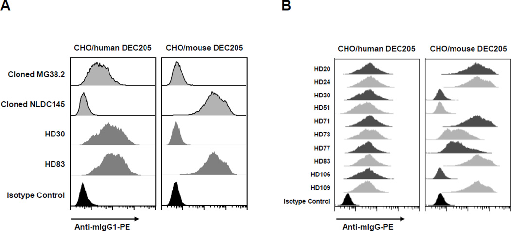
Figure 1. Reactivity of HD mAbs to human and mouse DEC205 on transfectant cells.
(A) Two of the 10 chosen HD mAbs, HD30 and HD83, were compared with anti-hDEC205 MG38.2 and anti-mDEC205 NLDC145 mAbs for their reactivities to human and mouse DEC205. CHO cells stably expressing hDEC205 or mDEC205 were stained with HD30, HD83, cloned MG38.2 (MG38.2 with the constant regions of mouse IgG1 heavy chains), or cloned NLDC145 (NLDC145 with the constant regions of mouse IgG1 heavy chains). All antibodies were used at 1 µg/ml followed by detection with PE-conjugated anti-mIgG1. Mouse IgG1 antibody was used as a negative isotype control.
(B) Reactivities of the 10 chosen HD mAbs to human and mouse DEC205 were compared by FACS analysis with CHO/hDEC205 and CHO/mDEC205 cells as in (A) but binding of each mAb to the cells was detected with PE-conjugated anti-mIgG.
3.2. Production of HD mAbs strongly reactive to hDEC205
After 3 DEC205 KO mice were immunized 4 times with hDEC205-hIgG1Fc protein, the mouse with a highest antibody titer against hDEC205 in serum was sacrificed to make the hybridomas. The supernatants from more than 1,700 hybridomas were screened by FACS with CHO/hDEC205 cells. Based on flow cytometric binding to cell surface hDEC205 (but not control CHO cells), over 400 hybridomas were identified to positively produce anti-hDEC205 mAbs. A total of 115 hybridomas, producing supernatants that showed strong binding to cell surface expressed hDEC205, were preserved in liquid nitrogen and named HD1 through HD115. For further characterization, we chose, cloned, and purified the mAbs from 10 HD hybridomas (Table 1), based on their superior anti-hDEC205 activities as a result of flow cytometric screen with CHO/hDEC205 cells.
Table 1. Summary of mouse Ig isotype, species reactivity, and epitope mapping for the 10 chosen HD mAbs.
Reactivity of anti-hDEC205 and anti-mDEC205 was determined on the basis of relative intensity of FACS staining on CHO/hDEC205 and CHO/mDEC205 cells (−, not detectable; +, weaker than anti-hDEC205 MG38.2 or anti-mDEC205 NLDC145; ++, similar to or stronger than MG38.2 or NLDC145). Reactivity of anti-monkey DEC205 was noted as positive (+) or negative (−) based on the result of immunohistochemical staining of monkey splenic tissue sections.
| mAb | Isotype | Anti-human DEC205 |
Anti-mouse DEC205 |
Anti-monkey DEC205 |
Epitope-mapped Subdomain |
|---|---|---|---|---|---|
| HD20 | IgG1, kappa | ++ | ++ | − | CR-FN |
| HD24 | IgG1, kappa | ++ | ++ | − | CR-FN |
| HD30 | IgG1, kappa | ++ | − | + | CRD1–2 |
| HD51 | IgG1, kappa | ++ | − | + | CRD1–2 |
| HD71 | IgG2b, kappa | ++ | ++ | + | CRD3–4 |
| HD73 | IgG1, kappa | ++ | + | + | CR-FN |
| HD77 | IgG1, kappa | ++ | + | + | CRD1–2 |
| HD83 | IgG1, kappa | ++ | ++ | + | CRD1–2 |
| HD106 | IgG1, kappa | ++ | − | + | CRD1–2 |
| HD109 | IgG1, kappa | ++ | ++ | + | CRD3–4 |
3.3. A significant number of HD mAbs also recognize mDEC205
The supernatants of the 115 HD hybridomas were also screened by FACS with CHO/mDEC205 cells. Although the majority of 115 HD supernatants stained hDEC205 but not mDEC205, more than 20 of the HD supernatants were able to recognize mDEC205 to variable extents. Out of the 10 chosen HD mAbs, 3 recognized only hDEC205 while the other 7 recognized hDEC205 plus mDEC205 to different extents (Table 1). As positive controls, we used anti-hDEC205 mAb MG38.2 and anti-mDEC205 mAb NLDC145. All 10 chosen HD mAbs showed stronger binding to hDEC205 than MG38.2 at the same concentrations (comparison of MG38.2 with HD30 and HD83 shown in Fig. 1A). The 5 HD mAbs (HD20, 24, 71, 83, 109) bound mDEC205 comparably to NLDC145 at the same concentrations (Figs. 1A and 1B).
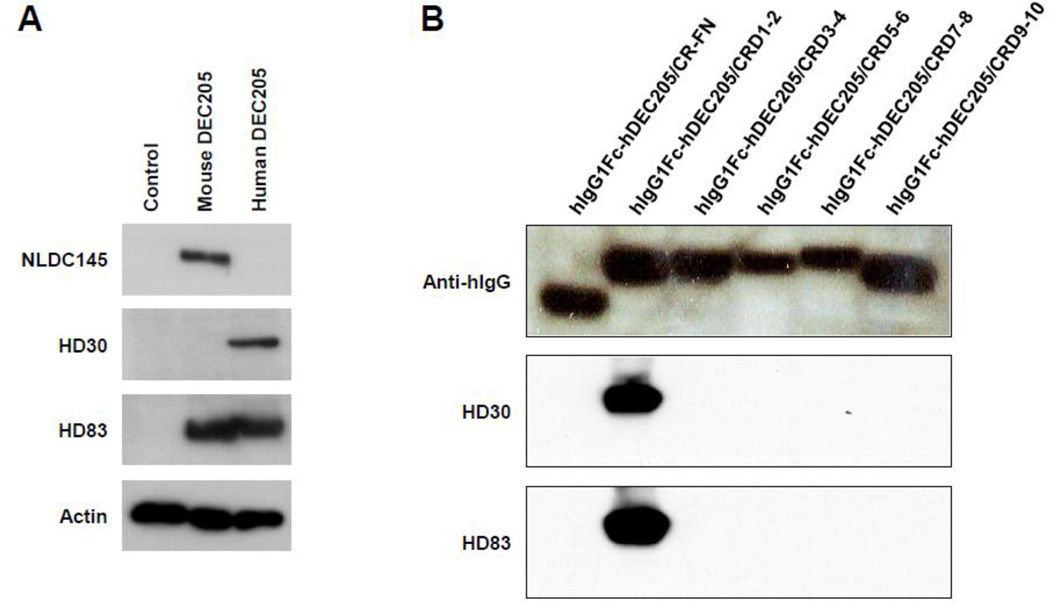
Some of the 115 HD mAbs were able to detect hDEC205 in western blot analysis, as shown for the western blots by HD30 (detecting only hDEC205) and HD83 (detecting both hDEC205 and mDEC205) in Fig. 2A. To determine the subdomain of hDEC205 where the epitopes of these HD mAbs reside, by western blot analysis, we expressed a series of deletion mutants of hDEC205 extracellular domain in fusion with human IgG1 Fc domain. As sown in Fig. 2B, although HD30 and HD83 recognize two different epitopes, the epitopes of both HD30 and HD83 were mapped on a same subdomain of hDEC205, i.e., carbohydrate recognition domains (CRDs) 1 and 2. We further mapped the epitope-containing subdomains for a number of anti-hDEC205 HD mAbs, and found that the epitopes of new HD mAbs could be mapped on a broad spectrum of hDEC205 extracellular subdomains. Out of the 10 chosen HD mAbs, 3 recognized epitopes in CR-FN subdomain, 5 in CRD1–2 subdomain, and 2 in CRD3–4 subdomain (Table 1).
Figure 2. Western blot analysis of anti-DEC205 mAbs.
(A) Control CHO, CHO/mDEC205, and CHO/hDEC205 cells were lysed and each lysate equivalent to 5 × 10⁵ cells was subjected to the western blot analyses by HD30, HD83, NLDC145, and anti-actin antibodies.
(B) A series of deletion constructs in the extracellular domain of hDEC205 were generated in fusion with hIgG1 Fc domain, and each deletion construct was expressed in 293T cells. Lysates of cells expressing different subdomains of hDEC205 were subjected to western blot analyses with HD30, HD83, or anti-hIgG.
3.4. Immunohistochemistry of DEC205 in human and monkey tissues by 10 chosen HD mAbs
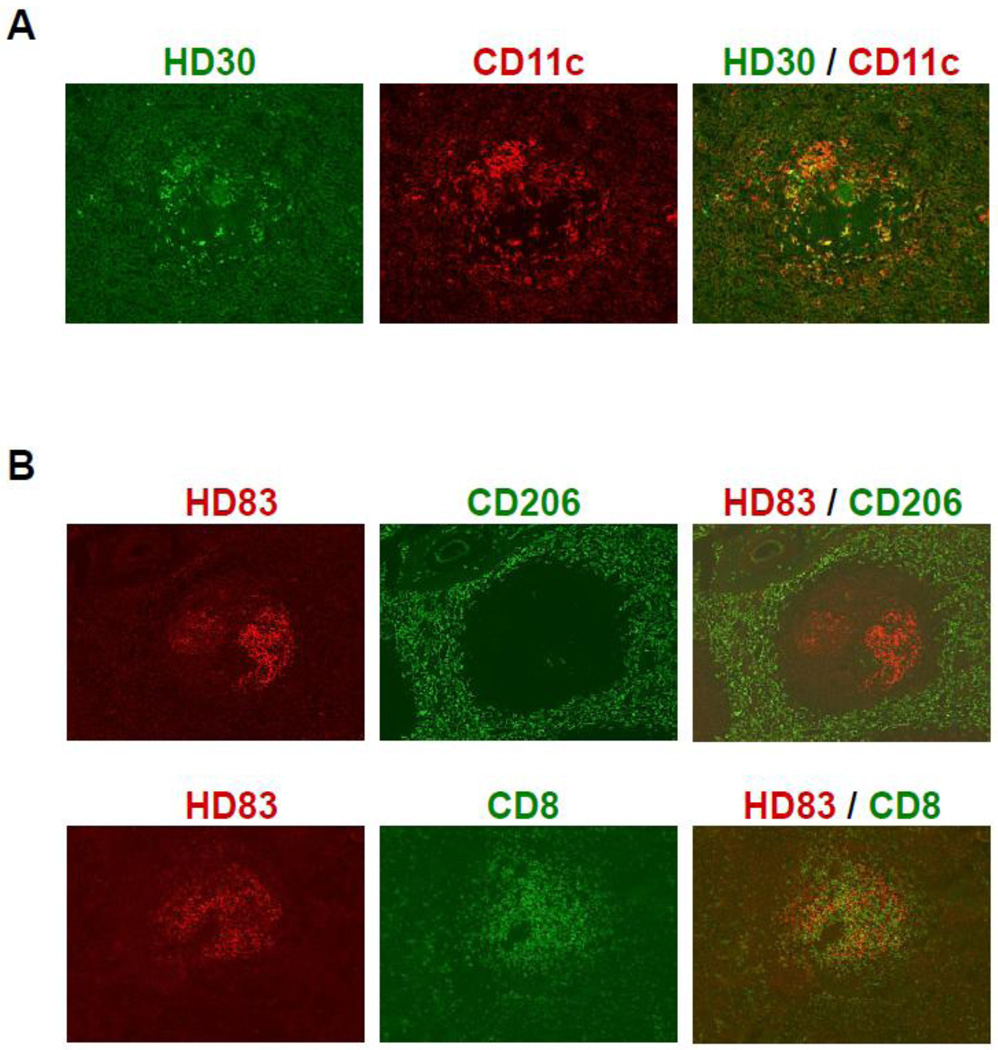
In our previous study, HD77 along with MG38.2 was successfully used to visualize the DEC205-expressing dendritic cells (DCs) in white pulp of human spleen (Pack et al., 2007). In addition to HD77, we tested the remaining of the 10 chosen HD mAbs and found that all 10 HD mAbs were similarly able to stain hDEC205 on CD11c+ DCs in human spleen (staining with HD30 shown in Fig. 3A). Then, we examined these 10 HD mAbs for their ability to stain DEC205 in monkey spleen. Out of the 10 chosen anti-hDEC205 mAbs, 8 HD mAbs could stain DEC205+ DCs in T-cell areas inside the white pulps of monkey spleen (staining with HD83 shown in Fig. 3B). HD20 and HD24 are those 2 mAbs unable to stain monkey DEC205 but they could bind both human and mouse DEC205 very efficiently (Fig. 1B).
Figure 3. Immunohistochemical stains of HD mAbs on the splenic tissues from human and monkey.
(A) Cells expressing DEC205 at high levels were detected in the white pulp of the human spleen by HD30. Double staining of human spleen sections was carried out with anti-DEC205 mAb HD30 (in green) and anti-CD11c antibody (DCs in red).
(B) HD83 stained DEC205+ cells in the T-cell areas of the white pulps in monkey spleen. Double staining of monkey spleen sections was carried out with anti-DEC205 mAb HD83 (in red) and antibodies to CD206 (red pulp macrophages in green; upper panel) or CD8 (T cells in green; lower panel).
3.5. Detection of DEC205+ DCs in rat tissues
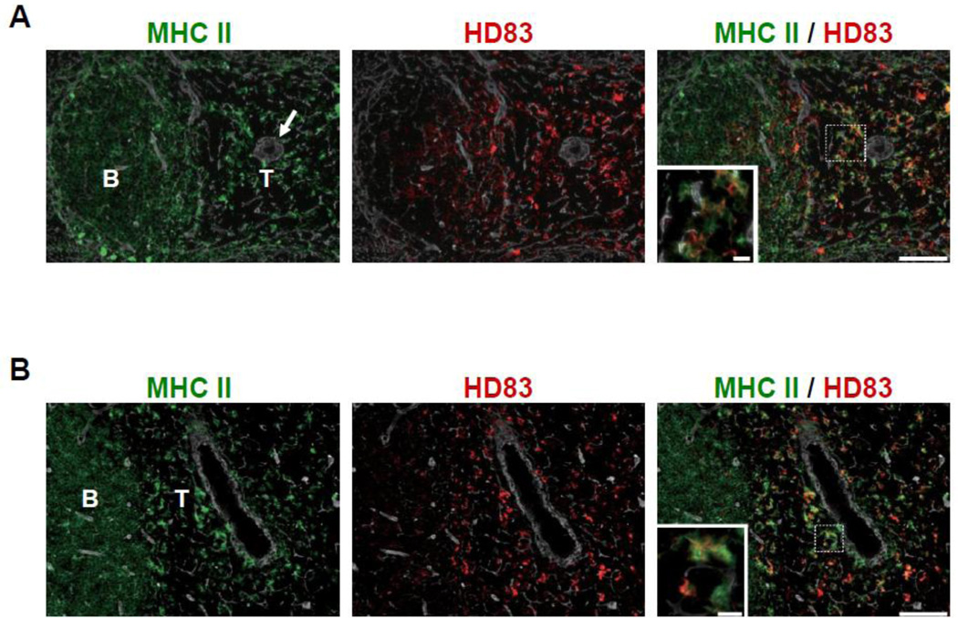
In addition to its anti-mDEC205 activity, HD83 was found to recognize DEC205 molecule from rat. Therefore, we attempted to visualize DEC205+ DCs in rat lymphoid tissues. When rat tissues were stained with anti-rat class II MHC in parallel, HD83 could label DEC205+ cells localized dominantly in the T-cell areas of rat spleen (Fig. 4A) and peripheral lymph node (Fig. 4B). As expected, these DEC205+ cells in rat lymphoid organs are in dendritic shape (high power magnification insets of Fig. 4A and 4B) indicating most of them are DCs.
Figure 4. HD83 stained DEC205+ DCs in the T-cell areas of rat lymphoid tissues.
(A) Rat spleen was stained triple-immunofluorescently for HD83 (red), class II MHC (green), and tissue framework (IV collagen, white). Inset shows dendritic shape of HD83+ class II MHC+ cells in high magnification. Arrow indicates central artery. B: B-cell area; T: T-cell area; Scale bar = 80 µm (10 µm in high magnification).
(B) Rat cervical lymph node was stained as in (A).
4. Discussion
In prior experiments to generate mAbs to human DEC205 in mice, the source of antigen was a small subdomain protein (30 kDa) at the extreme N-terminus of hDEC205, also known as cysteine rich (CR) domain and fibronectin type II (FN) domain (Guo et al., 2000). The hybridomas were also screened by ELISA with this CR-FN subdomain protein, resulting in only one anti-hDEC205 mAb, MG38.2 (Guo et al., 2000). To improve the yield of anti-hDEC205 mAb hybridoma production in mice, we expressed and purified a full-length extracellular domain protein (240 kDa) of hDEC205 from the stably transfected CHO cell culture (Cheong et al., 2010b). Then, we were able to generate 5 strong anti-hDEC205 mAbs in DEC205 wild type mice that express human immunoglobulin (Ig) genes, where this full-length extracellular domain protein of hDEC205 was used for immunization. All of those 5 anti-hDEC205 mAbs generated from the human Ig-expressing DEC205 wild type mice recognized only hDEC205 but not mDEC205 (Cheong et al., 2010b). In this report, we have employed DEC205 knockout (KO) mice in addition to the use of a full-length extracellular domain protein of hDEC205 as antigen. The hybridomas were screened by FACS with CHO cells expressing the full-length hDEC205 protein on the cell surface. Then, we were able to generate a large number of anti-hDEC205 HD mAbs, including mAbs cross-reactive to mDEC205.
It is known that the antibody responses against a protein that is highly conserved amongst different species are not easy to induce. In contrast to the conventional method of generating mAbs in wild type mice where one can only generate mAbs against the epitopes not conserved amongst species, our approach to make mAbs in KO mice with a full-length human homolog of the KO gene product as antigen has the potential to increase the yield of mAbs, including mAbs that are strongly reactive to conserved domains of an evolutionarily conserved protein. Therefore, this approach would produce a wide variety of mAbs against all epitopes available in the target protein whether or not the epitopes are conserved. Indeed, we generated a large number of anti-hDEC205 HD mAbs in this approach, a significant number of which were reactive to DEC205 from monkey, mouse, rat, or other mammals and to epitopes in a broad spectrum of extracellular subdomains of hDEC205. The wide cross-species reactivity of HD mAbs is not surprising because the sequences of DEC205 from various species (Guo et al., 2000; Gliddon et al., 2004; Nagasawa et al., 2010), including not only mammals but also birds and fishes, indicate the presence of numerous highly conserved stretches of identical amino acids in the extracellular domain.
A similar approach was taken to successfully generate strong mAbs to mouse DC-SIGN (mDC-SIGN) in a single trial, where a couple of strong anti-mDC-SIGN mAb hybridomas, i.e., MMD2 and MMD3 were obtained from the DC-SIGN KO mice immunized with a full-length extracellular domain protein of mDC-SIGN (Cheong et al., 2010a). Prior to employing DC-SIGN KO mice, a number of trials of immunizing wild type rats and hamsters with the same mDC-SIGN antigen had produced anti-mDC-SIGN mAbs with weaker binding activities than MMD2 and MMD3 (Cheong et al., 2010a). Unlike DEC205 gene, DC-SIGN has a sequence highly homologous to its family gene members, such as SIGN-R1 and SIGN-R3 in mice (Park et al., 2001). This might be the reason that only 2 strong anti-mDC-SIGN mAb hybridomas were generated from an immunized DC-SIGN KO mouse, compared to the large number (more than 115) of strong anti-hDEC205 HD mAb hybridomas obtained from an immunized DEC205 KO mouse in this study.
Different subsets of dendritic cells (DCs) with distinct function have been identified based on the molecular markers on surface (Steinman, 2012). DEC205+ DCs localized predominantly in T cell areas are critical in cross-priming CD8+ T cells and inducing Th1 immunity in mice, which have been demonstrated in high efficiency by targeting various antigens to DEC205+ DCs in mice (Trumpfheller et al., 2012). It will be important to expand this DEC205-targeted vaccine approach to clinical study using newly generated anti-hDEC205 mAbs. Especially, new anti-hDEC205 HD mAbs we generated can recognize DEC205 in different mammals, which should be useful for investigating the function of DEC205 and DEC205+ DCs in those mammals, as demonstrated here for rat DEC205+ DCs. In addition, we can now study the approach of targeting defined vaccine antigens to DEC205+ DCs in both humans and other animal models utilizing the same anti-hDEC205 HD mAbs.
Acknowledgments
We thank Frances Weis-Garcia and the Monoclonal Antibody Core Facility of the Rockefeller University and Memorial Sloan-Kettering Cancer Center for help with hybridoma production. We are grateful to Junko Sakumoto and members of Steinman Laboratory for their excellent support and valuable discussion. We were supported by NIH Grants to RMS (AI13013, AI40045, AI057158) and CGP (AI057158, AI093216).
Abbreviations
- CHO cells
Chinese hamster ovary cells
- CR domain
cysteine rich domain
- CRD
carbohydrate recognition domain
- DCs
dendritic cells
- FCS
fetal calf serum
- FN domain
fibronectin type II domain
- hDEC205
human DEC205
- Ig
immunoglobulin
- KO
knockout
- mAb
monoclonal antibody
- mDC-SIGN
mouse DC-SIGN
- mDEC205
mouse DEC205
- poly IC
polyriboinocinic polyribocytidylic acid
- SDS
sodium dodecyl sulfate
Footnotes
Publisher's Disclaimer: This is a PDF file of an unedited manuscript that has been accepted for publication. As a service to our customers we are providing this early version of the manuscript. The manuscript will undergo copyediting, typesetting, and review of the resulting proof before it is published in its final citable form. Please note that during the production process errors may be discovered which could affect the content, and all legal disclaimers that apply to the journal pertain.
References
- Bonifaz L, Bonnyay D, Mahnke K, Rivera M, Nussenzweig MC, Steinman RM. Efficient targeting of protein antigen to the dendritic cell receptor DEC-205 in the steady state leads to antigen presentation on major histocompatibility complex class I products and peripheral CD8+ T cell tolerance. J. Exp. Med. 2002;196:1627. doi: 10.1084/jem.20021598. [DOI] [PMC free article] [PubMed] [Google Scholar]
- Bonifaz LC, Bonnyay DP, Charalambous A, Darguste DI, Fujii S, Soares H, Brimnes MK, Moltedo B, Moran TM, Steinman RM. In vivo targeting of antigens to maturing dendritic cells via the DEC-205 receptor improves T cell vaccination. J. Exp. Med. 2004;199:815. doi: 10.1084/jem.20032220. [DOI] [PMC free article] [PubMed] [Google Scholar]
- Bozzacco L, Trumpfheller C, Huang Y, Longhi MP, Shimeliovich I, Schauer JD, Park CG, Steinman RM. HIV gag protein is efficiently cross-presented when targeted with an antibody towards the DEC-205 receptor in Flt3 ligand-mobilized murine DC. Eur. J Immunol. 2010;40:36. doi: 10.1002/eji.200939748. [DOI] [PMC free article] [PubMed] [Google Scholar]
- Cheong C, Matos I, Choi JH, Schauer JD, Dandamudi DB, Shrestha E, Makeyeva JA, Li X, Li P, Steinman RM, Park CG. New monoclonal anti-mouse DC-SIGN antibodies reactive with acetone-fixed cells. J. Immunol. Methods. 2010a;360:66. doi: 10.1016/j.jim.2010.06.006. [DOI] [PMC free article] [PubMed] [Google Scholar]
- Cheong C, Choi JH, Vitale L, He LZ, Trumpfheller C, Bozzacco L, Do Y, Nchinda G, Park SH, Dandamudi DB, Shrestha E, Pack M, Lee HW, Keler T, Steinman RM, Park CG. Improved cellular and humoral immune responses in vivo following targeting of HIV Gag to dendritic cells within human anti-human DEC205 monoclonal antibody. Blood. 2010b;116:3828. doi: 10.1182/blood-2010-06-288068. [DOI] [PMC free article] [PubMed] [Google Scholar]
- Gliddon DR, Hope JC, Brooke GP, Howard CJ. DEC-205 expression on migrating dendritic cells in afferent lymph. Immunology. 2004;111:262. doi: 10.1111/j.0019-2805.2004.01820.x. [DOI] [PMC free article] [PubMed] [Google Scholar]
- Guo M, Gong S, Maric S, Misulovin Z, Pack M, Mahnke K, Nussenzweig MC, Steinman RM. A monoclonal antibody to the DEC-205 endocytosis receptor on human dendritic cells. Hum. Immunol. 2000;61:729. doi: 10.1016/s0198-8859(00)00144-0. [DOI] [PubMed] [Google Scholar]
- Hawiger D, Inaba K, Dorsett Y, Guo M, Mahnke K, Rivera M, Ravetch JV, Steinman RM, Nussenzweig MC. Dendritic cells induce peripheral T cell unresponsiveness under steady state conditions in vivo. J. Exp. Med. 2001;194:769. doi: 10.1084/jem.194.6.769. [DOI] [PMC free article] [PubMed] [Google Scholar]
- Idoyaga J, Suda N, Suda K, Park CG, Steinman RM. Antibody to Langerin/CD207 localizes large numbers of CD8alpha+ dendritic cells to the marginal zone of mouse spleen. Proc. Natl Acad. Sci. U. S. A. 2009;106:1524. doi: 10.1073/pnas.0812247106. [DOI] [PMC free article] [PubMed] [Google Scholar]
- Jiang W, Swiggard WJ, Heufler C, Peng M, Mirza A, Steinman RM, Nussenzweig MC. The receptor DEC-205 expressed by dendritic cells and thymic epithelial cells is involved in antigen processing. Nature. 1995;375:151. doi: 10.1038/375151a0. [DOI] [PubMed] [Google Scholar]
- Kato M, McDonald KJ, Khan S, Ross IL, Vuckovic S, Chen K, Munster D, MacDonald KPA, Hart DNJ. Expression of human DEC-205 (CD205) multilectin receptor on leukocytes. Int. Immunol. 2006;18:857. doi: 10.1093/intimm/dxl022. [DOI] [PubMed] [Google Scholar]
- Kronin V, Wu L, Gong S, Nussenzweig MC, Shortman K. DEC-205 as a marker of dendritic cells with regulatory effects on CD8 T cell responses. Int. Immunol. 2000;12:731. doi: 10.1093/intimm/12.5.731. [DOI] [PubMed] [Google Scholar]
- Longhi MP, Trumpfheller C, Idoyaga J, Caskey M, Matos I, Kluger C, Salazar AM, Colonna M, Steinman RM. Dendritic cells require a systemic type I interferon response to induce CD4+ Th1 immunity with poly IC as adjuvant. J. Exp. Med. 2009;206:1589. doi: 10.1084/jem.20090247. [DOI] [PMC free article] [PubMed] [Google Scholar]
- Mahnke K, Guo M, Lee S, Sepulveda H, Swain SL, Nussenzweig M, Steinman RM. The dendritic cell receptor for endocytosis, DEC-205, can recycle and enhance antigen presentation via major histocompatibility complex class II-positive lysosomal compartments. J. Cell Biol. 2000;151:673. doi: 10.1083/jcb.151.3.673. [DOI] [PMC free article] [PubMed] [Google Scholar]
- Nagasawa K, Shikina S, Takeuchi Y, Yoshizaki G. Lymphocyte antigen 75 (Ly75/CD205) is a surface marker on mitotic germ cells in rainbow trout. Biol. Reprod. 2010;83:597. doi: 10.1095/biolreprod.109.082081. [DOI] [PubMed] [Google Scholar]
- Pack M, Trumpfheller C, Thomas D, Park CG, Granelli-Piperno A, Münz C, Steinman RM. DEC-205/CD205+ dendritic cells are abundant in the white pulp of the human spleen, including the border region between the red and white pulp. Immunology. 2007;123:438. doi: 10.1111/j.1365-2567.2007.02710.x. [DOI] [PMC free article] [PubMed] [Google Scholar]
- Park CG, Takahara K, Umemoto E, Yashima Y, Matsubara K, Matsuda Y, Clausen BE, Inaba K, Steinman RM. Five mouse homologues of the human dendritic cell C-type lectin, DC-SIGN. Int. Immunol. 2001;13:1283. doi: 10.1093/intimm/13.10.1283. [DOI] [PubMed] [Google Scholar]
- Steinman RM. Decisions About Dendritic Cells: Past, Present, and Future. Annu. Rev. Immunol. 2012;30:1. doi: 10.1146/annurev-immunol-100311-102839. [DOI] [PubMed] [Google Scholar]
- Trumpfheller C, Finke JS, López CB, Moran TM, Moltedo B, Soares H, Huang Y, Schlesinger SJ, Park CG, Nussenzweig MC, Granelli-Piperno A, Steinman RM. Intensified and protective CD4+ T cell immunity in mice with anti-dendritic cell HIV gag fusion antibody vaccine. J. Exp. Med. 2006;203:607. doi: 10.1084/jem.20052005. [DOI] [PMC free article] [PubMed] [Google Scholar]
- Trumpfheller C, Caskey M, Nchinda G, Longhi MP, Mizenina O, Huang Y, Schlesinger SJ, Colonna M, Steinman RM. The microbial mimic poly IC induces durable and protective CD4+ T cell immunity together with a dendritic cell targeted vaccine. Proc. Natl Acad. Sci. U. S. A. 2008;105:2574. doi: 10.1073/pnas.0711976105. [DOI] [PMC free article] [PubMed] [Google Scholar]
- Trumpfheller C, Longhi MP, Caskey M, Idoyaga J, Bozzacco L, Keler T, Schlesinger SJ, Steinman RM. Dendritic cell-targeted protein vaccines: a novel approach to induce T cell immunity. J. Intern. Med. 2012 doi: 10.1111/j.1365-2796.2011.02496.x. [DOI] [PMC free article] [PubMed] [Google Scholar]
- Zhou S, Ueta H, Xu XD, Shi C, Matsuno K. Predominant donor CD103+CD8+ T cell infiltration into the gut epithelium during acute GvHD: a role of gut lymph nodes. Int. Immunol. 2008;20:385. doi: 10.1093/intimm/dxm153. [DOI] [PubMed] [Google Scholar]


