SUMMARY
The laryngocele is an abnormal cystic dilatation of the saccule or appendix of the laryngeal ventricle, filled with air and communicating with the lumen of the larynx. When the neck of the laryngocele is obstructed, it becomes filled with mucus of the glandular secretion and is changed to a laryngomucocele. When this lesion becomes infected, a laryngopyocele is formed. The laryngocele is fairly rare and laryngopyocele occurs even more rarely. Overall, 39 cases of laryngopyocele have been reported in the world literature. Only in 4 cases was a laryngopyocele reported to have caused acute airway obstruction and only one case of internal laryngopyocele causing acute airway obstruction has been reported until now. This is the first case reported in the literature of an internal laryngopyocele in a female patient in a septic condition, which caused almost 100% obstruction of the airway. An emergency tracheotomy was performed in order to secure the airway. Computed tomography of neck was performed which revealed a cystic 29 mm hypodense mass extending from the right false vocal cord to the level of the epiglottis, narrowing the laryngeal cavity and causing an almost 100% airway obstruction. Laryngopyoceles may present with a rapid and alarming obstruction of the airway and, therefore, an urgent tracheotomy may be inevitable. It is an emergency case, in the field of otolaryngology, and should be included in the differential diagnosis of acute airway obstruction, especially when hoarseness, stridor and fever are present. Diagnosis requires a high index of suspicion for these lesions and scrupulous clinical and radiological evaluation. A computed tomography scan is critical in determining the nature and site of the lesion. The recommended treatment of laryngopyocele is immediate endoscopic drainage. Definitive management of laryngopyoceles is surgical excision which can be performed immediately after endoscopic drainage or some time thereafter.
KEY WORDS: Larynx, Laryngocele, Laryngopyocele, Airway obstruction
RIASSUNTO
Il laringocele è una dilatazione cistica del sacculo o appendice del ventricolo laringeo, piena di aria e comunicante con il lume laringeo. Quando il collo del laringocele si ostruisce, esso si riempie di muco e prende il nome di laringomucocele, la cui infezione porta alla formazione di un laringopiocele. Il laringocele è piuttosto raro e ancor di più lo è il laringopiocele. Finora sono stati descritti 39 casi di laringopiocele, e soltanto 4 di questi hanno determinato un'ostruzione acuta delle vie aeree. Solo in un caso l'ostruzione acuta delle vie aeree è stata causata da un laringopiocele interno. In considerazione della rarità dell'ostruzione acuta delle vie aeree da laringopiocele, si riporta per la prima volta in letteratura un caso di laringopiocele interno che ha determinato un'ostruzione pressoché totale delle vie aeree in una paziente in stato settico, tanto da rendere necessaria una tracheotomia d'urgenza. La tomografia computerizzata ha evidenziato una massa cistica ipodensa di 29 millimetri di diametro, estesa dalla corda vocale vera fino al livello dell'epiglottide. Il laringopiocele rappresenta un'emergenza nel campo dell'otorinolaringoiatria e dovrebbe essere incluso nella diagnosi differenziale di ostruzione acuta delle vie respiratorie, soprattutto quando sono presenti disfonia, stridore e iperpiressia. è necessaria pertanto un'attenta valutazione clinica, che non può prescindere dall'esecuzione di una tomografia computerizzata, essenziale per la definizione della natura e della sede della lesione. Il trattamento raccomandato per il laringopiocele è l'immediato drenaggio per via endoscopica, seguito da asportazione chirurgica per una risoluzione definitiva della patologia.
Introduction
Laryngocele is an abnormal cystic dilatation of the saccule or appendix of the laryngeal ventricle, filled with air and communicating with the lumen of the larynx 1. Virchow introduced the term laryngocele, in 1867, to describe an abnormal dilatation of the saccule forming an air sac 2 3. Based on location, three types of laryngocele have been described. The internal, the external and the combined or mixed laryngocele 4. When the neck of the laryngocele is obstructed, it becomes filled with mucus of glandular secretion and is altered to a laryngomucocele. When this lesion becomes infected, a laryngopyocele is formed.
A total of 39 cases of laryngopyocele have been reported in the world literature 1 3-9. Only one case of internal laryngopyoceles, causing acute airway obstruction, has been reported until now 9. In view of the rarity of the laryngopyoceles and the even rarer laryngopyocele that causes acute airway obstruction, it was decided that the report of another case would be worthwhile. Herewith, a rare case of an internal laryngopyocele is described in a 61-year-old female, in a septic condition which caused almost 100% airway obstruction.
Case report
A 61-year-old female presented to the emergency room, in respiratory distress, with a three-day history of sore throat, cough and odynophagia. She was a heavy smoker and had a past medical history of hypothyroidism and hypertension. Physical examination revealed marked respiratory distress with stridor, tachypnoea and hoarseness. Her temperature, on admission, was 38.3°C. Her white cell count was 23400 U /mm3 with 81.3% neutrophil leukocytes and C-Reactive Protein (CRP) was 17.8 mg/dl. Upon admission, the arterial blood gases on 21% O2 were PO2 82 mm Hg, PCO2 52 mm Hg and pH 7.32. On palpation of the neck, no mass was noted. Indirect flexible laryngoscopy demonstrated a large mass which originated in the right false vocal cord and caused an almost total obstruction of the airway. The mucosal surface of the mass was smooth.
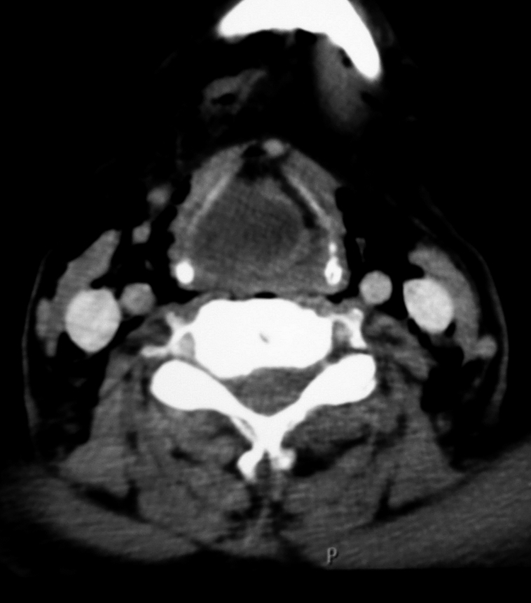
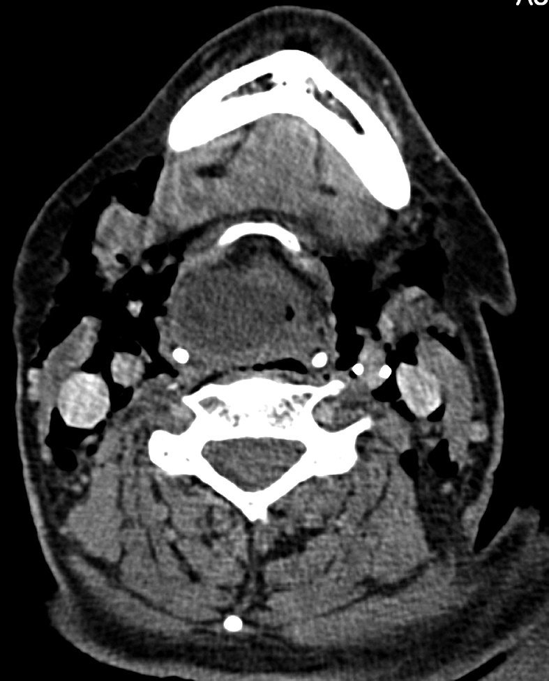
Waiting for the urgent computed tomography (CT) scan of the neck, she had a dramatic deterioration of dyspnoea and presented cyanosis. Since the large laryngeal mass prohibited intubation, an emergency tracheotomy was performed to secure a free airway. CT of the neck revealed a 29 mm low attenuation mass above the level of the true vocal folds which caused almost total obstruction of the airway (Figs. 1-2). Thickening of the walls was demonstrated and the lesion was confined within the larynx (Fig. 1). A diagnosis of internal laryngopyocele was made and confirmed with physical examination, indirect laryngoscopy and CT scan.
Fig. 1.

CT scan axial view demonstrating a supraglottic cystic hypodense mass with thickening of the wall. The mass causes an almost complete airway obstruction. It is confined to the larynx and a diagnosis of internal laryngopyocele was made.
Fig. 2.

CT scan image showing a large internal right-sided laryngopyocele at the level of hyoid bone. The mass causes an almost complete airway obstruction.
Direct laryngoscopy confirmed that the large laryngopyocele had caused an almost 100% obstruction and it originated in the right ventricle. No visible neoplasm was noted. The laryngopyocele was drained and a large amount of purulent material was removed and cultures were collected. The definitive surgery was scheduled at a later time. The patient was treated with cortisone and intravenous antibiotics, including ampicillin, clindamycin and ceftriaxone. Culture revealed the presence of Pseudomonas aeruginosa and, therefore, ceftriaxone was substituted with gentamycin. The fifth post-operative day despite medical advice and the fact that she still had the cannula, the patient signed out in order to be transported to her homeland.
Discussion
A laryngocele is an air-filled herniation of the saccule of the laryngeal ventricle which is in communication with the lumen of the larynx. The laryngeal ventricle is a fusiform fossa delimited by the true and the false vocal cord and extending from the thyroid notch to the arytenoid cartilage. The anterior part of the roof of the ventricle leads up into a blind pouch of the mucous membrane called the saccule or appendix. Embryologically, the saccule and ventricle of the larynx develop as a secondary outpouching from the laryngeal lumen towards the end of the second intrauterine month 3.
Several hundred laryngoceles have been reported in the English literature. A total of 39 cases of laryngopyocele have been reported in the world literature 1 3 4 6-10. Only in 4 of these cases had a laryngopyocele caused acute airway obstruction and only one case of internal laryngopyocele causing acute airway obstruction has been reported until now 9. The case described here is the first case reported in the English literature of a female patient in a septic condition, with an internal laryngopyocele causing acute airway obstruction (Table I).
Table I.
Laryngopyoceles as a cause of airway obstruction.
| Patient # | Age | Sex | Necessity of emergency tracheotomy | Laryngoscopic findings | Radiographic findings | Diagnosis |
|---|---|---|---|---|---|---|
| 1 | 59 | Male | Yes | Swelling of the left aryepiglottic fold and left false vocal cord | X-ray: large cavity with an air fluid level in the left neck Thawley et al. 10 |
Combined laryngopyocele |
| 2 | 57 | Female | No | A mass filling the right aryepiglottic fold and puriform fossa | X-ray: right sided neck mass displacing trachea to the left Weissler et al. 8 |
Combined laryngopyocele |
| 3 | 51 | Male | Yes | Diffuse swelling over the right false cord and aryepiglottic fold | X-ray: right sided neck mass and an air-fluid level Weissler et al. 8 |
Combined laryngopyocele |
| 4 | 34 | Male | No | A mass originating in the left false cord caused a near total airway obstruction | CT: 18 mm low-attenuation mass within the larynx that caused a significant airway obstruction Fredrickson et al. 9 |
Internal laryngopyocele |
| 5 | 61 | Female | Yes | A mass originating in the right false vocal cord, caused a near total airway obstruction | CT: 29 mm within larynx that caused an almost total airway obstruction In our case |
Internal laryngopyocele |
In the case presented here, the patient was in a septic condition, at the time of admission, based on her clinical picture and the laboratory results. Indirect flexible laryngoscopy demonstrated a large smooth mass which originated in the right false vocal cord and caused an almost total obstruction of the airway. The differential diagnosis of acute upper airway obstruction includes several entities, except laryngopyocele (Table II). Diagnosis of an internal laryngopyocele was made by correlating the history, the clinical picture and the laryngoscopic and the radiographic findings (Table II).
Table II.
Causes of acute upper airway obstruction in adults.
| Congential | Micrognathia Macroglossia |
| Infections | Retopharyngeal abcess Tuberculuous laryngitis Laryngopyocele Epiglottis |
| Traumatic | Intubation trauma Hematoma Facial fracture Laryngeal fracture Subglottic stenosis |
| Allergic/autoimmune | Rhinitis Sarcoidosis Wegener's disease Angioedema Asthma |
| Neoplastic | Nasopharyngeal carcinoma Epiglottis carcinoma Recurrent respiratory papillomatosis Haemangioma |
| Neurologic | Altered mental status Vocal fold paralysis Paralysis of respiratory muscles |
Laryngoceles are rare entities, occurring in only one per 2.5 million population per year in the UK 5. The sex incidence is 5:1 in favour of male sex and the maximum age of incidence is in the sixth decade 5 7. Based on location, three types of laryngoceles have been described, the external, the internal and the combined type. The external laryngocele presents clinically as a swelling in the neck, at the level of hyoid bone, anterior to the sternocleidomastoid muscle. During Valsava's manoeuvre, the swelling is increased and it becomes smaller on palpation. The internal and combined type, appear on laryngoscopy as a smooth swelling mass of the supraglottis.
The precise aetiology of laryngocele is unknown. Many authors have hypothesized that congenital or acquired factors may be responsible 2 5 10 13 14. It is believed that laryngoceles occur in subjects with congenitally dilated saccules. A less important role is played by the narrowness of the periventricular connective tissue and the weakening of the thyroaryepiglottic muscles 10 11. A congenital weakness or defect predisposes to the formation of laryngoceles under the influence of acquired factors 16 17. Factors that increase intra-glottic pressure such as professional trumpet playing, glass blowing, singing, straining at passing of stools, weight lifting and carcinoma of the larynx are considered to promote the development of laryngoceles 5 13 15 16 18.
The glandular serum-mucus secretion is evacuated through the ventricular opening. Some situations, such as chronic inflammations, laryngeal trauma or laryngeal carcinoma, lead to incomplete mechanical stenosis of the neck of the appendix 4. When the neck of the laryngocele is obstructed, the laryngocele becomes filled with mucus. If the mucus-filled laryngocele is infected, it is called a laryngopyocele 2 7 11. A review of the English literature showed that the most common type is the mixed laryngocele (44%), 30% were internal and 26% were external. Bilateral laryngoceles were found in 23% 12. Laryngopyocele occurs even more rarely. Approximately 8% of laryngoceles become infected and present as laryngopyoceles and most of these are of the combined variety 5. The most common bacteria isolated from laryngopyocele are Escherichia coli, haemolytic Streptococcus B, Staphylococcus Aureus and Pseudomonas Aeruginosa 4.
Laryngoceles are usually asymptomatic. The most frequent presenting symptom is hoarseness. Variable degrees of dyspnoea, dysphagia, cough and stridor may be present, depending on the dimension of the laryngocele. Laryngopyoceles may present with signs of rapidly progressive respiratory obstruction and/or an infected painful neck mass which may rapidly increase in size. The symptoms of laryngopyocele include hoarseness, dyspnoea, stridor, dysphagia, odynophagia, pain, sensation of a foreign body and fever 7 8. In the internal and combined forms, flexible nasolaryngoscopy can reveal a smooth mass of the vestibular fold, aryepiglottic fold and pyriform sinus which may displace the larynx to one side.
In the case presented here, after the emergency tracheotomy, computed tomography of the neck was performed which revealed a cystic 29 mm hypodense mass extending from the right false vocal cord to the level of the epiglottis, narrowing the laryngeal cavity and causing an almost 100% airway obstruction (Figs. 1-2). The lesion was confined within the larynx and it was diagnosed as an internal laryngopyocele. The cystic cavity was full of fluid and no air-fluid level was observed (Fig. 2).
Radiological evaluation includes neck ultrasound which may determine swelling dimensions and content and a CT scan that permits diagnosis. CT scan shows the characteristic intra-laryngeal and extra-laryngeal expansion and defines laryngopyocele content, the relationship with the laryngeal ventricle and thyroid membrane and the presence of a carcinoma 8 9 12. A contrast-enhanced CT scan can demonstrate signs of inflammation such as thickening of the walls or perimeter enhancement of the laryngocele and assist the differential diagnosis 12. In the differential diagnosis of laryngopyocele, it is necessary to take into consideration the saccular cyst, fluid filled laryngocele, branchial cysts, paraganglioma, schwannoma, and thyroglossal duct cysts which exist in the supraglottic area 7 9 19.
Laryngopyocele complications consist particularly in inhalation of the purulent material after cyst rupture, leading to acute respiratory damage. Therefore, the recommended treatment of laryngopyocele is immediate endoscopic drainage 9 21. The infection is treated with broad spectrum intravenous antibiotics and steroids. Additional surgery can be performed immediately after endoscopic drainage or at a later date. For the treatment of internal laryngopyocele, an endoscopic decompression with marsupialization is recommended. For external and combined laryngopyoceles, additional definitive surgery should be performed, via an external approach 9 21 22. This approach could be performed through a horizontal lateral cervicotomy, at the level of the thyroid membrane 4.
Conclusions
Laryngopyoceles are a rare complication of laryngoceles. They can present with rapid and alarming obstruction of the airway. Diagnosis requires a high index of suspicion, for these lesions, and careful clinical and radiological evaluation. Laryngopyoceles must be included in the differential diagnosis of acute airway obstruction, especially when hoarseness, inspiratory stridor and fever are present. A CT scan is essential in determining the nature and the site of the lesion. Aggressive antibiotic treatment and aspiration of the purulent content can avoid the need of emergency tracheotomy. The definitive management of laryngopyoceles is surgical excision.
References
- 1.Marcotullio D, Paduano F, Magliulo G. Laryngopyocele: an atypical case. Am J Otolaryngol. 1996;17:345–348. doi: 10.1016/s0196-0709(96)90023-x. [DOI] [PubMed] [Google Scholar]
- 2.DeSanto LW. Laryngocele, laryngeal mucocele and large saccular cysts: a developmental spectrum. Laryngoscope. 1974;84:1291–1296. doi: 10.1288/00005537-197408000-00003. [DOI] [PubMed] [Google Scholar]
- 3.Maharaj D, Fernandes CM, Pinto AP. Laryngopyocele (a report of two cases) J Laryngol Otol. 1987;101:838–842. doi: 10.1017/s002221510010283x. [DOI] [PubMed] [Google Scholar]
- 4.Cassano L, Lombardo P, Marchese-Ragona R, et al. Laryngopyocele: three new clinical cases and review of the literature. Eur Arch Otorhinolaryngol. 2000;257:507–511. doi: 10.1007/s004050000274. [DOI] [PubMed] [Google Scholar]
- 5.Stell PM, Maran AG. Laryngocele. J Laryngol Otol. 1975;89:915–924. doi: 10.1017/s0022215100081196. [DOI] [PubMed] [Google Scholar]
- 6.Ludwig A, Chilla R. Laryngopyocele. Rare case of relapsing cervical infections. HNO. 2010;58:313–316. doi: 10.1007/s00106-009-2057-2. [DOI] [PubMed] [Google Scholar]
- 7.Illum P, Nehen AM. Laryngopyocele with a report of two cases. J Laryngol Otol. 1980;94:211–218. doi: 10.1017/s0022215100088691. [DOI] [PubMed] [Google Scholar]
- 8.Weissler MC, Fried MP, Kelly JH. Laryngopyocele as a cause of airway obstruction. Laryngoscope. 1985;95:1348–1351. doi: 10.1288/00005537-198511000-00011. [DOI] [PubMed] [Google Scholar]
- 9.Fredrickson KL, D'Angelo AJ., Jr Internal laryngopyocele presenting as acute airway obstruction. Ear Nose Throat J. 2007;86:104–106. [PubMed] [Google Scholar]
- 10.Thawley SE, Bone RC. Laryngopyocele. Laryngoscope. 1973;83:362–368. doi: 10.1288/00005537-197303000-00006. [DOI] [PubMed] [Google Scholar]
- 11.Harrison DFN. The anatomy and physiology of the mammalian larynx. Cambridge: Cambridge University Press; 1995. pp. 90–92. [Google Scholar]
- 12.Hubbard C. Laryngocele – A study of five cases with reference to the radiologic features. Clin Radiol. 1987;38:639–643. doi: 10.1016/s0009-9260(87)80349-5. [DOI] [PubMed] [Google Scholar]
- 13.Macfie VE. Asympromatic laryngoceles in wind instrument bandsmen. Arch Otolaryngol. 1966;83:270–275. doi: 10.1001/archotol.1966.00760020272018. [DOI] [PubMed] [Google Scholar]
- 14.Upile T, Jerjes W, Sinapaul F. Laryngocele: a rare complication of surgical tracheostomy. BMC Surg. 2006;27:6–14. doi: 10.1186/1471-2482-6-14. [DOI] [PMC free article] [PubMed] [Google Scholar]
- 15.Prasad KC, Vijayalakshmi S, Prasad SC. Laryngoceles – presentations and management. Indian J Otolaryngol Head and Neck Surg. 2008;60:303–308. doi: 10.1007/s12070-008-0108-8. [DOI] [PMC free article] [PubMed] [Google Scholar]
- 16.Amin M, Maran AG. The aetiology of laryngocoele. Clin Otolaryngol Allied Sci. 1988;13:267–272. doi: 10.1111/j.1365-2273.1988.tb01130.x. [DOI] [PubMed] [Google Scholar]
- 17.Wright LD, Maguda TA. Laryngocele: case report and review of the literature. Laryngoscope. 1964;74:396–412. doi: 10.1288/00005537-196403000-00006. [DOI] [PubMed] [Google Scholar]
- 18.Hollinger LD, Barnes DR, Smid LJ. Laryngocele and saccular cysts. Ann Otol Rhinol Laryngol. 1978;87:675–685. doi: 10.1177/000348947808700513. [DOI] [PubMed] [Google Scholar]
- 19.Nazaroglu H, Ozates M, Uyar A, et al. Laryngopyocele: signs on computed tomography. Eur J Radiol. 2000;33:63–65. doi: 10.1016/s0720-048x(99)00079-0. [DOI] [PubMed] [Google Scholar]
- 20.Thome R, Thome DC, De la Cortina RAC. Lateral thyrotomy approach on the paraglottic space for laryngocele resection. Laryngoscope. 2000;110:447–450. doi: 10.1097/00005537-200003000-00023. [DOI] [PubMed] [Google Scholar]
- 21.Szware BJ, Kashima HK. Endoscopic management of combined laryngocele. Ann Otol Laryngol. 1997;106:556–559. doi: 10.1177/000348949710600704. [DOI] [PubMed] [Google Scholar]
- 22.Myssiorek D, Madnani D, Delacure MD. The external approach for submucosal lesions of the larynx. Otolaryngol Head Neck Surg. 2001;125:370–373. doi: 10.1067/mhn.2001.118690. [DOI] [PubMed] [Google Scholar]


