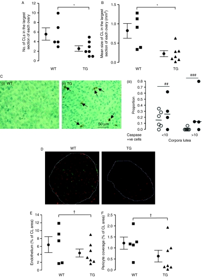
Figure 4.
Over-expression of VEGF165b in the mouse ovary results in changes in the corpus luteum (CL). Ovaries collected from mice at 0.5 dpc were fixed and stained by H&E to investigate CL structure. (A) The number of CL in the largest cross section of each ovary was counted. *P<0.05, t-test. (B) The total size of the CL in the largest cross section of each ovary was measured with ImageJ. *P<0.05, t-test. (C) Ovaries stained for cleaved caspase-3 were analysed for corpus luteum staining in WT (i) and TG (ii). Numbers of CL were counted (iii) according to the proportion with 1–10 (early regression) and >10 cleaved caspase-3-positive cells (late regression; ##P<0.01, ###P<0.001, Fisher's exact test on CL frequency). n=6 mice per group, 90 CL in WT, 45 in TG). (D) Ovaries collected from mice at 0.5 dpc were fixed and stained for microvessels using isolectin B4 (red) and pericytes with anti-NG2 antibody (green). (E) Blood vessel density was counted and expressed as microvascular density (microvessel covering area per mm2 of CL). (F) Pericytes covering area per unit area of CL were counted.,†P<0.05, t-test with Welch's correction.

