Abstract
Purpose
To describe the clinical outcomes and complications in a consecutive series of extreme lateral interbody fusion cases.
Methods
Retrospective cohort review of 97 consecutive patients from three centers with minimum 6-month follow-up (mean 12 months). Functional status was evaluated by preoperative and last follow-up Oswestry Disability Index score. Leg and back pain were evaluated by visual analog scales. Complications were recorded and permanent complications and neurological impairment was actively investigated at last follow-up.
Results
No permanent neurological impairment, vascular or visceral injuries were observed. Transient neurological symptoms presented in 7% of cases, all resolved within 1 month from surgery. Transient thigh discomfort was observed in 9%. Clinical success was recorded in 92% of cases.
Conclusions
Extreme lateral interbody fusion is a safe and effective technique for anterior interbody fusion.
Keywords: XLIF, Extreme lateral interbody fusion, Anterior lumbar interbody fusion, Spinal fusion, Outcomes, Complications
Introduction
Spinal fusion for treatment of patients with severe symptoms from disc disease, instability, deformity or stenosis has demonstrated to be successful and cost-effective [1–4]. Anterior fusion offers advantages in terms of diminishing damage to lumbar muscles, wider disc exposure, greater surface for fusion and optimal endplate preparation. Its disadvantages include potentially more severe complications (vascular, visceral, sexual) and difficulties from perivascular scarring for future revision approaches [5]. Endoscopic and minimally invasive anterior approaches have reduced morbidity with similar long-term results compared to open anterior surgery, but lack of reproducibility and are still burdened by high incidence of vascular and sexual complications [6]. Extreme lateral interbody fusion (XLIF) is an alternative technique to perform minimally invasive fusion at levels above L5. The availability of specific access instruments, directional EMG neuromonitoring devices and specifically designed implants makes the technique reproducible, biomechanically sound and clinically effective [7].
This study aims to analyze the early clinical experience with XLIF, regarding safety and clinical results in three European centers.
Methods
Design
Retrospective cohort study.
Patients
From March 2009 to April 2011, 97 patients were operated at three institutions by three senior surgeons (2 Orthopedists and 1 Neurosurgeon), all with more than 15 years of experience in spinal surgery. The cases included the learning curve on minimally invasive transpsoas lateral interbody fusion of all three surgeons.
Ninety-three patients were available at follow-up (95.9%). Two patients died of causes not related to spinal surgery (breast cancer and renal failure), two patients who lived in other countries could not be traced. One of these had a last 3-month follow-up documented, with a poor result. Mean follow-up was 12.1 months (6–28). Mean age was 59 years (27–85), 26% were male and 74% female (Table 1).
Table 1.
Description of the series
| Patients | ||
|---|---|---|
| Eligible | 97 | |
| Lost to follow-up | 4 | 2 dead (not related) |
| 2 living abroad (one with documented poor result at last 3-month follow-up | ||
| Follow-up | 12.1 months (mean); 6–28 | |
| Age | 59 years (mean); 27–85 | |
| Sex | 26% males; 74% females | |
| Number of levels treated | 1 level | 48 patients |
| 2 levels | 40 | |
| 3 levels | 8 | |
| 4 levels | 1 | |
| Highest level | T11–T12 | |
| Lowest level | L4–L5 | |
| Diagnoses | Degenerative disc disease | 78 patients |
| Degenerative scoliosis | 8 | |
| Postraumatic kyphosis (corpectomy) | 3 | |
| Pseudarthrosis following PSO | 2 | |
| Anterior column reconstruction following SPO | 2 | |
| Type of fixation | Bilateral pedicle screws (open or percutaneous) | 57 |
| Unilateral pedicle screws (most percutaneous) | 10 | |
| Translaminar facet screws | 2 | |
| Interspinous plate | 10 | |
| Interspinous elastic device | 1 | |
| Lateral plate | 3 | |
| Stand alone cages | 14 |
Number of levels instrumented via lateral access were 1 in 48 cases, 2 in 40 cases, 3 in 8 cases, and 4 in 1 case. The highest instrumented level was T11–T12 and the lowest one L4–L5.
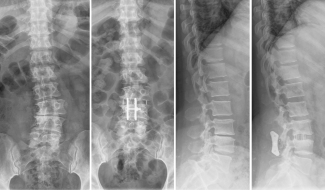
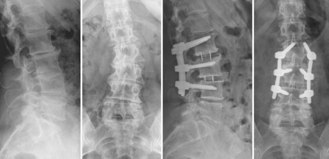
Diagnosis was mainly degenerative disc disease with or without stenosis or spondylolisthesis grade I. In two patients, anterior interbody fusion was performed after posterior correction of lumbar hypolordosis with Smith-Petersen osteotomies. Interbody fusion was implemented to increase stiffness and to reduce the risk of pseudartrhosis and implant failure. Other two patients with pseudarthrosis and loss of sagittal balance after pedicle substraction osteotomy (PSO) had XLIF procedure at disks below and over PSO, then posterior revision (in one case, with PSO at a new level). Eight patients had a diagnosis of lumbar degenerative scoliosis (Figs. 1, 2). Three patients had a lateral access corpectomy and reconstruction with an expandable cage for acute fracture or post-traumatic kyphosis.
Fig. 1.
De novo degenerative scoliosis. One level XLIF with coronal asymmetrical cage and posterior interlaminal plate fixation. Good disc height restoration and correction of segmental scoliosis at 6 months
Fig. 2.
De Novo degenerative scoliosis. Selective fusion of the apex with two-level XLIF and percutaneous pedicle screws (derotation of the apex through posterior instrumentation). Fair correction of the deformity, adjacent disc mobility is preserved. Good clinical result at 6 months
Outcomes
Clinical outcome was evaluated by intensity of low back and leg pain on VAS, and function measured with Oswestry Disability Index, both preoperative and at last follow-up. Postoperative complications were prospectively recorded, as was the presentation of thigh pain, thigh numbness, new neurological deficit, and surgical-related complications. Clinical success was considered to be achieved when the patient increased his functional ODI score by more than 12% or decreased his back pain VAS by more than three points.
Statistical analysis
Means were compared using the two-sided Student’s t test for paired or unpaired samples, as necessary. Statistical significance was set to p < 0.05.
Lateral access surgical technique
The XLIF procedure [8] was performed under general anesthesia with the patient in the lateral decubitus position on a radiolucent operating table with a break placed at the index level. Right or left decubitus was chosen based on preoperative evaluation of spinal, nerve and vascular anatomy. For procedures performed at L2-L3 or below, triggered EMG was used during surgery for safe identification of the position of roots of the lumbar plexus with an automatic surgeon driven system (Neurovision TM, Nuvasive, San Diego, CA, USA). Surface electrodes were placed at major dermatomes of the lower limbs to allow for recording of EMG signals during surgery. Avoiding the use of paralytic anesthetics allowed for reliable EMG monitoring of the lumbar plexus during both the approach through the psoas muscle and the procedure. Fluoroscopy was used to localize the diseased disc, and access was gained 90° off-midline through an approximately 3 cm incision. Blunt dissection was performed to access the psoas muscle with sequential dilators used to traverse the psoas muscle and access the lateral aspect of the disc space. Each dilator is integrated with a localized EMG stimulating field on the distal end, which was rotated during stimulation to provide 360° information on the position of motor nerves in the vicinity of the dilator. In addition, discrete EMG threshold responses are given for each response elicited, which provide information on the relative distance of motor nerves to the instrumentation, where a lower response threshold indicates closer nerve proximity compared to a higher response threshold [9, 10]. Once a corridor was made through the psoas muscle, the lateral disc space was accessed, and the retractor was placed over the final dilator, discectomy and disc space preparation were performed using standard techniques under direct visualization. Once disc preparation was complete, an intervertebral cage which spans the ring apophysis with a wide aperture (CoRoent XL TM, Nuvasive, San Diego, CA, USA), prefilled with graft material, was placed, resting on the strongest bone (that of the lateral aspects of vertebral endplates). Closure was performed with stitches in the external oblique fascial layer, subcutaneous layer, and skin with or without a drain. Video material illustrating the technique described has been previously published in the Open Operating Theatre series of the European Spine Journal [11].
Type of fixation
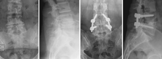
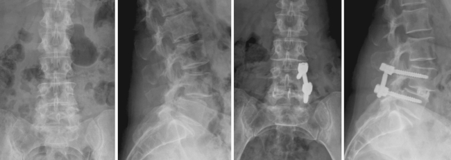
The XLIF procedure included stand alone interbody cage (14 patients), with 40–55 mm long cages, spanning the ring apophysis from lateral to lateral end of the disc space, and additional fixation was achieved with spinous process plate (n = 10) (Fig. 1), interspinous elastic device (n = 1), translaminar screws (n = 2), lateral plate (n = 3) bilateral pedicle screws (n = 57) (Fig. 3) or monolateral pedicle screws (n = 10) (Fig. 4). Cages were filled with autologous or homologous bone, or with osteoconductive materials (mainly calcium triphosphate granules), depending of surgeons’ choice.
Fig. 3.
One level degenerative disc disease without neurological symptoms. XLIF plus bilateral percutaneous pedicle-screw fixation. One-year postoperative films show interbody fusion with preservation of lordosis with excellent clinical result
Fig. 4.
L4-L5 degenerative spondylolisthesis. XLIF plus percutaneous unilateral pedicle-screw fixation. At 12 months, interbody fusion is evident with preservation of lordosis. Excellent clinical result
Results
Clinical outcomes
Mean intensity of back pain on Visual Analog Scale (VAS) was preoperatively 7.25 (4–10). Mean intensity of leg pain on VAS was preoperatively 5.8 (0–10). Mean Oswestry Disability Index (ODI) score was 51% (16–82%) (Table 2).
Table 2.
Clinical outcomes
| Outcome measures | Preoperative mean (min–max) | Postoperative mean (min–max) |
|---|---|---|
| Back pain (VAS) | 7.25 (4–10) | 2.8 (0–9)* |
| Leg pain (VAS) | 5.8 (0–10) | 2.1 (0–10)* |
| Oswestry Disability Index | 51 (16–82) | 23 (0–68)** |
* p < 0.01
** p > 0.001
At last follow-up, mean intensity of back pain on VAS was 2.8 (0–9). Mean intensity of leg pain on VAS was 2.1 (0–10). Mean ODI score was 23% (0–68%).
Mean improvement in back pain on VAS was 4.6 (−2 to 10, p < 0.01). Mean improvement in leg pain on VAS was 3.7 (−1 to 10, p < 0.01). Mean improvement in ODI score was 27.7% (−4 to 76%, p < 0.001).
Only eight patients (8.2%) had poor results, with neither pain VAS decrease greater than three points or functional ODI improvement lower than 12%.
Complications
Four patients (4.1%) had transient L4 weakness (resolved within 1 month), three patients (3.1%) had transient L4 hypoesthesia (resolved within 3 months). No patient had permanent neurological impairment. Transient crural discomfort lasting 1–4 weeks was observed in nine patients (9.2%) (Table 3).
Table 3.
Complications
| n | % | Comments | |
|---|---|---|---|
| Failed to improve | 8 | 8.2 | |
| Transient L4 weakness | 4 | 4.1 | All resolved in 1 month |
| Transient L4 hypoesthesia | 3 | 3.1 | All resolved in 3 months |
| Permanent neurological impairment | 0 | 0 | |
| Transient thigh symptoms | 9 | 9.2 | All resolved in 1–4 weeks |
| Subsidence of cage | 2 | 2.1 | One revised to larger implant |
| Deep iliac venous thrombosis | 1 | 1 | |
| Infection (posterior open wound) | 1 | 1 | Healed after debridement and i.v. antibiotics with implant preservation |
| Psoas hematoma | 1 | 1 | Resolved spontaneously |
Significant subsidence in stand-alone constructs was noted in two patients (2%), one of which had to be revised with a larger implant.
Other complications included deep venous thrombosis of the common iliac vein (n = 1), infection (n = 1) in a four level posterior open instrumentation and fusion, dural tear during posterior open decompression, resolved by primary repair (n = 1) psoas hematoma that resolved conservatively (n = 1) (1% each).
No patient needed conversion of the XLIF procedure to anterior open surgery. No ureter, bowel or vascular lesions were observed during retroperitoneal approach. No infection was observed in the lateral wound. One patient with pseudarthrosis and loss of sagittal balance after PSO who underwent single stage XLIF at two levels and posterior revision with new PSO had severe blood loss in the posterior procedure (>2,700 ml) with hemodinamic instability, needing multiple blood and plasma transfusions and intensive care. She presented cognitive loss with slow recovery in the following months, and subsequent death from dissemination of breast cancer.
Discussion
In this series, despite the wide range of diagnoses, indications and methods of additional fixation, clinical success rate at more than 6 months has been superior to 90%. The technique has shown to be safe (no vascular or visceral lesions have been observed, no wound infections in the anterior approach, no new permanent neurological deficit).
One criticism against the transpsoas lateral access is the incidence of thigh discomfort and numbness, reported to be as high as 74% [12]. In this multicenter study, including the learning curve of three surgeons, the observed incidence of transient neural complications or thigh discomfort/numbness has been 16%, with all cases resolved within 1 month. Previous experience in anterior surgery and posterior minimally invasive surgery, specific cadaver training on the technique have been felt by the authors to have an influence on safety and reproducibility of the approach. Accurate surgical technique with strict adherence to neuromonitoring protocol and delicate psoas dissection has been used in this series of patients.
One limitation of this study is the retrospective method, which could underestimate the incidence of less serious adverse events. Against this shortcoming is the fact that loss of follow up (two patients lost to final follow-up and two patients not evaluated due to death unrelated to the procedure) accounted for <5% of patients. Significant permanent complications (especially neurological or visceral complications) have been actively seeked for at last follow-up visit and are unlikely to be undereported.
Minimally invasive lateral transpsoas approach has been useful to address different pathologies from one level degenerative disc disease to elderly scoliosis or revision in complex sagittal imbalance cases. The authors feel that some of the more complex cases in this series (PSO, sagittal imbalance correction) could undergo a single stage combined approach that would have been difficult to justify with more aggressive anterior approach modalities.
Extreme lateral interbody fusion has been in this large series an effective and safe minimally invasive surgical method to treat miscellaneous lumbar and thoracolumbar spinal pathologies requiring spinal fusion.
Conflict of interest
None.
References
- 1.Wang MY, Cummock MD, Yu Y, Trivedi RA. An analysis of the differences in the acute hospitalization charges following minimally invasive versus open posterior lumbar interbody fusion. J Neurosurg Spine. 2010;12(6):694–699. doi: 10.3171/2009.12.SPINE09621. [DOI] [PubMed] [Google Scholar]
- 2.Glassman SD, Carreon LY, Djurasovic M, Dimar JR, Johnson JR, Puno RM, Campbell MJ. Lumbar fusion outcomes stratified by specific diagnostic indication. Spine J. 2009;9:13–21. doi: 10.1016/j.spinee.2008.08.011. [DOI] [PubMed] [Google Scholar]
- 3.Brantigan JW, Neidre A, Toohey JS. The Lumbar I/F Cage for posterior lumbar interbody fusion with the variable screw placement system: 10-year results of a Food and Drug Administration clinical trial. Spine J. 2004;4:681–688. doi: 10.1016/j.spinee.2004.05.253. [DOI] [PubMed] [Google Scholar]
- 4.Penta M, Fraser RD. Anterior lumbar interbody fusion. A minimum 10-year follow-up. Spine. 1997;22:2429–2434. doi: 10.1097/00007632-199710150-00021. [DOI] [PubMed] [Google Scholar]
- 5.Youssef JA, McAfee PC, Patty CA, Raley E, DeBauche S, Shucosky E, Chotikul L. Minimally invasive surgery: lateral approach interbody fusion: results and review. Spine. 2010;35:S302–S311. doi: 10.1097/BRS.0b013e3182023438. [DOI] [PubMed] [Google Scholar]
- 6.Brau SA. Mini-open approach to the spine for anterior lumbar interbody fusion: description of the procedure, results and complications. Spine J. 2002;2:216–223. doi: 10.1016/S1529-9430(02)00184-5. [DOI] [PubMed] [Google Scholar]
- 7.Rodgers WB, Gerber EJ, Patterson J. Intraoperative and early postoperative complications in extreme lateral interbody fusion: an analysis of 600 cases. Spine. 2011;36:26–32. doi: 10.1097/BRS.0b013e3181e1040a. [DOI] [PubMed] [Google Scholar]
- 8.Ozgur BM, Aryan HE, Pimenta L, Taylor WR. Extreme Lateral Interbody Fusion (XLIF): a novel surgical technique for anterior lumbar interbody fusion. Spine J. 2006;6:435–443. doi: 10.1016/j.spinee.2005.08.012. [DOI] [PubMed] [Google Scholar]
- 9.Tohmeh AG, Rodgers WB, Peterson MD. Dynamically evoked, discrete-threshold electromyography in the extreme lateral interbody fusion approach. J Neurosurg Spine. 2011;14:31–37. doi: 10.3171/2010.9.SPINE09871. [DOI] [PubMed] [Google Scholar]
- 10.Uribe JS, Vale FL, Dakwar E. Electromyographic monitoring and its anatomical implications in minimally invasive spine surgery. Spine. 2010;35:S368–S374. doi: 10.1097/BRS.0b013e3182027976. [DOI] [PubMed] [Google Scholar]
- 11.Berjano P, Lamartina C. Minimally invasive lateral transpsoas approach with advanced neurophysiologic monitoring for lumbar interbody fusion. Eur Spine J. 2011;20:1584–1586. doi: 10.1007/s00586-011-1997-x. [DOI] [PMC free article] [PubMed] [Google Scholar]
- 12.Anand N, Rosemann R, Khalsa B, Baron EM. Mid-term to long-term clinical and functional outcomes of minimally invasive correction and fusion for adults with scoliosis. Neurosurg Focus. 2010;28(3):E6. doi: 10.3171/2010.1.FOCUS09272. [DOI] [PubMed] [Google Scholar]


