Abstract
Purpose
Pre-curved peek rods to support posterior lumbar fusion have been available in the market since 4 years. Potential advantages using this new technology are increased load sharing on the anterior column promoting interbody fusion, reduced stress on bone–screw interface decreasing the rate of screw mobilization and, in the long term, reduced incidence of adjacent level disc degeneration.
Methods
The authors retrospectively reviewed 30 cases in which posterior fusion was supported by peek rods, analyzing early complications, rate of fusion and clinical outcome.
Results
At an average follow-up of 18 months, both clinical and radiographic results were satisfactory with only one case requiring surgical revision for a mechanical complication.
Conclusions
The semi-rigid systems can now be considered a viable option in the lumbar degenerative disease, although clinical evaluations are necessary in the longer term.
Keywords: Lumbar spine, Degenerative spine, Posterior fusion, Peek rods
Introduction
The posterior pedicular screw stabilization system with titanium rods has been used since many years, increasing the rate of fusion in the lumbar degenerative spine compared to surgical procedures without instrumentation [1–5]. Nevertheless, hardware failure following non-union has been reported in several studies, probably caused by the different stiffness between the bone and titanium [6]. The changes in load sharing in the spine following a titanium posterior fixation would possibly reduce the rate of interbody fusion by cages because of the stress shielding in the anterior column. Furthermore, degenerative changes in the disc adjacent to instrumented spine fusion have been observed by many authors and were often ascribed to the stiffness of the posterior instrumentation. Poly ether ether ketone (PEEK) was introduced in spine surgery more than 10 years ago, mainly for producing cages to support and promote interbody fusion and is still used in most institutions [7, 8]. Biomechanical studies [9, 10] comparing Ti alloy rods to the new PEEK rods posterior systems demonstrated that they provide, under the same primary stability, increased load sharing on the anterior column and lower stress on the bone–screw interface, possibly reducing the rate of the above-mentioned complications. Being PEEK translucent to X-rays, the new rods cause less artefacts on CT scan making radiologic follow-up easier [9, 11]. Despite the use of this new technology since 4 years, clinical reports have not been published yet.
Implant characters
PEEK rods come in sterile wrapping, pre-shaped in lordosis and with different length: from 40 to 130 mm. The rods are bulky compared to the titanium ones, their transverse section being egg shaped (7 × 6.35 mm). Because of this, derotation maneuvers are not available with this system; therefore, it is impossible to correct frontal plane deformities such as scoliosis “de novo”. Nevertheless, the elasticity of the material is partially compensated by the diameter of the rods, so that lumbar kyphosis that is not fixed can be quite easily corrected by the implant shape. Radiological landmarks are placed on both edges of the rod, making intraoperative control by image intensifier possible. Polyaxial screws are provided to hold the rods and the closing system consists of a self-breaking nut with a torque force applied significantly lower than that required for the titanium rods. Biomechanical tests by the manufacturer showed a reduced bone screw torque during flexion/extension by 28% relative to a 5.5-mm titanium rod [9]. Fatigue test similar to those used for titanium rods showed no mechanical failure after 10 million cycles and the stiffness of the PEEK rods was comparable to a 3.6-mm titanium rod. Since 1 year, also hydroxyapatite screws are available for the system as well as hybrid rods with a dynamic portion at one edge, and they also come in different degrees of lordosis. However, none of these was used in the patients of this study as a primary implant.
Materials and methods
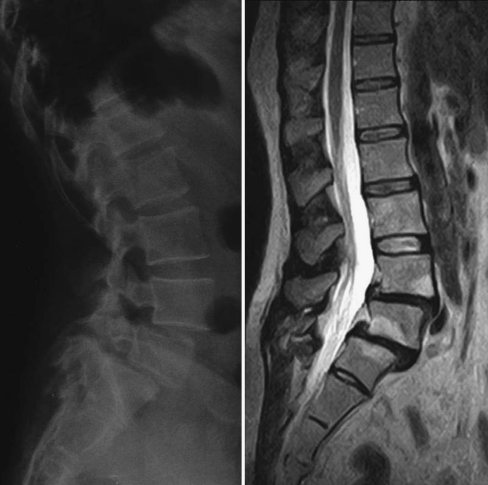
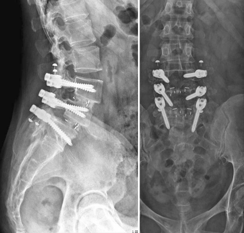
From October 2008 to September 2010, 30 patients with degenerative lumbar spine disease were operated. There were 13 males and 17 females with an average age of 61 years (from 31 to 80). Six patients suffered from multilevel spinal stenosis with claudication and required posterior laminectomy and multiple articular joint ablation; one had the same symptoms because of a segmental spinal stenosis. Seven patients presented with a symptomatic low-grade spondylolisthesis (3 isthmic and 4 degenerative) and two suffered from back pain due to degenerative disc changes. Ten patients had already been operated before (3 recurrent disc herniation, 1 interspinous device, 1 posterior fusion without instrumentation, 5 posterior fusion with instrumentation) and suffered from both sciatic and back pain. Interbody fusion with peek cages was performed in 22 cases (1 double level) (Figs. 1, 2). The average number of levels fused was 2.9 (from 2 to 5), and the screws were placed bilaterally at all levels except in one case of previous stabilization revision. Only autogenous bone graft by iliac crest and posterior vertebral arch, harvested from inside the operatory field, was used. Preoperative X-rays and MRI were available for all patients, and CT scan in 21. All patients were controlled after surgery at 1 month, 3 months, 6 months and 1 year. A 2 years postoperative control was available in 19 patients. Standard X-rays were taken at every postoperative control, while CT scan was taken only postoperatively at 6 months and 1 year. Fusion was considered achieved when a continuous layer of newly formed bone was clearly visible along the rods on CT scan multiplanar reconstructions, or in case of interbody fusion, when a clear bone bridging through the cages was detected.
Fig. 1.
L4–L5 and L5–S1 spondylolisthesis in a 45-year-old woman
Fig. 2.
Anteroposterior and lateral radiographs 2 years after posterior lumbar arthrodesis with PEEK rods and double-level interbody fusion
Results
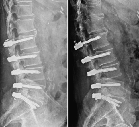
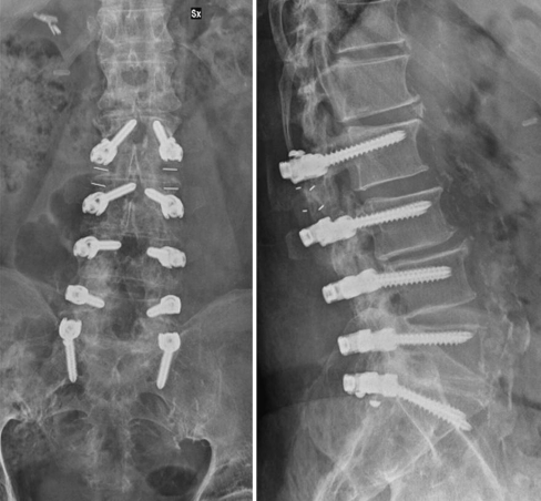
Overall, 84 screws and 60 rods were used. No screw misplacement was detected on the postop CT scan. There were no intraoperative complications except one dural tear, which was immediately repaired without clinical consequences. There were two early complications, one superficial wound dehiscence cured by local dressing and one deep infection treated by surgical debridement and antibiotic therapy. One patient showed cranial screw mobilization at the 8 months follow-up. Although the patient was asymptomatic, he underwent surgical revision using hydroxyapatite-coated screws at the proximal end of the instrumentation connected to a couple of hybrid peek rods with a dynamic segment at one edge (Figs. 3, 4). Compared to standard posterior instrumentations, radiological assessment of fusion was easier thanks to the absence of artefacts from the peek rods. In the group of 22 patients in whom anterior interbody cages were implanted, a clear fusion was visible in 18 at 6 months and in all of them at 12 months. In the eight patients receiving posterolateral autogenous grafting only, four were fused at 6 months and seven at 1 year. In three patients, the newly formed posterolateral bone layer remained incomplete even after 8, 16 and 18 months, respectively. One of them, as reported above, was re-operated because of screw mobilization. The reason for this failure was ascribed to a sagittal hypercorrection in a flat back patient, causing an increased mechanical stress on the bone screw interface. The other two showed neither clinical nor radiologic complications. The patient who suffered from a postoperative infection also reported sciatic pain, which disappeared after a foraminotomy performed during the surgical debridement. At the 18 months follow-up, this patient was still suffering from back pain. All the remaining patients showed improved clinical status and were satisfied with surgery at the 1-year follow-up.
Fig. 3.
Screw mobilization 8 months after operation
Fig. 4.
Revision with hydroxyapatite-coated screws and hybrid peek rods
Discussion
The effectiveness of posterior lumbar fixation in improving the rates of fusion has been well recognized [1–5]. In the last few years, the concept of dynamic stabilization has promoted the introduction of various devices for surgical treatment of degenerative spine disease. Although the theoretical advantage of traditional stiff instrumentations is still controversial, less rigid fixation applied to fusion can reduce stresses at the bone–screw interface and give additional load sharing onto the anterior column.
Recently “semi-rigid” implants with PEEK rods have become available to support fusion in the degenerative lumbar spine. The modulus of elasticity of this polymer is similar to that of bone (approximately, 17 GPa) [11]; this property can offer adequate rigidity for stimulating bone fusion, decreasing the stresses created by a titanium construct.
This is a report of our preliminary experience with the PEEK rod system. A long-term follow-up is necessary when dealing with a pathology normally showing progression in its natural history. Nevertheless, a short- and medium-term follow-up is sufficient to draw conclusions concerning early and medium-term complications and to verify if the main goal of the system, fusion, is achieved or not. For what concern the only mechanical complications recorded, this was ascribed to non-union and to an extreme mismatching between patient lordosis and rods’ pre-shaping. This eventually caused the cranial screw mobilization and pull-out. Fusion was achieved in a reasonable time in all patients with interbody cages, and this was the main target of the new load sharing feature and in 90% of the patient who underwent posterolateral fusion. Pre-shaped peek rods with different degrees of lordosis are now available in the market and, together with the new hydroxyapatite-coated screws, would probably further reduce the incidence of mechanical failure. Clinical outcome is encouraging, but requires a longer follow-up to be considered significant. Also, the effects on the adjacent discs should be evaluated in the long term.
Conflict of interest
None.
References
- 1.Schwab FJ, Nazarian DG, Mahmud F, et al. Effects of spinal instrumentation on fusion of the lumbosacral spine. Spine. 1995;20:2023–2028. doi: 10.1097/00007632-199509150-00014. [DOI] [PubMed] [Google Scholar]
- 2.Narayan P, Haid RW, Subach BR, et al. Effect of spinal disease on successful arthrodesis in lumbar pedicle screw fixation. J Neurosurg. 2002;97(3 Suppl):277–280. doi: 10.3171/spi.2002.97.3.0277. [DOI] [PubMed] [Google Scholar]
- 3.Asher MA, Carson WL, Hardacker JW, et al. The effect of arthrodesis, implant stiffness, and time on the canine lumbar spine. J Spinal Disord Tech. 2007;20:549–559. doi: 10.1097/BSD.0b013e31804c98e5. [DOI] [PubMed] [Google Scholar]
- 4.Kowalski RJ, Ferrara LA, Benzel EC. Biomechanics of bone fusion. Neurosurg Focus. 2001;10:E2. doi: 10.3171/foc.2001.10.4.3. [DOI] [PubMed] [Google Scholar]
- 5.Thomsen K, Christensen FB, Eiskjaer SP, et al. 1997 Volvo Award winner in clinical studies. The effect of pedicle screw instrumentation on functional outcome and fusion rates in posterolateral lumbar spinal fusion: a prospective, randomized clinical study. Spine. 1997;22:2813–2822. doi: 10.1097/00007632-199712150-00004. [DOI] [PubMed] [Google Scholar]
- 6.Wedemeyer M, Parent S, Mahar A, et al. Titanium versus stainless steel for anterior spinal fusions: an analysis of rod stress as a predictor of rod breakage during physiologic loading in a bovine model. Spine. 2007;32:42–48. doi: 10.1097/01.brs.0000251036.99413.20. [DOI] [PubMed] [Google Scholar]
- 7.Toth JM, Wang M, Estes BT, et al. Polyetheretherketone as a biomaterial for spinal applications. Biomaterials. 2006;27:324–334. doi: 10.1016/j.biomaterials.2005.07.011. [DOI] [PubMed] [Google Scholar]
- 8.Kurtz SM, Devine JN. PEEK biomaterials in trauma, orthopedic, and spinal implants. Biomaterials. 2007;28:4845–4869. doi: 10.1016/j.biomaterials.2007.07.013. [DOI] [PMC free article] [PubMed] [Google Scholar]
- 9.Ponnappan RK, Serhan H, Zarda B, Patel R, Albert T, Vaccaro AR. Biomechanical evaluation and comparison of polyetheretherketone rod system to traditional titanium rod fixation. Spine J. 2009;9(3):263–267. doi: 10.1016/j.spinee.2008.08.002. [DOI] [PubMed] [Google Scholar]
- 10.Galbusera F, Bellini CM, Anasetti F, Ciavarro C, Lovi A, Brayda-Bruno M. Rigid and flexible spinal stabilization devices: a biomechanical comparison. Med Eng Phys. 2011;33(4):490–496. doi: 10.1016/j.medengphy.2010.11.018. [DOI] [PubMed] [Google Scholar]
- 11.Wenz LM, Merritt K, Brown SA, Moet A, Steffee AD. In vitro biocompatibility of polyetheretherketone and polysulfone composites. J Biomed Mater Res. 1990;24:207–215. doi: 10.1002/jbm.820240207. [DOI] [PubMed] [Google Scholar]


