Abstract
Aims
The Community and Hospital Allied Network Glaucoma Evaluation Scheme (CHANGES) used accredited community-based optometrists with a special interest (OSIs) in glaucoma to monitor ocular hypertensive (OHT) patients under virtual supervision of the Hospital Glaucoma Service (HGS). The purpose of this paper was to report the outcomes of the first completed community-based visit.
Methods
Eligible patients underwent a glaucoma consultant-led clinical examination before transfer to CHANGES. Individualised intraocular pressure (IOP) and follow-up time interval targets were set for each patient. OSIs used applanation tonometry, slit-lamp biomicroscopy, automated visual field testing and digital optic disc photography. The hospital-based glaucoma team evaluated the data virtually. Patients were referred back to the HGS according to specific criteria.
Results
One hundred and sixty eight OHT patients were invited to attend their first OSI appointment. Of these, 144 attended their appointment (attendance rate 85.7%). Outcomes of 130 patients with complete data sets are reported. Sixteen patients (12.3%) were referred back to the HGS due to IOP above target, new visual field defects and/or optic nerve changes. The glaucoma consultant retained eight patients (6.1%) within the HGS on the basis of definite or probable glaucomatous conversion.
Conclusions
CHANGES freed up capacity within a busy HGS. However, improvements need to be made regarding non-attendance rates in the community. The relatively high one-year definite or probable conversion rate emphasises the importance of the comprehensive review of OHT patients and of hospital-led virtual supervision to maintain patient safety.
Keywords: ocular hypertension, community, monitoring, shared care, glaucoma
Introduction
The National Institute for Health and Clinical Excellence (NICE) published guidelines on the diagnosis and treatment of chronic open-angle glaucoma and ocular hypertension in April 2009.1 The response of the Association of Optometrists to advise their members to refer any patients with an intraocular pressure (IOP) above 21 mm Hg regardless of the method of measurement resulted in a significant increase in new referrals to hospital eye services in the United Kingdom. Options to deal with this increase included increased staffing levels or employing existing human resources such as optometrists in the evaluation and follow-up of such patients. The Community and Hospital Allied Network Glaucoma Evaluation Scheme (CHANGES) began in 2006 and involved trained optometrists with a special interest in glaucoma (OSIs) to assess patients suspected as having glaucoma in Cambridgeshire, UK (supervised by the Glaucoma Service of Hinchingbrooke Hospital NHS Trust). The design and safety of this scheme has been described in detail in an earlier publication.2
More recently, the shared-care phase of CHANGES (CHANGES-2) was launched, involving the monitoring of ocular hypertensive (OHT) patients in the community by accredited OSIs. In its first year of implementation in 2008, the scheme involved 29 OHT patients, the number increasing to 113 patients in 2009 and to 189 patients in 2010.
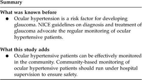
The purpose of this article is to describe the design and the activity of the CHANGES-2 scheme and to report the outcomes of community-shared monitoring of OHT patients and the conversion rate to probable or definite glaucoma.
Materials and methods
Since the introduction of CHANGES-2, all OHT patients (on or off ocular hypotensive treatment) were transferred to community-based monitoring after a glaucoma consultant-led clinical examination in the Hospital Glaucoma Service (HGS). The measurements obtained included: best corrected visual acuity (BCVA), visual field test (Humphrey VF SITA Fast Carl Zeiss Meditec, Dublin, CA, USA), Goldmann applanation tonometry (GAT, Haag-Streit, Bern, Switzerland), dilated slit-lamp biomicroscopy, and optic disc imaging using digital optic disc photography (Topcon NW6, Tokyo, Japan). A target IOP was set for each patient depending on individual risk factors for conversion to glaucoma. Target IOP was generally set at 28–30 mm Hg for untreated patients and 20–22 mm Hg for OHT patients on treatment. The time interval between planned monitoring visits in the community was also individualised and was set at 9 months for patients on treatment and 12 months for non-treated patients. Treatment eligibility and prescribed hypotensive agents were generally aligned with NICE guidelines for most patients.1 However, as quite a few patients had commenced treatment before the NICE guidelines were published, treatment for these patients did not necessarily follow the NICE recommendations.
A reminder letter was sent to patients a few weeks before the date their community-based OSI examination was due asking them to make an appointment. The OSI followed a pre-determined examination protocol which included the following: BCVA, GAT, 24-2 SITA Fast HVF, temporal limbal anterior chamber depth evaluation using the Van Herrick method,3 dilated slit-lamp biomicroscopy and optic nerve head imaging with digital optic disc photography.
Criteria for re-referral to the HGS included any of the following: a GAT-measured IOP exceeding target, a new VF defect which could be attributed to glaucoma, signs suspicious of glaucomatous structural change such as a disc haemorrhage, retinal nerve fibre layer defect or disc rim change (notching, thinning), or eyedrop intolerance for those patients on treatment. A standardised examination form was completed by the OSI for each patient and was sent to the HGS in addition to the digitised optic disc images in electronic form for virtual review. The hospital-based glaucoma team evaluated the data virtually for all patients, regardless of whether the OSI recommended referral or not. On the basis of the criteria mentioned above, a decision was made by the hospital glaucoma team as to whether the patient could safely continue to be monitored in the community or needed referral to the HGS for a consultant-led examination. Referred patients were offered an appointment with the HGS within 8 weeks from virtual review of the OSI-led examination results.
An analysis of the outcomes of the first community-based examination for all patients since the introduction of the scheme was performed to assess the practicality and efficacy of our shared scheme for monitoring OHT patients. The t-test and Fisher's exact test were used for statistical analysis using the SPSS program (version 16.0, SPSS, Chicago, IL, USA) and setting a P-value of <0.05 was used to indicate statistical significance.
Results
By the time of data collection (September 2010), 168 OHT patients (85 males, 83 females) had been invited for their first appointment with an accredited OSI. Of these, 144 attended their appointment, an attendance rate of 85.7%. Fourteen patients were excluded from further analysis due to incomplete data sets by the time data were collected, as their OSI-led examination results had not yet been received or reviewed by the hospital-based glaucoma team. Therefore, complete data sets from 130 OHT patients (69 males and 61 females, mean age 62.6±10 years) were analysed.
OHT patients had been under hospital-based monitoring for a median of 20 months (range 0–60 months) before transfer to CHANGES. Thirty-nine patients (30.0%) were on treatment for OHT at the time of transfer. After their first OSI-led examination, 114 OHT patients (87.7%) remained under monitoring in the community, whereas 16 patients (12.3%) were referred back to hospital for further evaluation by a glaucoma consultant. No patient required to be reviewed at the HGS before their OSI appointment was due.
The reasons for referral are summarised in Table 1.
Table 1. Reasons for referral of OHT patients to hospital glaucoma service.
| Reason for referral | No. of patients |
|---|---|
| IOP above target | 5a |
| New/Worse VF defect | 2 |
| Optic Disc changes | 1 |
| IOP + VF | 3 |
| IOP + disc changes | 2 |
| VF + disc changes | 3 |
| Total | 16 |
Abbreviations: IOP, intraocular pressure; VF, visual field.
One patient had stopped treatment due to side effects, and another was suspected as having occludable anterior chamber angles by the OSI.
In another 22 patients (17%) the OSI had concerns that did not fulfil the pre-determined criteria for referral, and following virtual assessment of the OSI findings by the hospital-based glaucoma team it was deemed that the patient need not be referred. No additional patients were suspected as possible converters by virtual review only.
Of the 16 referred patients, 8 were retained within the HGS on the basis of definite or probable glaucomatous conversion, a 1-year ‘conversion rate' of 6.1%. Seven patients were deemed stable by the glaucoma consultant and returned to CHANGES, whereas one patient who had stopped treatment due to side effects had her regimen changed and was re-discharged to community-based monitoring.
The mean age of ‘converters' was 65.5 years (SD 6.3), whereas that for ‘non-converters' was 62.5 years (SD 10.2) (independent samples t-test, P=0.407). Among ‘converters', 50.0% were females, whereas among ‘non-converters' 46.7% were females (Fisher's exact test, P>0.999). One (2.6%) of the treated OHT patients converted, whereas 7 (7.7%) of the untreated OHT patients converted (Fisher's exact test, P=0.434). The mean duration of OHT diagnosis for the converters was 20.8 months (range 14.2–30.9 months), whereas for non-converters was 20.6 months (range 8.9–40.4 months; independent samples t-test, P=0.964).
In 50 (38.5%) patients delay of more than 1 month between intended and actual OSI examination was noted (median delay among these 50 patients was 70 days, range 33–253 days).
At least three attempts were made on different days and at differing times4 to contact the 24 (14.3%) OHT patients who did not attend (DNA) their first OSI appointment. Six patients were either not contactable or declined conversation. Reasons for non-attendance were noted for the remaining 18 patients who were contacted (Table 2). No statistically significant differences were noted between all non-attenders and those who attended their OSI appointment with regard to age, sex, or ocular hypotensive treatment. However, there were 13 non-attenders who either did not answer/speak on the phone or stated that they had not made an appointment with an OSI despite having received a reminder letter (ie, were unwilling to attend) and they were significantly younger than those who attended (mean age of DNA patients 51 years vs 62 years for attendees, independent samples t-test, P<0.001).
Table 2. Reasons for not attending OSI appointment.
| Reasons | No. of patients (%) |
|---|---|
| Not contactable/refused to speak | 6 (25.0) |
| Did not receive reminder letter | 4 (16.7) |
| Deceased | 2 (8.3) |
| Lived or moved outside catchment area | 5 (20.8) |
| Did not make/cancelled appointment due to personal/family/work reasons | 7 (29.2) |
| Total | 24 (100.0) |
Discussion
In 1996 the Royal College of Ophthalmologists recommended shared care for stable OHT or glaucoma patients.5 Ten years later, in 2006, 66 shared schemes for referral refinement or monitoring glaucoma patients were reported to be in operation, 14 of which were community-based.6 Optometrists have a significant role in many of these schemes whether hospital- or community-based and it has been reported that specially trained optometrists can perform as reliably as clinicians when monitoring patients with OHT or stable glaucoma.7
Approximately 6% of OHT patients in this monitoring scheme converted to definite or probable glaucoma within 1 year and were then retained within the HGS. This relatively high 1-year conversion rate may have been influenced by the longer duration of the diagnosis for some of our OHT patients before transfer to CHANGES-2. In fact, 72 (55.4%) patients had been under HGS-based monitoring before transfer to CHANGES, therefore this case-mix contains OHT patients at variable timepoints in their natural history. The conversion rate emphasises the importance of a comprehensive review of such patients on a regular basis, which is usually impossible in a standard community optometrist practice, where often the equipment is not standardised to that of the hospital service. We are of the opinion that a HGS-supervised assessment of the OSI clinical information and the use of specific criteria for re-referral to HGS are essential to maintain patient safety and to reduce missed converters as all proformas are reviewed and any suspect cases can then be recalled. Interestingly, the overall referral rate would have been approximately 29%, that is, almost one out of every three OHT patients would have been referred back to the HGS if referrals had been left to the discretion of the OSI. This underlies the importance of a virtual review of all data collection as it enabled 22 referrals with queries raised by the OSI to be dismissed as ‘not needing referral to HGS' by the HGS, thereby avoiding unnecessary hospital visits.
The great majority of our OHT patients (88%) remained under community-based monitoring after their first OSI appointment, which demonstrates the importance of this scheme in freeing up capacity within a busy HGS. In comparison with the referral refinement phase of CHANGES,2 a similar first-attendance rate was noted in CHANGES-2 (85.7% in CHANGES-2 vs 87.7% in the referral refinement phase of CHANGES where low-risk glaucoma referrals were invited to make a first appointment with an OSI). However, it was noted that there was a significant delay in the actual compared with the intended OSI examination (a median delay of 70 days for 38.5% of patients); such delays may be attributed to the fact that patients were invited to make an appointment with an OSI instead of being sent an appointment date. Reminder letters to non-responders should improve non-attendance rates and delays in community-based appointments.
The most common reason for referral of patients to the HGS was found to be IOP above target, accounting for one third of referrals. In CHANGES-2, target IOP was individualised for each patient taking into account individual risk factors for conversion to glaucoma; untreated IOP targets were based on the increased relative risk for glaucoma associated with an IOP above 30 mm Hg8 and treated IOP targets were based on the findings of the Ocular Hypertension Treatment Study aimed for an IOP of less than 24 mm Hg and at least 20% less than baseline.9
Although this was not a primary aim of our study, a cost analysis of CHANGES-2 for the year 2010 suggested that CHANGES-2 costs were approximately 25% less than the costs that would have been incurred if the patients had been seen in the hospital setting, indicating that CHANGES could be a cost-effective alternative scheme for OHT monitoring.
In conclusion, CHANGES for OHT patients seems to be a safe scheme in that all data collected by the OSIs is virtually reviewed by a glaucoma-trained clinician, with no absolute reliance on the OSIs. It is an effective alternative pathway for monitoring OHT patients in the community, significantly improving capacity within HGS. Certain improvements need to be implemented with regard to non-attendance rates and delayed appointments to improve performance of the scheme.

Acknowledgments
We thank Malcolm Ward, the manager of the community practice, and the OSIs who participated in the CHANGES-2 scheme, namely Sally Bushby, Keziah Latham, and Sheila Urquhart. No financial support was received for this study.
The authors declare no conflict of interest.
Footnotes
Part of this work was presented at the Royal College of Ophthalmologists Annual Congress in Birmingham, 24-26 May 2011.
References
- NICE clinical guideline 85 Glaucoma: diagnosis and management of chronic open angle glaucoma and ocular hypertension 2009. Available at: http://www.nice.org.uk/Guidance/CG85 . (accessed May 2011). [PubMed]
- Bourne RRA, French KA, Chang L, Borman AD, Hingorani M, Newsom WD. Can a community optometrist-based referral refinement scheme reduce false-positive glaucoma hospital referrals without compromising quality of care? The Community and Hospital Allied Network Glaucoma Evaluation Scheme (CHANGES) Eye. 2010;24:881–887. doi: 10.1038/eye.2009.190. [DOI] [PubMed] [Google Scholar]
- Foster PJ, Devereux JG, Alsbirk PH, Lee PS, Uranchimeg D, Machin D, et al. Detection of gonioscopically occludable angles and primary angle closure glaucoma by estimation of limbal chamber depth in Asians: modified grading scheme. Br J Ophthalmol. 2000;84:186–192. doi: 10.1136/bjo.84.2.186. [DOI] [PMC free article] [PubMed] [Google Scholar]
- Wong SC, Chang L, Lee N. Partial booking: is it flawed. Qual Prim Care. 2003;11:117–122. [Google Scholar]
- The Royal College of Ophthalmologists, The Royal College of General Practitioners, and The College of Optometrists . Shared Care. RCOphth: London; 1996. [Google Scholar]
- Vernon SA, Adair A. Shared care in glaucoma: a national study of secondary care lead schemes in England. Eye. 2010;24:265–269. doi: 10.1038/eye.2009.118. [DOI] [PubMed] [Google Scholar]
- Spry PGD, Spencer IC, Sparrow JM, Peters TJ, Brookes ST, Gray S, et al. The Bristol Shared Care Glaucoma Study: reliability of community optometric and hospital eye service test measures. Br J Ophthalmol. 1999;83:707–712. doi: 10.1136/bjo.83.6.707. [DOI] [PMC free article] [PubMed] [Google Scholar]
- Sommer A, Tielsch JM, Katz J, Quigley HA, Gottsch JD, Javitt J, et al. Relationship between intraocular pressure and primary open angle glaucoma among white and black Americans. The Baltimore Eye Survey. Arch Ophthalmol. 1991;109:1090–1095. doi: 10.1001/archopht.1991.01080080050026. [DOI] [PubMed] [Google Scholar]
- Kass MA, Heuer DK, Higginbotham EJ, Johnson CA, Keltner JL, Miller JP, et al. The Ocular Hypertension Treatment Study: a randomized trial determines that topical ocular hypotensive medication delays or prevents the onset of primary open-angle glaucoma. Arch Ophthalmol. 2002;120:701–713. doi: 10.1001/archopht.120.6.701. [DOI] [PubMed] [Google Scholar]


