Abstract
We report the diagnosis and treatment of patients with retinal detachment and/or retinoschisis associated with optic nerve coloboma or morning glory syndrome. A retrospective review of patients with optic nerve coloboma or morning glory syndrome with associated retinal detachment or retinoschisis was conducted. For five patients (six eyes), we report the clinical findings, spectral domain optical coherence tomography (OCT) imaging, intraoperative findings, and treatment outcomes. OCT scans demonstrate a bilaminar structure of maculopathy, consisting of inner schisis-like changes and outer layer retinal detachment. In most cases, a retinal break was demonstrated within the optic disc defect with three-dimensional OCT imaging. Glial tissue was sometimes observed within the anomalous defect. Vitrectomy and resection of the tractional tissue in these cases produced good anatomical and visual outcomes. Retinal detachment spontaneously resolved in cases where traction was not present. Traction may contribute to the pathogenesis of retinal detachment associated with colobomatous optic disc anomalies, either directly or by creating a secondary retinal break. OCT imaging assists with understanding the contributing factors to retinal detachment in individual cases of colobomatous optic disc anomalies and can thereby assist with determining the most effective approach to management.
Keywords: retinal detachment, optic disc coloboma, morning glory syndrome, optic disc anomaly, pediatric retina
Introduction
Colobomatous optic disc anomalies, including optic disc colobomas and morning glory syndrome, are uncommon, and sometimes associated with retinal detachment and/or retinoschisis. In optic disc colobomas, the area of the optic disc is usually enlarged and the coloboma involves the inferior portion of the nerve. In morning glory syndrome, the optic nerve is located centrally and surrounded by a deeply excavated scleral defect. Fluid sometimes accumulates within and beneath the retina but the origin of the fluid remains unclear. The two possible sources are the cerebrospinal fluid1, 2, 3 or the vitreous cavity,4, 5, 6, 7, 8, 9, 10, 11 and tractional forces may have a role.2, 3, 5, 6 It also remains uncertain which treatment for these patients produce the best outcomes, as spontaneous resolution of retinal detachments may sometimes occur. Based on a study of a small number of eyes with peripapillary retinal detachment associated with optic disc coloboma or morning glory syndrome with optical coherence tomography (OCT), we believe that the mechanism causing retinal detachment in these eyes is similar.
Materials and methods
A retrospective case review was performed of patients who presented to Department of Ophthalmology, Columbia University Medical Center, with optic disc coloboma or morning glory syndrome and associated retinal detachment or retinoschisis between 2006 and 2011. Six eyes of five patients were identified. The medical records were reviewed for Snellen visual acuity, slit lamp, and funduscopic examinations. Fundus photographs were available in all cases. In all cases, spectral domain OCT (Cirrus, Zeiss, Dublin, CA, USA) imaging was available, and prior to 2008 time domain OCT scans (Stratus, Zeiss) only were available. A three-dimensional (3D) reconstruction of the OCT of the optic disc area was made using a software program, Camtasia Studio for Windows (TechSmith, Okemos, MI, USA). In three eyes, vitrectomy was done to correct the retinal detachment using vitrectomy, membrane peeling, endolaser, and gas tamponade. The intraoperative findings and treatment outcomes were also reviewed.
Results
Case 1
A 52-year-old female presented with an optic disc coloboma in the right eye with best-corrected visual acuity (BCVA) 20/30 in the right eye and finger counting in the left eye. She had a history of uveitic episodes bilaterally, and muscle surgery in the left eye at a young age for exotropia, possibly strabismic amblyopia. OCT of the right eye demonstrated retinoschisis but no retinal detachment, and no glial tissue at the disc or within the coloboma. The inferior peripapillary retinal pigment epithelium around the optic disc and coloboma was thin and irregular, suggestive of possible previous subclinical subretinal fluid that had spontaneously resolved. The coloboma was quite deep and its full depth could not be completely imaged by OCT. The retina lying within the coloboma was thin, shallowly detached, and a retinal break could not be definitely identified with 3D OCT reconstruction. The patient is being monitored.
Case 2
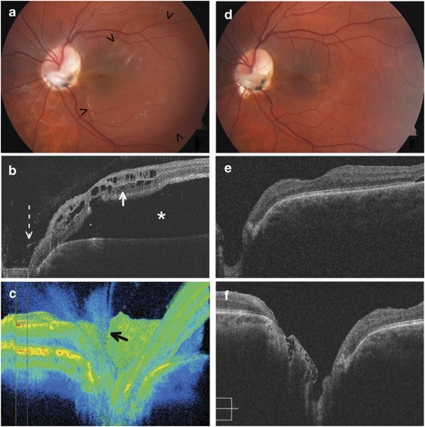
A 15-year-old male presented with an optic disc coloboma in the left eye, with BCVA 20/100. Thick preretinal glial tissue was present over the colobomatous defect and there was peripapillary retinal detachment involving the macula (Figure 1a). OCT confirmed retinal detachment and retinoschisis (Figure 1b). A 3D reconstruction demonstrated a dense vitreous insertion into the optic disc and coloboma (Figure 1c). The patient underwent vitrectomy, endolaser, and perfluoroethane gas injection. Intraoperatively, there was a firm attachment of the vitreous at the optic disc. The fibrous glial tissue was strongly adherent to the retina within the coloboma, and extended onto the retina at the margin of the coloboma. The tangential traction on the retina was reduced by separating the fibrous attachments to retina at the edge of the coloboma and debulking the glial tissue. A retinal break was suspected in thin retina within the coloboma. Postoperatively, BCVA in the left eye improved to 20/40 and imaging indicated resolution of the detachment (Figures 1d–f).
Figure 1.
Patient 2, left eye. (a) photograph at presentation, showing an anomalous optic disc with overlying glial tissue and retinal detachment involving the macula (arrowheads). (b) Preoperative OCT of the macula and temporal optic disc demonstrating retinal detachment (asterisk) and retinoschisis (white arrow), and glial tissue within the cup of the optic disc (broken arrow). (c) 3D OCT reconstruction of the optic nerve head demonstrating tissue within the disc cup exerting traction (black arrow). (d) Fundus photograph postoperatively demonstrating that glial tissue has been trimmed and the retina is flat, with only minor pigmentary disturbance. (e) OCT of the macula 7 months postoperatively demonstrating resolution of the retinal detachment and retinoschisis. (f) OCT of the optic disc 7 months postoperatively demonstrating the anomalous optic disc.
Case 3
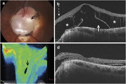
A 16-year-old boy presented with optic nerve coloboma, retinoschisis, and retinal detachment in the left eye (Figures 2a and b), with BCVA 20/125. His right eye had a small choroidal coloboma adjacent to the optic disc. In the left eye, 3D OCT displayed a retinal break (Figure 2c). This retinal break was not visible on conventional OCT line scans, but only visible from the 3D reconstruction. He underwent vitrectomy, and mechanical separation of the posterior hyaloid. Any cortical vitreous and adherent preretinal glial tissue (mainly located within the inferior margin of the coloboma) was separated and trimmed over the colobomatous area. Subretinal fluid was not drained, and light laser burns were placed using the diode endolaser under air at the retinal margin of the coloboma. Perfluoroethane gas tamponade was used. The retinal break that was identified on OCT scanning could not be visually confirmed because the white scleral tissue of the coloboma did not provide enough contrast against a thin atrophic retina. Postoperatively, BCVA improved to 20/40. OCT showed nearly complete resolution of the schisis and subretinal fluid by 9 months postoperatively (Figure 2d).
Figure 2.
Patient 3, left eye. (a) Fundus photograph of the optic disc on presentation, demonstrating an inferior coloboma (black arrow). A retinal break at the location of the arrow is difficult to visualise clinically. (b) OCT of the macula on presentation, demonstrating retinoschisis (asterisk) with subretinal fluid (white arrow) and retinal tissue extending into the optic disc cavity (broken arrow). (c) 3D OCT reconstruction of the optic disc demonstrating a retinal break in the cup of the disc (black arrow). (d) OCT of the macula 9 months postoperatively demonstrating marked reduction of the macular schisis and subretinal fluid.
Case 4
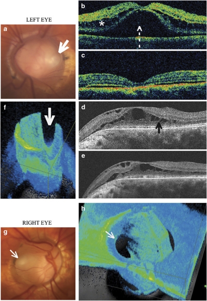
A 44-year-old female presented with morning glory optic discs in both eyes. She had reported intermittent episodes of blurred vision in the left eye that spontaneously improved over variable periods of time. The optic discs were enlarged in both eyes (Figures 3a and g), and in the left eye a localised retinal detachment with retinoschisis involving the macula was seen (Figure 3b). BCVA was 20/30 in the right eye and 20/400 in the left eye. With observation, BCVA in the left eye spontaneously improved to 20/60 within 2 months. OCT showed resolution of the retinoschisis and subretinal fluid (Figure 3c). At 6 years after presentation, BCVA was 20/60 in the right eye and 20/50 in the left eye. OCT showed new subretinal fluid in the right eye and recurrence of retinoschisis in the left eye (Figure 3d). At 7 years after presentation, BCVA was 20/50 bilaterally. The subretinal fluid in the right eye had decreased. Retinoschisis in the left eye persisted (Figure 3e). In each eye, there was a suggestion of an oval retinal break within thin detached retina that was pulled into the excavation of the morning glory optic disc. 3D OCT of both optic discs demonstrated retinal breaks but did not show evidence of any vitreous or glial tissue (Figures 3f and h).
Figure 3.
(Top) Patient 4, left eye. (a) Fundus photograph showing colobomatous optic disc with morning glory configuration with vessels emerging radially from the edge of the disc, and a retinal break (broad white arrow). (b) OCT of macula at presentation showing both retinoschisis (asterisk) and retinal detachment (broken arrow). (c) OCT of macula 2 months after observation shows spontaneous resolution of the retinoschisis and subretinal fluid. (d) OCT of the macula 6 years after initial presentation demonstrating recurrence of retinoschisis (black arrow). (e) OCT of the macula 7 years after initial presentation demonstrating persistent retinoschisis. (f) 3D OCT reconstruction of the left optic disc showing a retinal break (broad white arrow). (Bottom) Patient 4, right eye. (g) Photograph of the right optic disc showing a morning glory optic disc with a retinal break (narrow white arrow). (h) 3D OCT reconstruction of the right optic disc demonstrating the retinal break (narrow white arrow).
Case 5
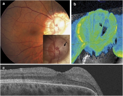
A 49-year-old female presented in September 1999 with a retinal detachment in the right eye. She was told of an optic disc anomaly at age 12 years, and a serous detachment of the macula was observed in 1997, which spontaneously resolved. Subsequently, she noted a field defect and was found to have a large retinal detachment, involving the nasal and inferior retina. Her BCVA was 20/25 and a retinal detachment was seen extending from 0130 to 0700 hours, with the fluid extending to the equator nasally and to the ora serrata inferiorly. There was a morning glory optic disc (Figure 4a) with thin elevated retina within the nasal portion of the defect, and glial tissue extending on the adjacent retina nasally. No peripheral retinal breaks were found. A pneumatic retinopexy was attempted and after 2 days, the retina completely flattened. Laser photocoagulation using krypton wavelength was applied at the margin of the morning glory defect from 0100 to 0730 hours. However, 1 week later the retinal detachment recurred and vitrectomy was done. The posterior hylaoid was separated and membranes within the morning glory defect were removed, but during fluid–air exchange subretinal fluid could not be drained internally at the optic disc. Gas was used for tamponade and diode laser photocoagulation was placed completely around the optic disc. The retina remained attached postoperatively, and after cataract extraction in 2001, the BCVA was 20/30. The patient agreed to return for OCT evaluation in January 2009. The spectral domain OCT revealed a deeply excavated scleral defect around the optic disc (Figure 4b). An oval retinal break in thin detached retina within the nasal side of the morning glory defect was found on 3D reconstruction (Figure 4b). The retina was completely attached (Figures 4a and c), with 20/20 BCVA.
Figure 4.
Patient 5, right eye. (a) Fundus photograph of the macula 10 years postoperatively, demonstrating resolution of the retinal detachment. Inset, optic disc photograph, with a break in the thin overlying retinal tissue (black arrow). (b) OCT 3D reconstruction of the optic nerve head, showing a deeply excavated defect and an oval retinal break in the retina within the morning glory defect (black arrow). (c) OCT imaging demonstrating a completely attached retina after vitrectomy with separation of the posterior hyaloid, membrane removal, gas tamponade and laser photocoagulation.
Discussion
Optic disc colobomas and the morning glory syndrome are uncommon. Retinal detachment may develop in up to one-third of patients with morning glory syndrome and spontaneous resolution has also been observed.12 Various theories have been advanced regarding the origin of the subretinal fluid seen in these eyes—cerebrospinal fluid,1, 2, 3 or fluid from the vitreous space.4, 5, 6, 7, 8, 9, 10, 11 This issue remains a subject of controversy. In both optic disc colobomas and morning glory syndrome, the peripapillary retina extends into the anomalous peripapillary scleral defect. The retinal tissue within the defect has been observed to be thinner, incompletely developed, and atrophic. Sometimes the retina is detached in this area. Retinal breaks have been observed in the peripapillary retina,5, 6, 7, 8, 9 but because the breaks were observed in some eyes undergoing reoperation for retinal detachment, the exact origin remains uncertain.
We found the use of 3D reconstruction of OCT images helpful in the assessment of these optic disc anomalies. The vitreous in young eyes, such as these, is usually transparent and clear, and thus is also difficult, if not impossible, to appreciate on clinical examination. Specifically, the 3D image allowed a qualitative assessment of the degree of vitreous condensation and adhesion in the area of the optic disc. Although still not possible to determine the degree of traction, the density of the vitreous strands could be better visualised on 3D scans compared with two-dimensional scans. 3D reconstruction allows rotation of the viewing perspective, improving the ability to identify retinal breaks within the coloboma. By comparing the images on 3D reconstruction to other scans, we were careful to be certain that the breaks identified were not artifacts from the limitation of OCT imaging. In this series of eyes, retinal breaks were identified using 3D OCT reconstruction of the optic disc imaging in four of the six eyes. In one eye (case 3), the break could not be visualised clinically or intraoperatively because of low contrast between the white scleral tissue in the coloboma and the thin retina. In the eye where the retinal break could not be visualised with OCT (case 2), intraoperatively a small break was seen in atrophic retina within the coloboma after removal of some overlying glial tissue. Thus, a total of five of six eyes (83%) were observed to be associated with a retinal break within the scleral defect.
Three eyes required vitrectomy for management of the retinal detachment associated with the optic disc anomaly. In all cases, the posterior hyaloid required mechanical separation, and seemed strongly adherent, perhaps because of the young age of the patients. In addition, these eyes had a thick layer of fibro-glial tissue over the coloboma and extending onto the retina. Separating the adhesion of this tissue and trimming it seemed to be effective in relieving the traction around the retinal break. It was not possible to peel the tissue completely from the thin retina overlying the coloboma. The risk of causing a retinal break within the optic nerve defect in the thin retina overlying the coloboma is high, and care was taken not to pull too firmly and cause any new retinal breaks centrally. The use of laser endophotocoagulation at the retinal margin of the colobomatous defect seems to provide an effective barrier to the passage of fluid into the subretinal space. It is suggested that the long wavelength of the diode laser (840 nm) may be safer by reducing the damage to the inner retina caused by photocoagulation. In these eyes, the resolution of the subretinal fluid and retinoschisis often took up to 12 months postoperatively.
It is interesting to have observed two eyes in one patient that had retinal detachments, which had spontaneously reabsorbed (case 4). In these eyes, the preoperative OCT did not demonstrate any condensation of vitreous or glial membranes within the defect of the optic nerve. It seems that the absence of vitreous or glial tissue causing traction on the retina within the defect, that retinal detachment is less likely to occur, may remain subclinical, or may also spontaneously resolve. We also conclude that the presence of glial tissue with the coloboma or morning glory in deeply excavated disc increases the likelihood of subsequent retinal detachment, and if surgery is required, the glial tissue should be removed or trimmed so that traction on the retina can be released.
In all cases, the visual outcomes improved as the retinal detachment and retinoschisis resolved. In one eye followed for over 10 years, the visual acuity has been stabilized without recurrence of retinal detachment.
Acknowledgments
This work was supported by an unrestricted grant to the Department of Ophthalmology, Columbia University from Research to Prevent Blindness, Inc., and Dr Gregory-Roberts was supported by a grant from the Endeavour Awards, Australia.
The authors declare no conflict of interest.
References
- Meirelles RL, Aggio FB, Costa RA, Farah ME. STRATUS optical coherence tomography in unilateral colobomatous excavation of the optic disc and secondary retinoschisis. Graefes Arch Clin Exp Ophthalmol. 2005;243:76–81. doi: 10.1007/s00417-004-0956-1. [DOI] [PubMed] [Google Scholar]
- Chang S, Haik BG, Ellsworth RM, St Louis L, Berrocal JA. Treatment of total retinal detachment in morning glory syndrome. Am J Ophthalmol. 1984;97:596–600. doi: 10.1016/0002-9394(84)90379-9. [DOI] [PubMed] [Google Scholar]
- Cennamo G, de Crecchio G, Iaccarino G, Forte R, Cennamo G. Evaluation of morning glory syndrome with spectral optical coherence tomography and echography. Ophthalmology. 2010;117:1269–1273. doi: 10.1016/j.ophtha.2009.10.045. [DOI] [PubMed] [Google Scholar]
- Bartz-Schmidt KU, Heimann K. Pathogenesis of retinal detachment associated with morning glory disc. Int Ophthalmol. 1995;19:35–38. doi: 10.1007/BF00156417. [DOI] [PubMed] [Google Scholar]
- Coll GE, Chang S, Flynn TE, Brown GC. Communication between the subretinal space and the vitreous cavity in the morning glory syndrome. Graefes Arch Clin Exp Ophthalmol. 1995;233:441–443. doi: 10.1007/BF00180949. [DOI] [PubMed] [Google Scholar]
- Ho CL, Wei LC. Rhegmatogenous retinal detachment in morning glory syndrome pathogenesis and treatment. Int Ophthalmol. 2002;24:21–24. doi: 10.1023/a:1014498717741. [DOI] [PubMed] [Google Scholar]
- Harris MJ, De Bustros S, Michels RG, Joondeph HC. Treatment of combined traction-rhegmatogenous retinal detachment in the morning glory syndrome. Retina. 1984;4:249–252. doi: 10.1097/00006982-198400440-00007. [DOI] [PubMed] [Google Scholar]
- Akiyama K, Azuma N, Hida T, Uemura Y. Retinal detachment in morning glory syndrome. Ophthalmic Surg. 1984;15:841–843. [PubMed] [Google Scholar]
- Von Fricken MA, Dhungel R. Retinal detachment in the morning glory syndrome: pathogenesis and management. Retina. 1984;4:97–99. doi: 10.1097/00006982-198400420-00004. [DOI] [PubMed] [Google Scholar]
- Yamakiri K, Uemura A, Sakamoto T. Retinal detachment caused by a slitlike break within the excavated disc in morning glory syndrome. Retina. 2004;24:652–653. doi: 10.1097/00006982-200408000-00032. [DOI] [PubMed] [Google Scholar]
- Ho TC, Tsai PC, Chen MS, Lin LL. Optical coherence tomography in the detection of retinal break and management of retinal detachment in morning glory syndrome. Acta Ophthalmologica Scandinavica. 2006;84:225–227. doi: 10.1111/j.1600-0420.2005.00589.x. [DOI] [PubMed] [Google Scholar]
- Haik BG, Greenstein SH, Smith ME, Abramson DH, Ellsworth RM. Retinal detachment in the morning glory anomaly. Ophthalmology. 1984;91:1638–1647. doi: 10.1016/s0161-6420(84)34103-3. [DOI] [PubMed] [Google Scholar]


