Abstract
Purpose
To evaluate orbital blood flow velocities and optic nerve diameter with Doppler and gray-scale sonography in patients with acute unilateral optic neuritis (ON).
Methods
Orbital Doppler and gray-scale sonography was performed in 46 eyes of 23 patients aged 19- to 47-years with acute unilateral ON. ON was diagnosed by an ophthalmologist on the basis of clinical presentation, presence of decreased visual acuity and assessment of visual evoked potentials. The peak systolic velocity (PSV) and end-diastolic velocity (EDV), as well as the resistance index (RI) and pulsatile index (PI) of the ophthalmic artery (OA), central retinal artery (CRA), posterior ciliary arteries (PCAs) and optic nerve diameter were measured in both eyes. We compared results from affected and unaffected eyes using the paired t-test. The area under the receiver operating characteristic (ROC) curves was used to assess the diagnosis of ON based on measured blood flow parameters of the OA, CRA and PCAs and optic nerve diameter.
Results
The mean (standard deviation) optic nerve diameter in eyes with ON was 4.1 (0.8) mm, which was significantly larger than the 3.0 (0.4) mm diameter measured in unaffected control eyes (p < 0.001). There were no differences in average PSV, EDV, RI, or PI of the OA and CRA between affected and unaffected eyes (p > 0.05). The mean RI in the PCAs was slightly lower in the eyes with ON than in the contralateral eyes (0.60 vs. 0.64, p < 0.05). The area under the ROC curves indicated that optic nerve diameter was the best parameter for the diagnosis of ON.
Conclusions
Optic nerve diameter was related to ON, but orbital blood flow parameters were not.
Keywords: Blood flow parameters, Doppler sonography, Ophthalmic artery, Optic neuritis
Optic neuritis (ON), an immune-mediated inflammatory disorder of the optic nerve, causes loss of vision usually due to swelling and destruction of the myelin sheath around the optic nerve. It is characterized by sudden partial or complete loss of vision, dyschromatopsia, pain with or without optic disc swelling and afferent papillary defect in asymmetric or unilateral cases.
It has been suggested that various vascular factors may be involved in the pathogenesis of ON [1-7]. Nerve sheath thickening and inflammatory processes may cause changes in blood flow velocity and resistance in orbital vessels. There have also been conflicting reports of impaired hemodynamics in the orbital vessels of patients with ON [4,5,7-11]. In some studies orbital blood flow velocities were increased [4,5,7,11], whereas others remained unchanged [8,10] or even decreased [9,12]. Accurate assessment of orbital blood flow is critical in understanding the dysregulation that occurs during ON.
Blood is mainly supplied to the optic nerve by the ophthalmic artery (OA) via the central retinal artery (CRA) and posterior ciliary arteries (PCA), both of which divide into multiple branches. The CRA originates in the OA and enters the optic nerve approximately 7.5 mm behind the ocular bulb. The PCA are also supplied with blood by the OA and they divide into multiple branches to supply the pial arteries. These arteries have a diameter of around 0.2 mm and form the pial network that adheres to the optic sheath, which also contributes to the vascularization of the optic nerve [13].
Colour Doppler imaging (CDI) is one of the most widely used and well-established techniques for assessing ocular blood flow velocities in the retrobulbar vessels. This is a non-invasive, painless imaging method with highly reproducible procedures. Estimation of the orbital blood flow velocity from the CDI of the OA, CRA, and PCA is a technique that offers great potential in this field [14].
Given the conflicting results in previous studies, we examined a group of patients with unilateral ON and compared the orbital blood flow velocity, resistance index (RI), pulsatile index (PI) and optic nerve diameter in the affected eye with those for the unaffected eyes using colour Doppler and gray-scale sonography.
Materials and Methods
Patients
Twenty-three consecutive, previously untreated patients with acute unilateral ON were recruited from the ophthalmology outpatient clinics of Isfahan University of Medical Sciences, Iran between October 2009 and February 2010. These included four men and 19 women, with a mean (standard deviation [SD]) age of 27.2 (7.0) years, ranging from 19 to 47 years. The inclusion criterion was typical clinical presentation of acute unilateral ON before the age of 50 years and exclusion criteria were bilateral ON, recurrent ON and any disease or anomaly of the contralateral eye. Pregnant or nursing women were also excluded. No patient had a history of any major systemic diseases, including cardiovascular disease, arterial hypertension, hyperlipidemia or diabetes mellitus. All patients underwent neurologic and ophthalmologic examinations including visual acuity assessment, direct ophthalmoscopy and measurement of visual evoked potentials (VEP).
The tenets of the Declaration of Helsinki were followed, institutional ethical committee approval was granted, patients were informed about the Doppler method; and an informed consent form was signed by each participant.
Ascertainment of optic neuritis
ON was diagnosed by an expert ophthalmologist (AD). Cases of acute unilateral ON were identified according to the degree of decrease in visual acuity, impaired perimetry findings, slight swelling of the optic disc and facultative retro- or parabulbar pain, afferent papillary defect and delayed VEP responses. The contralateral eye in all patients was un-affected by clinical signs of ON before the study began. However, we could not exclude the possibility that subclinical damage to the contralateral optic nerve may have occurred, although the VEPs in these eyes were normal. Multiple sclerosis (MS) was presumed to be the cause of ON in 18 patients, with no obvious cause in the remaining five.
The best corrected visual acuity of the affected eyes varied from counting fingers to 20 / 20; the best corrected visual acuity of unaffected eyes was at least 20 / 20 in all cases. The systolic and diastolic blood pressures measured during blood flow velocity investigations did not exceed 130 and 85 mmHg, respectively. Estimated intrabulbar pressure did not exceed 18.3 mmHg.
Sonography
Orbital Doppler and gray-scale sonography was performed within one to seven days of presentation, prior to the initiation of corticosteroids treatment. CDI of the eye was performed in all individuals by two expert sonographers (MK and MR) using a colour Doppler unit and a 7.5- to 10-MHz linear-array transducer (model G-60; Siemens, Erlangen, Germany). The nerve diameter was measured on both sides, with the unaffected nerve serving as a control. A difference in nerve diameter of 0.3 mm or more, compared with the contralateral side, was defined as a sign of nerve thickening [15]. The sonographers were unaware of which side was involved for each patient. The patients were examined in the supine position in order to avoid any pressure on the eye. Sterile coupling gel was applied to their closed eyelids, with the examiner's hand resting on the orbital margin to minimize pressure on the globe, and real-time gray-scale and colour-flow images were obtained [16]. Both orbits of all patients were examined. We obtained peak systolic velocity (PSV) and end-diastolic velocity (EDV) measurements in the OA, CRA and PCAs of both orbits and used these to calculate the vascular resistance (expressed by the RI and PI) using the formulas RI = (PSV - EDV) / PSV [17] and PI = (PSV - EDV / Vmean, where Vmean = 1/3 (PSV - EDV) + EDV [18], in all patients. Signals from short PCA can be located in the lateral and medial sections of the eyeball and since numerous branches of short PCAs were available for assessment, only the medial branch was chosen for analysis in this study.
Statistical analysis
On the basis of an estimated standard deviation of 1.5 and accounting for pair-wise comparisons, we determined that 23 patients would be required to yield 80% power to detect (with a two-sided alpha of 0.05) a 1.0 mm mean difference in optic nerve diameter. PSV, EDV, RI, and PI obtained from the OA, CRA, PCAs, and the optic nerve diameter in the orbit with ON were compared with those from the contralateral orbit without ON using a paired Student's t-test. The utility of optic nerve diameter, PSV, EDV, RI, and PI in diagnosing ON was examined by analysis of the area under receiver operating characteristic (ROC) curves, with sensitivity was plotted as a function of 1- specificity. Areas under the ROC curves were compared using the algorithm developed by DeLong et al. [19]. Analyses were performed on a PC using SPSS ver. 18 (SPSS Inc., Chicago, IL, USA). All tests for statistical significance were two-tailed and performed assuming a type I error probability <0.05.
Results
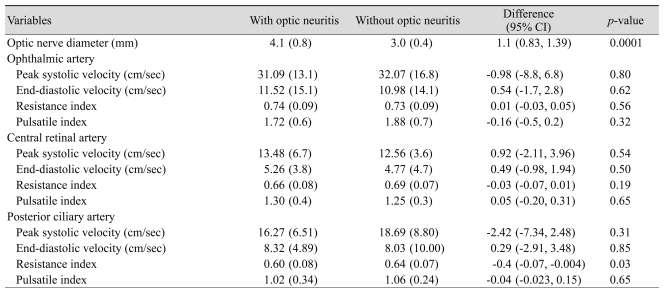
ON involved the right eye in 12 patients (52.2%) and the left eye in the remaining 11 (47.8%) patients. Of the 23 eyes with ON, 20 (86.9%) showed a thickening of the affected optic nerve. Only three eyes with ON had a difference in nerve diameter less than 0.3 mm. The increase in diameter of the affected nerve compared with the unaffected contralateral nerve averaged (SD) 1.1 (0.6) mm (range, 0.1 to 2.2 mm). When the cut-off value for nerve pathology was set at a nerve diameter of ≥4 mm, 15 (65.2%) of the affected nerves and 100% of the unaffected nerves were correctly assessed. The mean (SD) values of parameters obtained from CDI measurements in the OA, CRA, PCAs and optic nerve in the 23 pairs of eyes with and without ON are shown in Table 1. As expected, those eyes with ON had a higher mean optic nerve diameter. The mean (SD) optic nerve diameter was 4.1 (0.8) mm for eyes with ON and 3.0 (0.4) mm for eyes without ON (p < 0.001). There were no statistically significant differences between eyes with and without ON in terms of PSV, EDV, RI, and PI in OA and CRA measurements. The mean RI in the PCAs was slightly lower in eyes with ON than in control eyes (0.60 vs. 0.64, p < 0.05).
Table 1.
Means (SD) of characteristics measured in the ophthalmic, central retinal, posterior ciliary arteries, and optic nerve in 23 pairs of eyes with and without optic neuritis
The difference in the mean of the variables between eyes with optic neuritis and those without optic neuritis.
SD = standard deviation; CI = confidence interval.
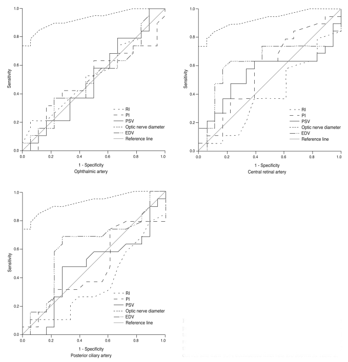
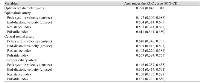
The areas under the ROC curves for the occurrence of ON for orbital blood flow velocities and resistance indices of OA, CRA, PCAs, and optic nerve diameter are shown in Fig. 1 and Table 2. The area under the ROC curve was 0.928 (95% CI [confidence interval]: 0.843, 0.101) for optic nerve diameter. Optic nerve diameter was a significant predictor of ON (p < 0.001). The areas under the ROC curves were 0.497 (95% CI: 0.306, 0.688) for PSV, 0.504 (95% CI: 0.314, 0.695) for EDV, 0.534 (95% CI: 0.345, 0.722) for RI and 0.503 (95% CI: 0.311, 0.695) for PI in OA. None of the blood flow parameters were significantly higher in ON eyes. PSV, EDV, RI, and PI of the OA, CRA, and PCAs covered a similar area. The EDV of the CRA and PCAs had areas slightly but not significantly larger than that of the other blood flow parameters.
Fig. 1.
Receiver operating characteristic (ROC) curves for the peak systolic velocity (PSV), end-diastolic velocity (EDV), resistance index (RI), and pulsatile index (PI) of ophthalmic, central retinal and posterior ciliary arteries and optic nerve diameter for the diagnosis of optic neuritis. The estimated area under the ROC curves and their 95% confidence intervals are shown in Table 2.
Table 2.
Area under the ROC curve (95% CI) of optic nerve diameter and blood flow parameters of the ophthalmic, central retinal and posterior ciliary arteries
ROC = receiver operating characteristic; CI = confidence interval.
Discussion
In this study we found no significant difference in PSV, EDV, or PI of the OA, CRA, and PCAs between eyes with and without ON. The mean RI in the PCA was slightly lower in the eyes with ON than in the contralateral eyes. The results confirms the reliability of optic nerve diameter in the diagnosis of ON. Thickening of the optic nerve results from the inflammation in ON and has been described previously [4,20]. The observed thickening is likely to be a major cause of the initial loss of visual acuity [4]. The OA enters through the optic canal, together with the optic nerve. The enlarged optic nerve in ON compresses the OA within the optic canal and this compression may contribute to the impairment of orbital hemodynamics.
In this study, we observed a slightly lower RI in PCA. The anterior optic nerve derives its blood supply from the PCA. PSV and EDV are both dependent on the Doppler angle. The RI is angle-independent and provides a good measurement by which to quantify vascular resistance in circulation, particularly in tortuous vessels like the PCA.
Few studies have performed CDI to assess orbital blood flow velocities and optic nerve diameter in ON; these were usually of limited sample size and the results produced were inconsistent. The inconsistency may be explained in part by differences in patient characteristics, the cause of ON, definitions of ON, blood flow velocity measurements and the amount of time elapsed between the acute attack and examination. While differences in hemodynamic parameters over the course of the disease may partly account for the range of results seen in the literature, it is much more likely that the main component of variance in ON Doppler studies results from differences in instrumentation and technique. Large foot-print transducers, in the 7.5- to 10.0-MHz range, with poor lateral resolution, placed directly on the closed eyelid at the orbital ridge while not specifically avoiding the lens will not yield optimal measurements. Elvin et al. [4] evaluated only the resistive indices of the CRA; they found resistive indices increased in the affected side and attributed the difference to nerve swelling and the resultant resistance to flow. The velocities were not measured and the OA was not evaluated. Our findings also differ from those of Karaali et al. [5], who found that PSV, EDV, and RI in the OA are increased in patients with acute ON although the velocities and resistive indices of the CRA in affected and normal eyes did not differ significantly. Hradilek et al. [11] also reported that PSV, PI, and RI in the OA are increased in patients with acute ON although the EDV in affected and normal eyes did not differ significantly. However, these changes do not persist over a long period. Furthermore, Pache et al. [9] reported a significant reduction in the PSV and EDV of the OA, PCAs, and CRA in patients with MS compared with healthy controls. Modrzejewska et al. [12] also reported statistically significant diminishing blood flow velocity parameters in the eyeball arteries of patients with both MS and ON. They did not observe any changes in vascular resistance indices when compared with the control group. Orbital blood flow velocities were measured several months after ON onset. The absence of abnormalities in vascular resistance could be due to a comparatively long passage of time between the clinical manifestation of ON and the study. Similar to our findings, Akarsu et al. [8] reported that in patients with MS, the mean PSV, EDV, and RI in the OA of eyes with ON were not significantly different from those in unaffected fellow eyes and healthy control eyes. The mean EDV in the CRA was lower and the mean resistivity indices in the CRA and PCA were higher in eyes with ON than in control eyes. Goh et al. [10] reported results similar to ours. They found that orbital hemodynamics in all retrobulbar vessels did not significantly differ from normal controls in patients with compressive, inflammatory, toxic or hereditary optic neuropathy. Our findings indicate that optic nerve diameter is the most reliable and practical predictor of ON.
The pathophysiology of ON has not been fully described, but thickening of the nerve in association with demyelination and inflammation may compress the blood vessels within the optic canal. Alternatively, vasospasm due to an increased plasma level of endothelin-1 [9], a potent vasoconstrictor, may result in vasospasm and vascular dysregulation. This could cause an increase in resistance to flow in the artery and through ischemia lead to eventual exoplasmic stasis and visual loss [21]. Evaluation with gray-scale sonography of the optic nerve and CDI of orbital hemodynamics in patients with acute ON, combined with proof of decreased RI in PCAs may facilitate an accurate diagnosis.
One limitation of this study is that blood flow measurements were only performed in the contralateral eyes of the patients without an additional control group. However, as values of orbital blood flow velocity are variables within the healthy population, any differences in values between the orbit affected by ON and the unaffected orbit are highly significant. Use of the contralateral eye as an internal control therefore seems practical since CDI measurement is known to be influenced by cardiac output and the patient's stress level [22]. Moreover, ON swelling may influence CDI readings to an unknown degree independent of changes in blood flow due to the altered scattering properties of tissue. Additionally, Akarsu et al. [8] demonstrated that there was a similarity in blood flow velocities between contralateral unaffected eyes and healthy control eyes. Further studies are required to fully clarify this issue. None of the patients were being treated with systemic or topical corticosteroids. The patients in our series were under 50 years old, an age at which glaucoma is uncommon and intraocular pressure was normal in all patients. An experimental study by Guthoff et al. [23] showed that an increase in intraorbital pressure leads to a decrease in CRA flow. Great care was thus taken to apply as little pressure as possible to the patient's eye during CDI examination. Even though the study included the thorough examination of 23 pairs of eyes, the findings should be interpreted with caution due to the relatively small sample size.
CDI measurements were performed by expert sonographers. Although we have not carried out any special reliability studies, CDI measurements of the eye vessels [23] and between the right and left eyes [24] have been shown to be reproducible over time.
In conclusion, our findings indicates that gray-scale sonography is a non-invasive and repeatable diagnostic method, which reveals significant optic nerve thickening on the side affected by ON and could play a role in disease diagnosis. No unilateral differences were observed in the average PSV, EDV, RI, or PI of the OA and CRA. The reduced RI in PCAs may indicate disturbances in retinal and choroidal circulation in patients with ON. Further studies with larger groups of patients are needed with age- and sex-matched control groups in order to better understand the role of retrobulbar hemodynamics in the pathogenesis of ON.
Acknowledgements
We thank Professor R. B. Jones, K. Shashok, and Sarah Griffin-Mason (Author AID in the Eastern Mediterranean) for improving the English usage in the manuscript.
Footnotes
No potential conflict of interest relevant to this article was reported.
References
- 1.Acevedo AR, Nava C, Arriada N, et al. Cardiovascular dysfunction in multiple sclerosis. Acta Neurol Scand. 2000;101:85–88. doi: 10.1034/j.1600-0404.2000.101002085.x. [DOI] [PubMed] [Google Scholar]
- 2.Flachenecker P, Wolf A, Krauser M, et al. Cardiovascular autonomic dysfunction in multiple sclerosis: correlation with orthostatic intolerance. J Neurol. 1999;246:578–586. doi: 10.1007/s004150050407. [DOI] [PubMed] [Google Scholar]
- 3.Speciale L, Sarasella M, Ruzzante S, et al. Endothelin and nitric oxide levels in cerebrospinal fluid of patients with multiple sclerosis. J Neurovirol. 2000;6(Suppl 2):S62–S66. [PubMed] [Google Scholar]
- 4.Elvin A, Andersson T, Soderstrom M. Optic neuritis: Doppler ultrasonography compared with MR and correlated with visual evoked potential assessments. Acta Radiol. 1998;39:243–248. doi: 10.1080/02841859809172188. [DOI] [PubMed] [Google Scholar]
- 5.Karaali K, Senol U, Aydin H, et al. Optic neuritis: evaluation with orbital Doppler sonography. Radiology. 2003;226:355–358. doi: 10.1148/radiol.2262011915. [DOI] [PubMed] [Google Scholar]
- 6.Akarsu C, Bilgili MY. Color Doppler imaging in ocular hypertension and open-angle glaucoma. Graefes Arch Clin Exp Ophthalmol. 2004;242:125–129. doi: 10.1007/s00417-003-0809-3. [DOI] [PubMed] [Google Scholar]
- 7.Hradilek P, Zapletalova O, Dolezil D, Skoloudik D. Acute optic ne uritis in multiple sclerosis: evaluation of hemodynamics in the ophthalmic artery with colour Doppler imaging. Neuro-ophthalmology. 2005;29:161–164. [Google Scholar]
- 8.Akarsu C, Tan FU, Kendi T. Color Doppler imaging in optic neuritis with multiple sclerosis. Graefes Arch Clin Exp Ophthalmol. 2004;242:990–994. doi: 10.1007/s00417-004-0948-1. [DOI] [PubMed] [Google Scholar]
- 9.Pache M, Kaiser HJ, Akhalbedashvili N, et al. Extraocular blood flow and endothelin-1 plasma levels in patients with multiple sclerosis. Eur Neurol. 2003;49:164–168. doi: 10.1159/000069085. [DOI] [PubMed] [Google Scholar]
- 10.Goh KY, Kay MD, Hughes JR. Orbital color Doppler imaging in nonischemic optic atrophy. Ophthalmology. 1997;104:330–333. doi: 10.1016/s0161-6420(97)30315-7. [DOI] [PubMed] [Google Scholar]
- 11.Hradilek P, Stourac P, Bar M, et al. Colour Doppler imaging evaluation of blood flow parameters in the ophthalmic artery in acute and chronic phases of optic neuritis in multiple sclerosis. Acta Ophthalmol. 2009;87:65–70. doi: 10.1111/j.1755-3768.2008.01195.x. [DOI] [PubMed] [Google Scholar]
- 12.Modrzejewska M, Karczewicz D, Wilk G. Assessment of blood flow velocity in eyeball arteries in multiple sclerosis patients with past retrobulbar optic neuritis in color Doppler ultrasonography. Klin Oczna. 2007;109:183–186. [PubMed] [Google Scholar]
- 13.Erdogmus S, Govsa F. Topography of the posterior arteries supplying the eye and relations to the optic nerve. Acta Ophthalmol Scand. 2006;84:642–649. doi: 10.1111/j.1600-0420.2006.00673.x. [DOI] [PubMed] [Google Scholar]
- 14.Baxter GM, Williamson TH. Color Doppler imaging of the eye: normal ranges, reproducibility, and observer variation. J Ultrasound Med. 1995;14:91–96. doi: 10.7863/jum.1995.14.2.91. [DOI] [PubMed] [Google Scholar]
- 15.Dees C, Buimer R, Dick AD, Atta HR. Ultrasonographic investigation of optic neuritis. Eye (Lond) 1995;9(Pt 4):488–494. doi: 10.1038/eye.1995.113. [DOI] [PubMed] [Google Scholar]
- 16.Williamson TH, Harris A. Color Doppler ultrasound imaging of the eye and orbit. Surv Ophthalmol. 1996;40:255–267. doi: 10.1016/s0039-6257(96)82001-7. [DOI] [PubMed] [Google Scholar]
- 17.Planiol T, Pourcelot L, Itti R. The carotid and cerebral circulations. Advances in its study by external physical methods. Principles, normal recordings, adopted parameters. Nouv Presse Med. 1973;2:2451–2456. [PubMed] [Google Scholar]
- 18.Gosling RG, King DH. Arterial assessment by Doppler-shift ultrasound. Proc R Soc Med. 1974;67(6 Pt 1):447–449. [PMC free article] [PubMed] [Google Scholar]
- 19.DeLong ER, DeLong DM, Clarke-Pearson DL. Comparing the areas under two or more correlated receiver operating characteristic curves: a nonparametric approach. Biometrics. 1988;44:837–845. [PubMed] [Google Scholar]
- 20.Gerling J, Janknecht P, Hansen LL, Kommerell G. Diameter of the optic nerve in idiopathic optic neuritis and in anterior ischemic optic neuropathy. Int Ophthalmol. 1997;21:131–135. doi: 10.1023/a:1026422819404. [DOI] [PubMed] [Google Scholar]
- 21.McLeod D, Marshall J, Kohner EM. Role of axoplasmic transport in the pathophysiology of ischaemic disc swelling. Br J Ophthalmol. 1980;64:247–261. doi: 10.1136/bjo.64.4.247. [DOI] [PMC free article] [PubMed] [Google Scholar]
- 22.Mostbeck GH, Gossinger HD, Mallek R, et al. Effect of heart rate on Doppler measurements of resistive index in renal arteries. Radiology. 1990;175:511–513. doi: 10.1148/radiology.175.2.2183288. [DOI] [PubMed] [Google Scholar]
- 23.Guthoff RF, Berger RW, Winkler P, et al. Doppler ultrasonography of the ophthalmic and central retinal vessels. Arch Ophthalmol. 1991;109:532–536. doi: 10.1001/archopht.1991.01080040100037. [DOI] [PubMed] [Google Scholar]
- 24.MacKenzie F, De Vermette R, Nimrod C, et al. Doppler sonographic studies on the ophthalmic and central retinal arteries in the gravid woman. J Ultrasound Med. 1995;14:643–647. doi: 10.7863/jum.1995.14.9.643. [DOI] [PubMed] [Google Scholar]


