

A 5-year-old boy weighing 8 kg presented with recurrent pulmonary infections, occasional nonbilious vomiting, intermittent cough, and failure to thrive since birth. On physical examination, vital signs were normal, but on auscultation left-sided fine crackles were found. The child was anemic, other blood biochemistry levels were within the normal range. A chest X-ray and computed tomography (CT) revealed bronchiectasis localized to the left hemithorax and a radiolucent air column within a distended esophagus. Other causes of chronic pulmonary diseases, such as cystic fibrosis, congenital immune deficiencies and primary ciliary dyskinesia were excluded. A barium swallow demonstrated a dilated esophagus with an air-fluid level and sudden narrowing at the distal end [Figure 1]. Esophageal manometry showed uncoordinated, low-amplitude peristalsis of the esophageal body and the lower sphincter pressure was normal.
Figure 1.
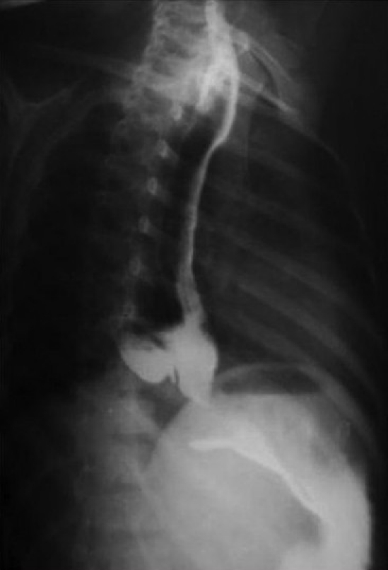
A barium swallow showing pneumoesophagus and a dilated esophagus with a narrowed cardioesophageal junction
QUESTION
Q1. What is the diagnosis?
ANSWER
The patient underwent rigid bronchoscopy, which revealed a 2 × 2 mm defect on the posterior wall of the trachea about 3 cm above the carina. The fistulous opening was catheterized with a ureteric catheter and left there as a guide. The neck was explored through a right transverse cervical incision. The fistula was identified, ligated, and excised. The trachea and the esophagus were repaired in a transverse and vertical fashion, respectively. The patient was discharged on pantoprazole, antibiotics and advice for chest physiotherapy.
H-type tracheoesophageal fistula (H-type TEF) is characterized by a clinical triad of paroxysmal coughing precipitated by feeding, gaseous distention of the gastrointestinal tract, and pneumonitis.[1] Although mostly diagnosed in infancy, some cases diagnosed in adulthood have also been reported.[2] Esophageal dysmotility may be a spectrum of congenital TEF anomalies or acquired due to distension.[2,3] So in delayed cases of H-type TEF dysmotility, dilated esophagus and associated respiratory infection mimics achalasia clinically and radiologically.[2,4]
H-type TEF is a rare anomaly comprising only 4% of tracheoesophageal anomalies,[5] whereas achalasia occurs in 5% of children younger than 15 years.[2] Seventy percent of H-type TEF occurs at or above the level of the second thoracic vertebra and extends in an oblique course like the letter N, from the posterior wall of the trachea to the anterior wall of the esophagus, and this is the reason why routine barium swallows done in the erect posture often miss it.[5] On barium swallow the air easily passed from the trachea to the esophagus, producing air-filled dilated esophagus (pneumoesophagus) with a normal lower esophageal sphincter.
The cineesophagography with manometry can distinguish both more precisely, as contrary to H-TEF, in achalasia there is incoordinate relaxation of the lower esophageal sphincter with contraction of the body of esophagus. Direct sagittal CT, HRCT, three-dimensional CT, and virtual bronchoscopy are the most sensitive to detect H-type TEF.[5]
There are no definite guidelines established for the management of this rare situation yet. Most authors, except Boybeyi et al., had done cardiomyotomy with or without fundoplication with fistula repair in the same sitting. It seems to be reasonable as gastroesophageal reflux disease has an incidence of 10% in H-type TEF.[1–3,6]
Footnotes
Source of Support: Nil
Conflict of Interest: None declared.
REFERENCES
- 1.Ng J, Bartram J, Antao B, Everard M, Shawis R. H-type tracheoesophageal fistula masquerading as achalasia cardia in a 13-year-old child. J Paediatr Child Health. 2006;42:215–6. doi: 10.1111/j.1440-1754.2006.00833.x. [DOI] [PubMed] [Google Scholar]
- 2.Olivet RT, Payne WS. Congenital H-type tracheoesophageal fistula complicated by achalasia in an adult: Report of a case. Mayo Clin Proc. 1975;50:464–8. [PubMed] [Google Scholar]
- 3.Stephens RW, Lingeman RE, Lawson LJ. Congenital tracheoesophageal fistulas in adults. Ann Otol Rhinol Laryngol. 1976;85:613–7. doi: 10.1177/000348947608500508. [DOI] [PubMed] [Google Scholar]
- 4.LaSalle AJ, Andrassy RJ, Ver Steeg K, Ratner I. Congenital tracheoesophageal fistula without esophageal atresia. J Thorac Cardiovasc Surg. 1979;78:583–8. [PubMed] [Google Scholar]
- 5.Le SD, Lam WW, Tam PK, Cheng W, Chan FL. H-type tracheo-oesophageal fistula: Appearance on three-dimensional computed tomography and virtual bronchoscopy. Pediatr Surg Int. 2001;17:642–3. doi: 10.1007/s003830100012. [DOI] [PubMed] [Google Scholar]
- 6.Boybeyi O, Köse M, Ersöz DD, Haliloglu M, Karnak I, Senocak ME. Achalasia-like findings in a case with delayed diagnosis of H-type tracheoesophageal fistula. Pediatr Surg Int. 2008;24:965–9. doi: 10.1007/s00383-008-2192-y. [DOI] [PubMed] [Google Scholar]