Fig. 4.
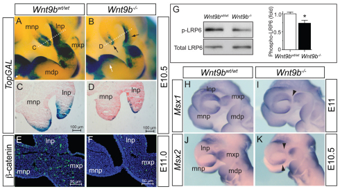
Disruption of WNT/β-catenin signaling in Wnt9b–/– embryos. (A-D) TopGAL expression detected by colorimetric staining of β-galactosidase activity in wild-type and Wnt9b–/– mouse embryos. Black arrows indicate the NPs where significantly reduced TopGAL expression was observed in Wnt9b–/– embryos. There was no reduction of TopGAL expression in the MdP (white arrow in B). Dotted white lines in A and B indicate the plane of sections shown in C and D, respectively. Decreased TopGAL expression was apparent in both surface ectoderm and mesenchyme of the NPs (C,D). (E,F) Immunofluorescent staining of active β-catenin protein (green) in the facial processes at E11.0. (G) Western blot analysis of phosphorylated LRP6 in NP and MxP tissue lysates. Total LRP6 level provided a protein loading control. Signal levels from the triplicate samples were quantified using ImageJ and are presented as mean±s.e.m. *P<0.05, Student’s t-test. (H-K) Msx1 and Msx2 expression in E10.5-11 embryos was determined by whole-mount in situ hybridization. Arrowheads indicate the reduced expression domains in Wnt9b–/– embryos. mdp, mandibular process of branchial arch 1; lnp, lateral nasal process; mnp, medial nasal process; mxp, maxillary process.

