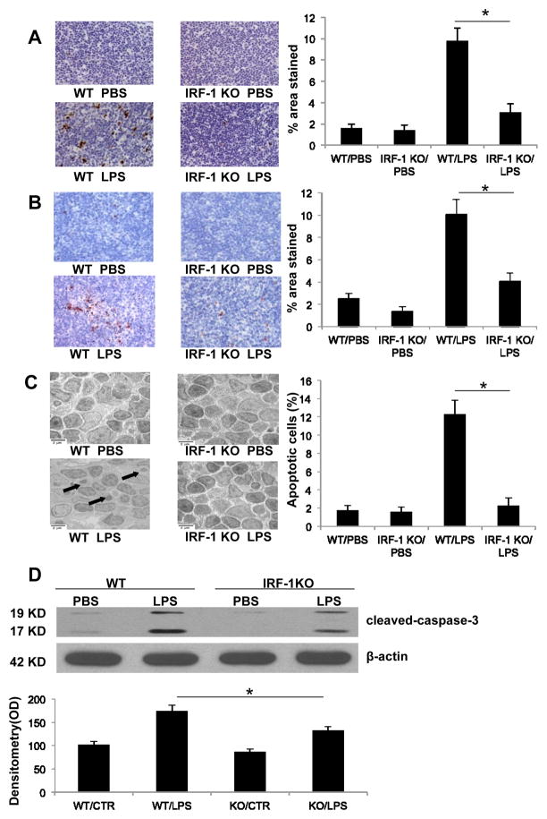
Figure 2. IRF-1 KO mice have decreased apoptosis in splenocytes following LPS exposure.
Permanent blocks of splenic tissue obtained from WT and IRF-1 KO mice 16 h following PBS or LPS (20 mg/kg) injection were sectioned and (A) TUNEL staining or (B) cleaved caspase-3 immunohistochemistry staining was performed (magnitude X 200). Positive staining was presented as percentage of stained area over total area (% area stained). (C) Splenic tissue obtained from WT and IRF-1 KO mice 16 h following PBS or LPS (20 mg/kg) injection was imaged by transmission electron microscope (magnitude X 5000). Arrow points to apoptotic bodies. Percentage of apoptotic cells among total cells was used for apoptosis quantification. (D) WT and IRF-1 KO mice were injected with PBS or LPS (20 mg/kg) for 16 h and splenocytes were isolated. Caspase-3 cleavage was analyzed by Western blot. *, P<0.05; results are representative of 3 separate independent experiments.

