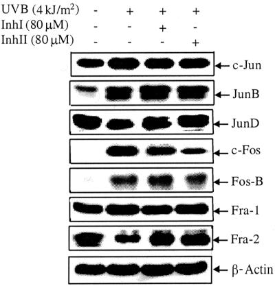
Figure 6.
Various AP-1 protein compositional patterns after UVB stimulation and Inh I or Inh II treatment, assessed by Western blotting. Cl 41 cells were cultured and treated and Western blotting was carried out as indicated in the text. JunD and Fra-2 proteins were attenuated after UVB treatment and increased again when cells were treated with Inh I or Inh II before UVB. c-Jun, JunB, c-Fos, FosB, and Fra-1 proteins were increased after UVB exposure and inhibited when cells were pretreated with either Inh I or Inh II before UVB treatment. Western immunoblotting was performed three times with samples from various cell preparations. Similar results were obtained each time and a representative result is shown. β-Actin was used as internal control to ensure equal protein loading.

