Abstract
Oysters, as a major group of marine bivalves, can tolerate a wide range of natural and anthropogenic stressors including heat stress. Recent studies have shown that oysters pretreated with heat shock can result in induced heat tolerance. A systematic study of cellular recovery from heat shock may provide insights into the mechanism of acquired thermal tolerance. In this study, we performed the first network analysis of oyster transcriptome by reanalyzing microarray data from a previous study. Network analysis revealed a cascade of cellular responses during oyster recovery after heat shock and identified responsive gene modules and key genes. Our study demonstrates the power of network analysis in a non-model organism with poor gene annotations, which can lead to new discoveries that go beyond the focus on individual genes.
Introduction
Oysters are a major group of marine bivalves which represent about 8,000 species worldwide [1]. They usually inhabit coastal shallow waters and estuaries, and like other marine ectotherms they can tolerate a wide range of natural and anthropogenic stressors such as thermal fluctuation, anoxia, osmotic change, and a variety of toxicants. Among environmental factors, temperature has long been recognized as a key factor that can potentially influence all physiological processes in marine ectotherms [2]. Oysters can experience rapid and dramatic temperature fluctuations during diurnal/tidal cycles (up to 10∼20°C within a few hours) and seasonal changes (from 0 to 35∼40°C) [3]. It has been shown that thermal tolerance is rather a complex physiological trait which requires the initiation of coordinated cellular responses to thermal stress [4]. Many oyster studies focused on understanding of cellular responses during heat stress [5], [6], whereas the recovery process after heat shock has been less studied. Recent studies have shown that oysters recovered from sublethal heat stress could be more resistant to subsequent thermal stress (i.e. acquired thermal tolerance) [7]. However, most of these studies focused on heat shock proteins (HSPs) or HSP-related proteins [4], [7], [8]. A systematic study of cellular recovery from heat shock would provide new insights into the mechanism of acquired thermal tolerance. Lang et al. [9] have recently performed the transcriptome profiling of selectively bred Pacific oyster (Crassostrea gigas) families during recovery after heat shock (RHS) using an oyster cDNA microarray containing 13,752 features [10]. Although they identified ∼110 candidate genes that showed differential expression patterns during RHS, their analytical procedure basically represents a gene-centric approach that focuses on individual genes with high statistical significance. Such approach ignores gene interactions and might suffer from lack of sufficient contextual information for generating scientifically sound hypotheses.
Recent developments in statistical genomics provide a foundation for a shift from the gene-centric to a network- or module-centric approach in microarray data analysis [11]. In addition to determining the roles of individual genes, network analysis enables researchers to study cells as a complex network of biochemical factors. Although network analysis has been widely used in gene expression studies of human and model organisms [12]–[14], little effort has been devoted to expanding its application to the less-studied non-model organisms such as oysters.
Here we present the first network analysis of oyster transcriptome by reanalyzing microarray datasets from Lang et al. [9] to identify gene modules and candidate key genes responsible for oyster RHS.
Results and Discussion
Network construction and identification of an RHS-responsive module
In the study of Lang et al. [9], transcriptome profiling was performed using an oyster cDNA microarray for Pacific oyster families that were sampled at different recovery times (1, 3, 6 and 24 hours) after heat shock (40°C for 1 hour). Because barely little difference of gene expression was observed among families in their study, microarray data from these families were combined to increase the power for detection of coexpression patterns in the network analysis.
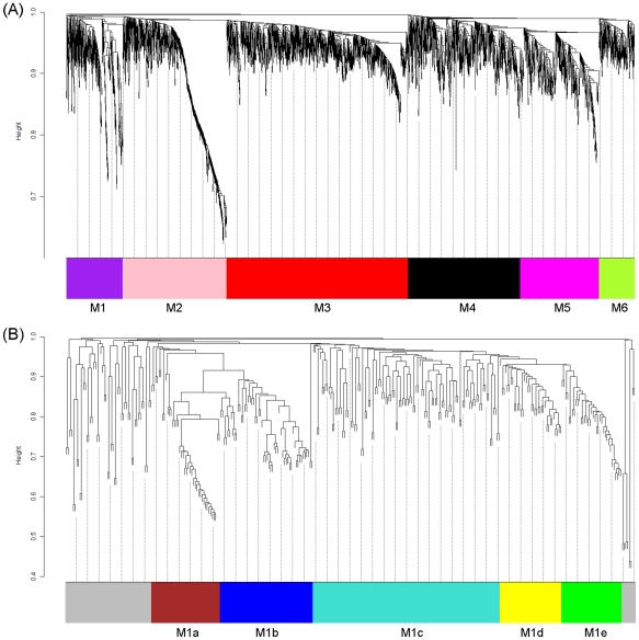
Network analysis was performed using a recently developed weighted gene co-expression network analysis (WGCNA) method [15] that enables identification of transcriptional modules and key/driver genes within modules based on gene-to-gene correlations across all microarray samples. A total of 60 samples (12 oyster samples×5 time points) were used for calculation of gene-to-gene correlations. The oyster cDNA microarray contained 13,752 probes, of which 3,362 probes representing 1,668 genes passed the quality filter and were included in the network construction. In total, six modules (M1∼M6) containing almost all expressed genes were identified with module size ranging from 211 to 1,075 (Figure 1, Table S1). It is worth mentioning that for genes that had probe replicates, 96% of the replicate probes were assigned to the same module colors, suggesting the high reproducibility of the microarray data as well as the high reliability of network construction. The analysis of variance (ANOVA) revealed that 336 probes were differentially expressed (FDR<0.05) among sampling times (T0, T1, T3, T6 and T24), accounting for ∼10% of all expressed genes. To identify modules responsive to heat stress, enrichment analysis of differentially expressed genes (DEGs) was performed for each module using a hypergeometric test. It turned out that only M1 was significantly enriched with DEGs (p = 4e-128). Of the 332 probes in this module, 193 (58%) were DEGs.
Figure 1. Network analysis of the oyster gill transcriptome during recovery after heat shock (RHS).
(A) and subnetwork analysis of the RHS-responsive module M1 (B). Dendrograms are produced by average linkage hierarchical clustering of genes on the basis of topological overlap (see Methods for details). Modules of coexpressed genes are labelled in unique colors. Unassigned genes are labelled in grey.
M1 subnetwork
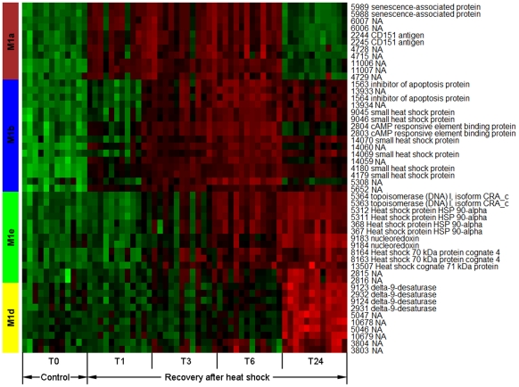
In order to gain a better understanding of coexpression patterns in M1, a subnetwork was constructed for M1. As shown in Figure 1, M1 was composed of five submodules (M1a∼M1e). Hub genes (i.e. top 15% genes with high intramodular connectivity) in M1 only distributed in four submodules (M1a, M1b, M1d and M1e). A heat map was constructed for visualization of coexpression patterns of these hub genes in the 4 submodules (Figure 2). Hub genes in M1a showed elevated expression at T1, T3 and T6 after heat shock. Two hub genes in this module were annotated; one was SAG (senescence-associated protein) and the other was CD151 (cluster of differentiation 151). Activation of SAG indicates the induction of cellular senescence [16], [17]. It has been shown that senescence and apoptosis can compete with each other in an exclusive way, and senescence can proceed when apoptosis is inhibited [18]. A recent study has revealed that activation of CD151 can facilitate the inhibition of apoptosis possibly through regulation of Bax and Bcl-2 genes [19]. Besides CD151, M1a contained other annotated DEGs such as CDH1 (Cadherin-1), activation of which is also associated with inhibition of apoptosis [20]. Therefore, coexpression patterns observed in M1a may indicate the ongoing transition from apoptosis to cellular senescence. At T24, gene expression in M1a went back to the normal level, possibly suggesting the termination of cellular senescence.
Figure 2. Heat map visualization of coexpression patterns of hub genes in submodules M1a, M1b, M1d and M1e.
T0 represents an oyster control without heat treatment, whereas time points T1, T3, T6 and T24 represent recovery time (hour) after heat shock. Probe IDs and their associated gene annotations are shown on the right of the heat map. Red, up-regulation; Green, down-regulation.
Subsequent to M1a, hub genes in M1b showed responsive expression at T3, T6 and T24 after heat shock. IAP (inhibitor of apoptosis protein), CREB (cAMP response element-binding protein) and sHSPs (small heat shock proteins) are annotated hub genes in this module. IAP blocks apoptosis at the core of the apoptotic machinery by inhibiting effector caspases [21]. Interaction between IAP and CREB was demonstrated in a previous study showing that CREB can regulate the promoter activity of IAP by binding to the IAP's enhancer sequence [22]. In response to heat stress, sHSPs can stabilize protein conformation, prevent aggregation and thereby maintain the non-native proteins in a competent state for subsequent refolding, which is achieved by other HSPs/chaperones (e.g. HSP70 and HSP90) [23]. In addition, sHSPs also play an important role in inhibition of apoptosis [24]–[26]. It has been proposed that HSP-mediated regulation of the apoptotic pathways probably constitutes a fundamental protective mechanism that decreases cellular sensitivity to damaging events to allow cells to escape the otherwise inevitable engagement of apoptosis [27]. Therefore, M1b is enriched with genes functioning in inhibition of apoptosis as well as stabilization of protein conformation.
M1e contained hub genes with increased expression at T6 and T24, indicating a later response during RHS than M1a and M1b. HSP70 and HSP90 are dominant hub genes in this module, which are well known as molecular chaperones that help in the refolding of misfolded proteins and assist in their elimination if they become irreversibly damaged. Elevated expression of TOP1 (topoisomerase I) was observed in this module, which involves in regulation of DNA supercoiling that might be accumulated during rapid induction of the heat-shock genes [28]. It has been shown that TOP1 plays an important role in the acquisition of thermotolerance probably by preventing inhibition of further transcription of HSPs caused by hyper negative supercoiling [29]. Expression of NRX (nucleoredoxin) was also increased in this module. NRXs are a novel member of thioredoxin family. Members of the thioredoxin family have been shown to function as facilitators and regulators of protein folding [30]. Taken together, it seems that M1e is enriched with protein-refolding associated genes.
M1d was composed of hub genes with increased expression only at T24. ACOD (delta-9-desaturase) is the only annotated hub gene in this module, which functions in conversion of saturated fatty acids to monounsaturated fatty acids. Up-regulation of ACOD indicated the increase of unsaturation of lipid membrane (i.e. increased membrane fluidity) during RHS. Cell membranes are known to be a highly sensitive monitor of the most diverse environmental changes. The unsaturation level of membranes is involved in the transduction of thermal stress into cellular signals, thus affecting the general stress-response mechanisms [31]. The increase of membrane fluidity can elevate the sensitivity of cells to heat [32], implying that when oysters are subject to subsequent exposure to heat stress, the cells might respond at a temperature lower than the original threshold, causing the repair system to function faster than previously. This speculation may well explain the previous observations that oysters pretreated with heat shock can result in induced heat tolerance [7]. It has also been shown that desaturase activation or hyper-induction plays an important role in the response to heat stress in certain thermotolerant yeast and bacterial strains [33], [34].
Network visualization of the four submodules revealed a cascade of cellular responses (M1a→M1b→M1e→M1d) during RHS. As shown in Figure 3, inter-modular interactions only occurred between adjacent modules. For example, M1b is the only module that interacts with M1a, but no interactions between M1a and the other two modules. According to Figure 3, the scenario of cellular responses during oyster RHS is likely to be as follows: (i) after heat shock, cellular senescence was induced accompanied by inhibition of apoptosis (M1a); (ii) sHSPs were expressed to stabilize protein conformation (M1b) and facilitate further protein refolding by HSP70 and HSP90 (M1e); and (iii) then increase of membrane fluidity was induced, which possibly enhanced the sensitivity of cells to subsequent heat stress.
Figure 3. Network visualization of submodule interactions.
Each node represents a hub gene. Hub genes derived from the same submodule are labelled in the same color. Hub genes are connected by an edge if the topological overlap between them is more than 0.13. Interactions (i.e. edges) between modules are labelled in grey.
In addition, network analysis enables identification of key genes responsible for module interactions. One advantage of the coexpression network analysis is that it does not rely on the information of gene annotation for identification of key genes in a module, thus providing the opportunity for identification of novel candidate genes in non-model species with poorly characterized genome. For example, from a network perspective, the unannotated gene (probe IDs 14059 and 14060) in M1b seems to be an important hub gene that is responsible for interactions between M1a and M1b. However, this gene could be possibly overlooked by the traditional analysis methods since no annotation has been assigned to this gene. Network analysis also revealed that NRX and ACOD are important hub genes that maintain the interactions between M1e and M1d. Although there is no documented direct interaction between these two genes, it might be worthy of further investigation due to the vital role of ACOD in maintaining the fluidity of lipid membrane.
In summary, we performed the first network analysis of oyster transcriptome by reanalyzing microarray datasets from Lang et al. [9]. Network analysis revealed a cascade of cellular responses during oyster RHS and identified responsive gene modules and candidate key genes. Our study demonstrates the power of network analysis in a non-model organism with poor gene annotations, which can lead to new exciting discoveries that go beyond the focus on individual genes.
Materials and Methods
Ethics Statement
Not applicable. Our research did not involve human participants or samples.
Microarray data acquisition
The vsn-transformed microarray data from Lang et al. [9] were downloaded from the Gene Expression Ominibus website (http://www.ncbi.nlm.nih.gov/projects/geo; Series GSE12070, GSM304764∼GSM304823).
Probe annotation
In order to increase the probe annotation rate, 206,388 ESTs and 1,080,743 raw reads from a 454 sequencing of Crassostrea gigas transcriptome project (SRA accession no. SRX032364 and SRX032365) were downloaded from NCBI databases and then assembled using the Newbler v2.3 program (Roche) with default parameter settings. The probes that can be unambiguously mapped to the assembled isotigs were annotated by BlastX the corresponding isotigs against the Nr and SwissProt databases with an e-value threshold of 1E-6. For the remaining probes, they were directly compared against the Nr and SwissProt databases using BlastX with the same e-value threshold. To increase the computational speed, BlastX searches were limited to the first 20 significant hits for each query with non-characterized matching entries (e.g. hypothetical genes/proteins) excluded. Gene names were assigned to each probe based on the best Blast hit, and the corresponding information was provided in Table S1.
Microarray data pre-processing
The oyster cDNA microarray contains 13,752 probes, of which 3,362 passed the previously defined signal-intensity filter [9] and were included in the subsequent analysis. Outlier values for each gene were removed based on the Grubbs' test (p<0.05).
Gene network construction
Gene network was constructed using the R package WGCNA following the procedure described in [35]. Here we chose a power of eleven so that the resulting networks exhibited approximate scale-free topology (model fitting index R∧2 = 0.71). Next, all genes were hierarchically clustered based on dissimilarity measure of topological overlap which measures inter-connectedness for a pair of genes [15]. The resulting gene dendrogram was used for module detection using the Dynamic Tree Cut method (minimum module size = 80 and cutting height = 0.995) [36]. Fine cutting (minimum module size = 30 and cutting height = 0.997) was further performed for the module of interest.
Identification of RHS-responsive modules
Differential expression analysis among sampling times was conducted for each probe using analysis of variance (ANOVA). To account for multiple tests, false discovery rate (FDR) was calculated using the qvalue package [37]. Only probes with q<0.05 were considered to be differentially expressed. To identify the RHS-responsive module, overrepresentation analysis of DGEs was performed for each module using a hypergeometric test (p<0.05).
Hub gene selection and visualization
Hub genes refer to highly connected genes in a module [15]. They can be determined by calculating the intramodular connectivity Kin, which is a measure of a gene's connection strength to other genes in a module. In this study, top 15% genes with high Kin were considered as hub genes for a given module. Coexpression patterns and interactions of hub genes were visualized using the heat map and Cytoscape [38], respectively.
Supporting Information
Combined results of probe annotation, WGCNA analysis and differential expression analysis.
(XLS)
Footnotes
Competing Interests: The authors have declared that no competing interests exist.
Funding: Financial support for this work was provided by National Natural Science Foundation of China (31172384), National High Technology Research and Development Program of China (2012AA10A401), Public Science and Technology Research Funds Projects of Ocean (201205031), and Program for New Century Excellent Talents in University (NCET-10-0761). The funders had no role in study design, data collection and analysis, decision to publish, or preparation of the manuscript.
References
- 1.Boss KJ. Mollusca. In: Parker SP, editor. Synopsis and classification of living organisms. Volume 1. New York: McGraw-Hill; 1982. pp. 945–1166. [Google Scholar]
- 2.Kinne O. The effects of temperature and salinity in marine and brackish water animals. J Temp Oceanogr Mar Biol Annu Rev. 1963;1:301–340. [Google Scholar]
- 3.Ivanina AV, Taylor C, Sokolova IM. Effects of elevated temperature and cadmium exposure on stress protein response in eastern oysters Crassostrea virginica (Gmelin). Aquat Toxicol. 2009;91:245–254. doi: 10.1016/j.aquatox.2008.11.016. [DOI] [PubMed] [Google Scholar]
- 4.Hamdoun AM, Cheney DP, Cherr GN. Phenotypic plasticity of HSP70 and HSP70 gene expression in the Pacific oyster (Crassostrea gigas): implications for thermal limits and induction of thermal tolerance. Biol Bull. 2003;205:160–169. doi: 10.2307/1543236. [DOI] [PubMed] [Google Scholar]
- 5.Meistertzheim AL, Tanguy A, Moraga D, Thébault MT. Identification of differentially expressed genes of the Pacific oyster Crassostrea gigas exposed to prolonged thermal stress. FEBS J. 2007;274:6392–6402. doi: 10.1111/j.1742-4658.2007.06156.x. [DOI] [PubMed] [Google Scholar]
- 6.Farcy E, Voiseux C, Lebel JM, Fiévet B. Transcriptional expression levels of cell stress marker genes in the Pacific oyster Crassostrea gigas exposed to acute thermal stress. Cell Stress Chaperon. 2009;14:371–380. doi: 10.1007/s12192-008-0091-8. [DOI] [PMC free article] [PubMed] [Google Scholar]
- 7.Shamseldin A, Clegg JS, Friedman CS, Cherr GN, Pillai MC. Induced thermotolerance in the Pacific oyster, Crassostrea gigas. J Shellfish Res. 1997;16:487–491. [Google Scholar]
- 8.Clegg JS, Uhlinger KR, Jackson SA, Cherr GN, Rifkin E, et al. Induced thermotolerance and the heat-shock protein-70 family in the Pacfic oyster Crassostrea gigas. Mol Mar Biol Biotechnol. 1998;7:21–30. [Google Scholar]
- 9.Lang RP, Bayne CJ, Camara MD, Cunningham C, Jenny MJ, et al. Transcriptome profiling of selectively bred Pacific oyster Crassostrea gigas families that differ in tolerance of heat shock. Mar Biotechnol. 2009;11:650–668. doi: 10.1007/s10126-009-9181-6. [DOI] [PMC free article] [PubMed] [Google Scholar]
- 10.Jenny MJ, Chapman RW, Mancia A, Chen YA, McKillen DJ, et al. A cDNA microarray for Crassostrea virginica and C. gigas. Mar Biotechnol. 2007;9:577–591. doi: 10.1007/s10126-007-9041-1. [DOI] [PubMed] [Google Scholar]
- 11.Lee WP, Tzou WS. Computational methods for discovering gene networks from expression data. Brief Bioinform. 2009;10:408–423. doi: 10.1093/bib/bbp028. [DOI] [PubMed] [Google Scholar]
- 12.Lee I, Date SV, Adai AT, Marcotte EM. A probabilistic functional network of yeast genes. Science. 2004;306:1555–1558. doi: 10.1126/science.1099511. [DOI] [PubMed] [Google Scholar]
- 13.Wille A, Zimmermann P, Vranová E, Fürholz A, Laule O, et al. Sparse graphical Gaussian modeling of the isoprenoid gene network in Arabidopsis thaliana. Genome Biol. 2004;5:R92. doi: 10.1186/gb-2004-5-11-r92. [DOI] [PMC free article] [PubMed] [Google Scholar]
- 14.Oldham MC, Konopka G, Iwamoto K, Langfelder P, Kato T, et al. Functional organization of the transcriptome in human brain. Nat Neurosci. 2008;11:1271–1282. doi: 10.1038/nn.2207. [DOI] [PMC free article] [PubMed] [Google Scholar]
- 15.Zhang B, Horvath S. A general framework for weighted gene co-expression network analysis. Stat Appl Genet Mol Biol. 2005;4:Article17. doi: 10.2202/1544-6115.1128. [DOI] [PubMed] [Google Scholar]
- 16.Chen GH, Huang LT, Yap MN, Lee RH, Huang YJ, et al. Molecular characterization of a senescence-associated gene encoding cysteine proteinase and its gene expression during leaf senescence in sweet potato. Plant Cell Physiol. 2002;43:984–991. doi: 10.1093/pcp/pcf125. [DOI] [PubMed] [Google Scholar]
- 17.Yap MN, Lee RH, Huang YJ, Liao CJ, Chen SC. Molecular characterization of a novel senescence-associated gene SPA15 induced during leaf senescence in sweet potato. Plant Mol Biol. 2003;51:471–481. doi: 10.1023/a:1022334820332. [DOI] [PubMed] [Google Scholar]
- 18.Rebbaa A, Zheng X, Chou PM, Mirkin BL. Caspase inhibition switches doxorubicin-induced apoptosis to senescence. Oncogene. 2003;22:2805–2811. doi: 10.1038/sj.onc.1206366. [DOI] [PubMed] [Google Scholar]
- 19.Liu T, Lui Z, Hu L, Zeng K, Gao B. Effects of recombined adeno-associated virus mediated CD151 gene on myocyte apoptosis and Bax/Bcl-2 proteins late after myocardial infarction. J Clin Cardiol. 2010;26:212–215. [Google Scholar]
- 20.Liu W, Li W, Fujita T, Yang Q, Wan Y. Proteolysis of CDH1 enhances susceptibility to UV radiation-induced apoptosis. Carcinogenesis. 2008;29:263–272. doi: 10.1093/carcin/bgm251. [DOI] [PubMed] [Google Scholar]
- 21.Fulda S, Debatin KM. Targeting inhibitor of apoptosis proteins (IAPs) for diagnosis and treatment of human diseases. Recent Pat Anticancer Drug Discov. 2006;1:81–89. doi: 10.2174/157489206775246539. [DOI] [PubMed] [Google Scholar]
- 22.Dong Z, Nishiyama J, Yi X, Venkatachalam MA, Denton M, et al. Gene promoter of apoptosis inhibitory protein IAP2: identification of enhancer elements and activation by severe hypoxia. Biochem J. 2002;364:413–421. doi: 10.1042/BJ20011431. [DOI] [PMC free article] [PubMed] [Google Scholar]
- 23.Wang W, Vinocur B, Shoseyov O, Altman A. Role of plant heat-shock proteins and molecular chaperones in the abiotic stress response. Trends Plant Sci. 2004;9:244–252. doi: 10.1016/j.tplants.2004.03.006. [DOI] [PubMed] [Google Scholar]
- 24.Kamradt MC, Lu M, Werner ME, Kwan T, Chen F, et al. The small heat shock protein alpha B-crystallin is a novel inhibitor of TRAIL-induced apoptosis that suppresses the activation of caspase-3. J Biol Chem. 2005;280:11059–11066. doi: 10.1074/jbc.M413382200. [DOI] [PubMed] [Google Scholar]
- 25.Havasi A, Li Z, Wang Z, Martin JL, Botla V, et al. Hsp27 inhibits Bax activation and apoptosis via a phosphatidylinositol 3-kinase-dependent mechanism. J Biol Chem. 2008;283:12305–12313. doi: 10.1074/jbc.M801291200. [DOI] [PMC free article] [PubMed] [Google Scholar]
- 26.Pasupuleti N, Matsuyama S, Voss O, Doseff AI, Song K, et al. The anti-apoptotic function of human αA-crystallin is directly related to its chaperone activity. Cell Death Dis. 2010;1:e31. doi: 10.1038/cddis.2010.3. [DOI] [PMC free article] [PubMed] [Google Scholar]
- 27.Beere HM. “The stress of dying”: the role of heat shock proteins in the regulation of apoptosis. J Cell Sci. 2004;117:2641–2651. doi: 10.1242/jcs.01284. [DOI] [PubMed] [Google Scholar]
- 28.Liu LF, Wang JC. Supercoiling of the DNA template during transcription. Proc Natl Acad Sci USA. 1987;84:7024–7027. doi: 10.1073/pnas.84.20.7024. [DOI] [PMC free article] [PubMed] [Google Scholar]
- 29.Tse-Dinh YC, Qi H, Menzel R. DNA supercoiling and bacterial adaptation: thermotolerance and thermoresistance. Trends Microbiol. 1997;5:323–326. doi: 10.1016/s0966-842x(97)01080-9. [DOI] [PubMed] [Google Scholar]
- 30.Berndt C, Lillig CH, Holmgren A. Thioredoxins and glutaredoxins as facilitators of protein folding. Biochim Biophys Acta. 2008;1783:641–650. doi: 10.1016/j.bbamcr.2008.02.003. [DOI] [PubMed] [Google Scholar]
- 31.Chatterjee MT, Khalawan SA, Curran BP. Cellular lipid composition influences stress activation of the yeast general stress response element (STRE). Microbiology. 2000;146:877–884. doi: 10.1099/00221287-146-4-877. [DOI] [PubMed] [Google Scholar]
- 32.Hidvégi EJ, Yatvin MB, Dennis WH, Hidvégi E. Effect of altered membrane lipid composition and procaine on hyperthermic killing of ascites tumor cells. Oncology. 1980;37:360–363. doi: 10.1159/000225470. [DOI] [PubMed] [Google Scholar]
- 33.Guerzoni ME, Ferruzzi M, Sinigaglia M, Criscuoli GC. Increased cellular fatty acid desaturation as a possible key factor in thermotolerance in Saccharomyces cerevisiae. Can J Microbiol. 1997;43:569–576. doi: 10.1139/m97-080. [DOI] [PubMed] [Google Scholar]
- 34.Guerzoni ME, Lanciotti R, Cocconcelli PS. Alteration in cellular fatty acid composition as a response to salt, acid, oxidative and thermal stresses in Lactobacillus helveticus. Microbiology. 2001;147:2255–2264. doi: 10.1099/00221287-147-8-2255. [DOI] [PubMed] [Google Scholar]
- 35.Ponomarev I, Wang S, Zhang L, Harris RA, Mayfield RD. Gene coexpression networks in human brain identify epigenetic modifications in alcohol dependence. J Neurosci. 2012;32:1884–1897. doi: 10.1523/JNEUROSCI.3136-11.2012. [DOI] [PMC free article] [PubMed] [Google Scholar]
- 36.Langfelder P, Horvath S. WGCNA: an R package for weighted correlation network analysis. BMC Bioinformatics. 2008;9:559. doi: 10.1186/1471-2105-9-559. [DOI] [PMC free article] [PubMed] [Google Scholar]
- 37.Storey JD. A direct approach to false discovery rates. J Roy Stat Soc Ser B. 2002;64:479–498. [Google Scholar]
- 38.Smoot ME, Ono K, Ruscheinski J, Wang PL, Ideker T. Cytoscape 2.8: new features for data integration and network visualization. Bioinformatics. 2011;27:431–432. doi: 10.1093/bioinformatics/btq675. [DOI] [PMC free article] [PubMed] [Google Scholar]
Associated Data
This section collects any data citations, data availability statements, or supplementary materials included in this article.
Supplementary Materials
Combined results of probe annotation, WGCNA analysis and differential expression analysis.
(XLS)


