Abstract
Oral lichen planus (OLP) is a chronic inflammatory disease that affects the mucus membrane of the oral cavity. It is a T-cell mediated autoimmune disease in which the cytotoxic CD8+ T cells trigger apoptosis of the basal cells of the oral epithelium. Several antigen-specific and nonspecific inflammatory mechanisms have been put forward to explain the accumulation and homing of CD8+ T cells subepithelially and the subsequent keratinocyte apoptosis. A wide spectrum of treatment modalities is available, from topical corticosteroids to laser ablation of the lesion. In this review, we discuss the various concepts in the pathogenesis and current treatment modalities of OLP.
Keywords: Apoptosis, autoimmune, basal keratinocytes, corticosteroids, oral lichen planus
INTRODUCTION
Lichen planus is a chronic inflammatory disease that affects the skin and the mucus membrane. Oral lichen planus (OLP), the mucosal counterpart of cutaneous lichen planus, presents frequently in the fourth decade of life and affects women more than men in a ratio of 1.4:1.[1] The disease affects 1–2% of the population.[2,3] It is seen clinically as reticular, papular, plaque-like, erosive, atrophic or bullous types. Intraorally, the buccal mucosa, tongue and the gingiva are commonly involved although other sites may be rarely affected.[4] Oral mucosal lesions present alone or with concomitant skin lesions. The skin lesions present as violaceous flat-topped papules in ankles, wrist, and genitalia, but characteristically the facial skin is spared.
The etiology and pathogenesis of OLP has been the focus of much research, and several antigen-specific and nonspecific inflammatory mechanisms have been put forward to explain the pathogenesis. Although mostly palliative, a spectrum of treatment modalities is in practice, from topical application of steroids to laser therapy. In this review, we discuss the recent concepts in the pathogenesis and current treatment modalities of OLP.
PATHOGENESIS
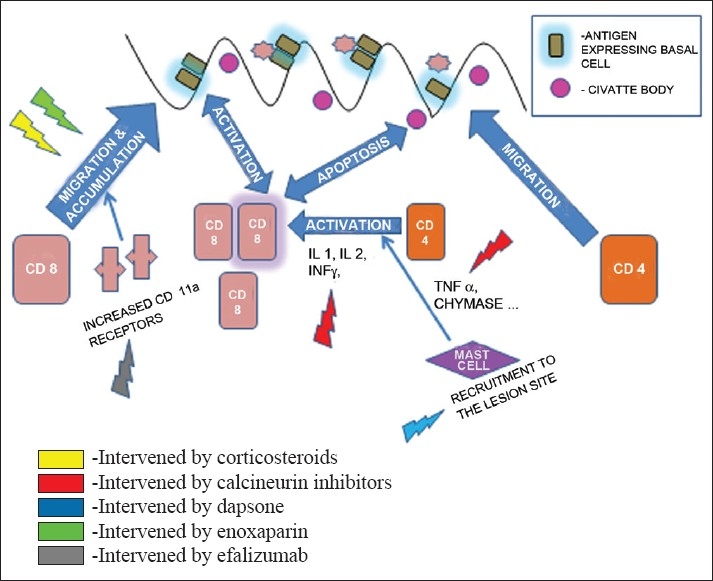
OLP is a T-cell mediated autoimmune disease in which the auto-cytotoxic CD8+ T cells trigger apoptosis of the basal cells of the oral epithelium.[5] An early event in the disease mechanism involves keratinocyte antigen expression or unmasking of an antigen that may be a self-peptide or a heat shock protein.[1,6] Following this, T cells (mostly CD8+, and some CD4+ cells) migrate into the epithelium either due to random encounter of antigen during routine surveillance or a chemokine-mediated migration toward basal keratinocytes.[1] These migrated CD8+ cells are activated directly by antigen binding to major histocompatibility complex (MHC)-1 on keratinocyte or through activated CD4+ lymphocytes. In addition, the number of Langerhan cells in OLP lesions are increased along with upregulation of MHC-II expression; subsequent antigen presentation to CD4+ cells and Interleukin (IL)-12 activates CD4 + T helper cells which activate CD8+ T cells through receptor interaction, interferon γ (INF – γ) and IL-2. The activated CD8+ T cells in turn kill the basal keratinocytes through tumor necrosis factor (TNF)-α, Fas–FasL mediated or granzyme B activated apoptosis.[1,6]
A CYTOKINE-MEDIATED LYMPHOCYTE HOMING MECHANISM
Attraction of the lymphocytes to the epithelium–connective tissue interface has also been proposed to be due to cytokine-mediated upregulation of adhesion molecules on endothelial cells and concomitant expression of receptor molecules by circulating lymphocytes. In OLP, there is increased expression of the vascular adhesion molecules (CD62E, CD54, CD106) by the endothelial cells of the subepithelial vascular plexus.[7] The infiltrating lymphocytes express reciprocal receptors (CD11a) to these vascular adhesion molecules. This supports the above-explained hypothesis that the cytokine-mediated lymphocyte homing mechanism plays an important role in the pathogenesis of lichen planus. Some of the cytokines that are responsible for the upregulation of the adhesion molecules are: TNF-α, IFN-γ and IL-1. These are derived from the resident macrophages, Langerhans cells, lymphocytes and the overlying keratinocytes themselves, thus setting up a vicious cycle.[7]
The normal integrity of the basement membrane is maintained by a living basal keratinocyte due to its secretion of collagen 4 and laminin 5 into the epithelial basement membrane zone. In turn, keratinocytes require a basement membrane derived cell survival signal to prevent the onset of its apoptosis. Apoptotic keratinocytes are no longer able to perform this function, which results in disruption of the basement membrane. Again, a non-intact basement membrane cannot send a cell survival signal. This sets in a vicious cycle which relates to the chronic nature of the disease.[1,8]
The matrix metalloproteinases (MMP) are principally involved in tissue matrix protein degradation. MMP- 9, which cleaves collagen 4, along with its activators is upregulated in OLP lesional T cells, resulting in increased basement membrane disruption.[9]
RANTES (Regulated on Activation, Normal T-cell Expressed and Secreted) is a member of the CC chemokine family which plays a critical role in the recruitment of lymphocytes and mast cells in OLP. CCR1, CCR3, CCR4, CCR5, CCR9 and CCR10, which are cell surface receptors for RANTES, have been identified in lichen planus.[1,10] The recruited mast cell undergoes degranulation under the influence of RANTES, which releases chymase and TNF-α. These substances upregulate RANTES secretion by OLP lesional T cells. This again sets in a vicious cycle which relates to the chronic nature of the disease.[10]
60% mast cells have been found to be degranulated in OLP compared to 20% in normal mucosa. Mast cell degranulation releases a range of pro-inflammatory mediators such as TNF-α, chymase and tryptase. TNF-α upregulates the expression of endothelial cell adhesion molecules (CD62E, CD54 and CD106) in OLP, which is required for lymphocyte adhesion to the luminal surfaces of blood vessels and subsequent extravasation and stimulates RANTES secretion from T cells. Chymase, a mast cell protease, is a known activator of MMP-9, leading to basement membrane disruption in OLP.[1,9]
Weak expression of transforming growth factor (TGF)-β1 has been found in OLP. TGF-β1 deficiency may predispose to autoimmune lymphocytic inflammation. The balance between TGF-β1 and IFN-γ determines the level of immunological activity in OLP lesions. Local overproduction of IFN-γ by CD4+ T cells in OLP lesions downregulates the immunosuppressive effect of TGF-β1 and upregulates keratinocyte MHC class II expression and CD8+ cytotoxic T-cell activity.[1,8]
HEPATITIS C VIRUS INFECTION AND ORAL LICHEN PLANUS
Epidemiological evidences from more than 90 controlled studies worldwide strongly suggest that Hepatitis C Virus (HCV) may be an etiologic factor in OLP. The association seems to be prevalent in Southern Europe, Japan and USA. However, countries with highest prevalence of HCV report negative or nonsignificant associations suggesting that the LP–HCV association cannot be explained on the basis of high prevalence in population alone. In OLP, HCV replication has been reported in the epithelial cells from mucosa of LP lesions by reverse transcription/polymerase chain reaction or in-situ hybridization; also, HCV-specific CD4 and CD8 lymphocytes were reported in the subepithelial band. These probably suggest that HCV-specific T lymphocytes may play a role in the pathogenesis of OLP. The characteristic band like lymphocytic infiltrate might thus be directed toward HCV infected cells. Whether HCV infected patients have increased risk of developing OLP or patients with OLP have enhanced risk of developing HCV infection is yet to be answered. The putative pathogenetic link between OLP and HCV still remains controversial and needs a lot of prospective and interventional studies for a better understanding.[11]
DIFFERENTIAL DIAGNOSIS
The diagnosis of reticular lichen planus can often be made based on the clinical findings alone. Interlacing white striae appearing bilaterally on the posterior buccal mucosa is often pathognomonic. Difficulties arise often when there is superimposed candidal infection which masquerades the classic reticular pattern and in eliciting the erosive and erythematous forms of OLP. The differential diagnosis can include cheek chewing/frictional keratosis, lichenoid reactions, leukoplakia, lupus erythematosus, pemphigus, mucus membrane pemphigoid, erythematous candidiasis and chronic ulcerative stomatitis. Lichenoid drug reactions are usually unilateral in distribution, accompanied by a history of new drug intake. The most reliable method to diagnose lichenoid drug reactions is to note if the reaction resolves after the offending drug is withdrawn, and returns if the patient is challenged again. Dental restorative material induced lichenoid reactions can be identified when OLP like lesions are confined to areas of the oral mucosa in close contact or proximity to restorative materials, usually amalgam. A positive patch test, a strong clinical correlation of proximity of a restoration and biopsy suggestive of diffuse lymphocytic infiltrate rather than a subepithelial band favor a diagnosis of oral lichenoid reactions. Clinically, lesions of lupus erythematosus (LE) most often resemble erosive lichen planus but tend to be less symmetrically distributed. The keratotic striae of LE are much more delicate and subtle than Wickham's striae and show a characteristic radiation from the central focus. Biopsy of LE shows a characteristic perivascular infiltrate.
Erosive or atrophic types that usually affect the gingiva should be differentiated from pemphigoid, as both may have a desquamative clinical appearance. Both pemphigus and pemphigoid occur as solitary erythematous lesions and are not associated with any white striae. This can aid in clinical differential diagnosis as erosive and atrophic forms of OLP usually show concomitant reticular form. Peeling of the epithelium from the epithelium–connective tissue junction on slight lateral pressure in nonaffected area (Nikolsky's sign) differentiates it from erosive and erythematous forms of lichen planus. A biopsy from the perilesional tissue can diagnose pemphigus or pemphigoid, which show intraepithelial or subepithelial split histologically. In some cases, erythema multiforme (EM) can resemble bullous lichen planus, but EM is more acute and generally involves the labial mucosa. Chronic ulcerative stomatitis (CUS) is an immune-mediated disorder affecting the oral mucosa which clinically and histopathologically resembles lichen planus. Diagnosis of CUS is based on direct immunofluorescence studies where autoantibodies are directed against p63 in the basal and parabasal layers of the epithelium. These lesions have to be differentiated from lichen planus because CUS does not respond to corticosteroid therapy and has to be treated using antimalarial drugs.[12]
RECENT CONCEPTS IN TREATMENT
Corticosteroids have been the mainstay of management of OLP; yet, other modalities like calcineurin inhibitors, retinoids, dapsone, hydroxychloroquine, mycophenolate mofetil and enoxaparin have contributed significantly toward treatment of the disease.Analysis of current data on pathogenesis of the disease suggests that blocking IL-12, IFN-γ, TNF-α, RANTES, or MMP-9 activity or upregulating TGF-β1 activity in OLP may be of therapeutic value in the future.[1,13]
Corticosteroids
These are the most commonly used group of drugs for the treatment of OLP.[14] The rationale behind their usage is their ability to modulate inflammation and immune response. They act by reducing the lymphocytic exudate and stabilizing the lysosomal membrane.[15] Topical midpotency corticosteroids such as triamcinolone acetonide, high-potent fluorinated corticosteroids such as fluocinonide acetonide, disodium betamethasone phosphate, and more recently, superpotent halogenated corticosteroids such as clobetasol are used based on the severity of the lesion. The greatest disadvantage in using topical corticosteroids is their lack of adherence to the mucosa for a sufficient length of time. Although trials were done using topical steroids along with adhesive base, no study shows their superiority when compared to steroids without the base (carboxymethyl cellulose).[16] However, the same study also recommends the usage of adhesive paste used for dentures, which contains only inactive ingredients as a vehicle to carry the topical application. This has shown excellent bioadhesive properties, due to its high molecular weight (above 100,000) and the flexibility of the polymeric chain. Small and accessible erosive lesions located on the gingiva and palate can be treated by the use of an adherent paste in a made-to-measure tray (custom tray), which allows for accurate control over the contact time and ensures that the entire lesional surface is exposed to the drugs.[17] Patients with widespread forms of OLP are prescribed high-potent and superpotent corticosteroids mouthwashes and intralesional injections. Long-term use of topical steroid can lead to the development of secondary candidiasis which necessitates antifungal therapy.[15] The potential tachyphylaxis and adrenal insufficiency is high when using superpotent steroids like clobetaso l, especially when used for a longer period of time. Systemic corticosteroids are reserved for recalcitrant erosive or erythematous LP where topical approaches have failed. Systemic prednisolone is the drug of choice, but should be used at the lowest possible dosage for the shortest duration (40–80 mg for 5–7 days).[14]
OTHER IMMUNOSUPPRESSANTS AND IMMUNOMODULATORY AGENTS
Calcineurin inhibitors
Calcineurin is a protein phosphatase which is involved in the activation of transcription of IL-2, which stimulates the growth and differentiation of T-cell response.[18] In immunosuppressive therapy, calcineurin is inhibited by cyclosporine, tacrolimus and pimecrolimus. These drugs are called calcineurin inhibitors.
Cyclosporine
Cyclosporine, a calcineurin inhibitor, is an immunosuppressant used widely in post-allogenic organ transplant to reduce the activity of patient's immune system. This selectively suppresses T-cell activity, the main reason for transplant rejection, and hence enhances the uptake of the foreign organ. Cyclosporine binds to the cytosolic protein cyclophilin of immunocompetent lymphocytes, especially T-lymphocytes. This complex of cylosporine and cyclophilin inhibits calcineurin, which under normal circumstances induces the transcription of IL-2. They also inhibit lymphokine production and IL release, leading to a reduced function of effector T-cells. Cyclosporine is used as a mouth rinse or topically with adhesive bases in OLP. However, the solution is prohibitively expensive and should be reserved for highly recalcitrant cases of OLP. Systemic absorption is very low.[14] It is known to cause dose-related gum hyperplasia which reduces when the drug is withdrawn.
Tacrolimus
Tacrolimus, also a calcineurin inhibitor, is a steroid-free topical immunosuppressive agent approved for the treatment of atopic dermatitis. It is 10–100 times as potent as cyclosporine and has greater percutaneous absorption than cyclosporine. It has been successfully used in recalcitrant OLP cases. This substance is produced by Streptomyces tsukubaensis and belongs to the macrolide family. The immunosuppressive action of tacrolimus is similar to that of cyclosporine, although it has a greater capacity to penetrate the mucosa. It inhibits the first phase of T-cell activation, inhibiting the phosphatase activity of calcineurin. Burning sensation is the commonest side effect observed; relapses of OLP after cessation have also been observed. The US Food and Drug Administration has recently issued a potential cancer risk from the prolonged use of tacrolimus and has recommended its use for short periods of time and not continuously.[14,18]
Pimecrolimus
Pimecrolimus inhibits T-cell activation by inhibiting the synthesis and release of cytokines from T cells. Pimecrolimus also prevents the release of inflammatory cytokines and mediators from mast cells. 1% topical cream of pimecrolimus has been successfully used as treatment for OLP. Pimecrolimus has significant anti-inflammatory activity and immunomodulatory capabilities with low systemic immunosuppressive potential.[19,20]
Retinoids
Topical retinoids such as tretinoin, isotretinoin and fenretinide, with their immunomodulating properties, have been reported to be effective in OLP. Reversal of white striae can be achieved with topical retinoids, although effects may only be temporary. Systemic retinoids have been used in cases of severe lichen planus with variable degree of success. The positive effects of retinoids should be weighed against their rather significant side effects like cheilitis, elevation of serum liver enzymes and triglyceride levels and teratogenicity.[8,21]
Dapsone
As an antibacterial agent, dapsone inhibits bacterial synthesis of dihydrofolic acid and hence is used in the treatment of leprosy. When used for the treatment of skin diseases, it probably acts as an anti-inflammatory agent by inhibiting the release of chemotactic factors for mast cells.[22] The most common untoward effect of dapsone is hemolysis of varying degree, which is dose related and develops in almost every individual administered 200–300 mg of oral dapsone daily. Glucose-6-phosphate dehydrogenase (G6PD) deficiency can increase the risk of hemolytic anemia or methemoglobinemia in patients receiving dapsone. Screening for G6PD deficiency is required before prescribing dapsone. Hypersensitivity reaction to dapsone called Dapsone reaction is frequent in patients receiving multiple drug therapy. The symptoms of rash, fever and jaundice generally occur within the first 6 weeks of therapy and can be ameliorated by corticosteroid therapy.[23]
Mycophenolates
Originally used to treat psoriasis, mycophenolic acid (now reformulated as mycophenolate mofetil) has been reintroduced in dermatological medicine. Being a very well-tolerated immunosuppressive drug used in organ transplant, it has been successfully used to treat severe cases of OLP. Mycophenolates are quite expensive and effective with long-term usage.[24]
Low-dose, low molecular weight heparin (enoxaparin)
Low-dose heparin devoid of anticoagulant properties inhibits T lymphocyte heparanase activity which is crucial in T-cell migration to target tissues. This promises to be a simple, effective and safe treatment for OLP when injected subcutaneously as it has no side effects.[25]
Efalizumab
It is a recombinant humanized monoclonal antibody which is used as an immunosuppressant in the treatment of psoriasis. Efalizumab, a monoclonal antibody to CD11a, binds to this adhesion molecule and causes improvement in OLP by decreased activation and trafficking of T lymphocytes. In vitro studies of mononuclear cells in OLP have demonstrated a decrease of 60% in migration by peripheral blood mononuclear cells after pretreatment with anti-CD11a antibodies. It is administered once a week as a subcutaneous injection. It is currently an approved drug for the treatment of plaque psoriasis.[26] Figure 1 gives a schematic representation of the probable sites of action of drugs based on their property in OLP.
Figure 1.

Proposed sites of action based on property of drugs in oral lichen planus
NON-PHARMACOLOGICAL MODALITIES
PUVA therapy
This non-pharmacologic approach uses photochemotherapy with 8-methoxypsoralen and long wave ultraviolet light (PUVA). Psoralens are compounds found in many plants, which make the skin temporarily sensitive to UV radiation. Methoxypsoralen is given orally, followed by administration of 2 hours of UV radiation intraorally in the affected sites. It has been successfully used in the treatment of severe cases of OLP.[27] Two major disadvantages of PUVA therapy include the adverse effects of nausea and dizziness secondary to psoralen and 24-hour photosensitivity when this medicine is taken orally. Also, dosimetry can be difficult within the complicated geometry of the mouth, because PUVA is usually administered on skin over large, open surfaces.[28]
Photodynamic therapy
Photodynamic therapy (PDT) is a technique that uses a photosensitizing compound like methylene blue, activated at a specific wavelength of laser light, to destroy the targeted cell via strong oxidizers, which cause cellular damage, membrane lysis, and protein inactivation. PDT has been used with relative success in the field of oncology, notably in head and neck tumors. PDT is found to have immunomodulatory effects and may induce apoptosis in the hyperproliferating inflammatory cells which are present in psoriasis and lichen planus. This may reverse the hyperproliferation and inflammation of lichen planus.[29]
Laser therapy
In patients who are suffering from painful erosive OLP and are unresponsive to even topical superpotent corticosteroids, surgical management using cryosurgery and different types of laser have also been tried. A 980-nm Diode laser,[30] CO2 laser evaporation,[31] biostimulation with a pulsed diode laser using 904-nm pulsed infrared rays[32] and low-dose excimer 308-nm laser with UV-B rays have been tried.[28] All types of laser destroy the superficial epithelium containing the target keratinocytes by protein denaturation. A deeper penetrating beam like the diode laser destroys the underlying connective tissue with the inflammatory component along the epithelium. The few studies documented show a lot of promise, but their effectiveness is yet to be proven.
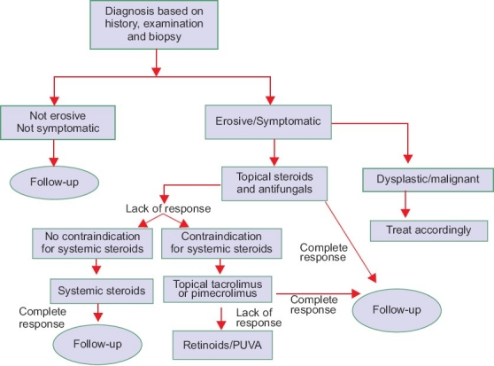
No therapy for OLP is completely curative; the goal of treatment for symptomatic patients is palliation. The following [Figure 2] simple systematic protocol will aid in effective treatment.
Figure 2.

Protocol/algorithm for treatment of oral lichen planus
Relief can be achieved in a majority of cases through topical application of corticosteroids, with or without the combination of other immunomodulators. Very rarely does the condition necessitate systemic therapy. Laser therapy and other recent modalities are tried as the final remedy.
CONCLUSION
OLP is a very common oral dermatosis and one of the most frequent mucosal pathosis encountered by dental practitioners. It is imperative that the lesion is identified precisely and proper treatment be administered at the earliest. A proper understanding of the pathogenesis of the disease becomes important for providing the right treatment.
Footnotes
Source of Support: Nil
Conflict of Interest: None declared.
REFERENCES
- 1.Sugerman PB, Savage NW, Walsh LJ, Zhao ZZ, Zhou XJ, Khan A, et al. The pathogenesis of oral lichen planus. Crit Rev Oral Biol Med. 2002;13:350–65. doi: 10.1177/154411130201300405. [DOI] [PubMed] [Google Scholar]
- 2.Bouquot JE, Gorlin RJ. Leukoplakia, lichen planus, and other oral keratoses in 23,616 white Americans over the age of 35 years. Oral Surg Oral Med Oral Pathol. 1986;61:373–81. doi: 10.1016/0030-4220(86)90422-6. [DOI] [PubMed] [Google Scholar]
- 3.Scully C, Beyli M, Ferreiro MC, Ficarra G, Gill Y, Griffiths M, et al. Update on oral lichen planus:etiopathogenesis and management. Crit Rev Oral Biol Med. 1998;9:86–122. doi: 10.1177/10454411980090010501. [DOI] [PubMed] [Google Scholar]
- 4.Ismail SB, Kumar SK, Zain RB. Oral lichen planus and Lichenoid reactions; Etiopathogenesis, diagnosis, management and malignant transformation. J Oral Sci. 2007;49:89–106. doi: 10.2334/josnusd.49.89. [DOI] [PubMed] [Google Scholar]
- 5.Eversole LR. Immunopathogenesis of oral lichen planus and recurrent aphthous stomatitis. Semin Cutan Med Surg. 1997;16:284–94. doi: 10.1016/s1085-5629(97)80018-1. [DOI] [PubMed] [Google Scholar]
- 6.Zhou XJ, Sugarman PB, Savage NW, Walsh LJ, Seymour GJ. Intra-epithelial CD8+ T cells and basement membrane disruption in oral lichen planus. J Oral Pathol Med. 2002;31:23–7. doi: 10.1046/j.0904-2512.2001.10063.x. [DOI] [PubMed] [Google Scholar]
- 7.Regezi JA, Dekker NP, MacPhail LA, Lozada-Nur F, McCalmont TH. Vascular adhesion molecules in oral lichen planus. Oral Surg Oral Med Oral Pathol Oral Radiol Endod. 1996;81:682–90. doi: 10.1016/s1079-2104(96)80074-6. [DOI] [PubMed] [Google Scholar]
- 8.Lodi G, Scully C, Carozzo M, Griffiths M, Sugerman PB, Thongprasom K. Current controversies in oral lichen planus: Report on an international consensus meeting. Part 1. Viral infections and etiopathogenesis. Oral Surg Oral Med Oral Pathol Oral Radiol Endod. 2005;100:40–51. doi: 10.1016/j.tripleo.2004.06.077. [DOI] [PubMed] [Google Scholar]
- 9.Zhou XJ, Sugerman PB, Savage NW, Walsh LJ. Matrix metalloproteinases and their inhibitors in oral lichen planus. J Cutan Pathol. 2001;28:72–82. doi: 10.1034/j.1600-0560.2001.280203.x. [DOI] [PubMed] [Google Scholar]
- 10.Zhao ZZ, Sugerman PB, Zhou XJ, Walsh LJ, Savage NW. Mast cell degranulation and the role of T cell RANTES in oral lichen planus. Oral Dis. 2001;7:246–51. [PubMed] [Google Scholar]
- 11.Carrozzo M, Thorpe R. Oral lichen planus – a review. Minerva Stomatol. 2009;58:519–37. [PubMed] [Google Scholar]
- 12.Neville BW, Damm DD, Allen CM, Bouquot JE. 3rd Ed. India: Elsevier; 2009. Oral and MaxilloFacial Pathology; pp. 782–90. [Google Scholar]
- 13.Roopashree MR, Gondhalekar RV, Shashikanth MC, George J, Thippeswamy SH, Shukla A. Pathogenesis of oral lichen planus-a review. J Oral Pathol Med. 2010;39:729–34. doi: 10.1111/j.1600-0714.2010.00946.x. [DOI] [PubMed] [Google Scholar]
- 14.Eisen D, Carrozzo M, Bagan Sebastian JV, Thongprasom K. Oral lichen planus: Clinical features and management. Oral Dis. 2005;11:338–49. doi: 10.1111/j.1601-0825.2005.01142.x. [DOI] [PubMed] [Google Scholar]
- 15.Vincent SD, Fotos PG, Baker KA, Williams TP. Oral lichen planus: The clinical, historical, and therapeutic features of 100 cases. Oral Surg Oral Med Oral Pathol. 1990;70:165–71. doi: 10.1016/0030-4220(90)90112-6. [DOI] [PubMed] [Google Scholar]
- 16.Lo Muzio L, della Valle A, Mignogna MD, Pannone G, Bucci P, Bucci E, et al. The treatment of oral aphthous ulceration or erosive lichen planus with topical clobetasol propionate in three preparations: A clinical and pilot study on 54 patients. J Oral Pathol Med. 2001;30:611–7. doi: 10.1034/j.1600-0714.2001.301006.x. [DOI] [PubMed] [Google Scholar]
- 17.Gonzalez-Moles MA, Ruiz-Avila I, Rodriguez-Archilla A, Morales-Garcia P, Mesa-Aguado F, Bascones-Martinez A, et al. Treatment of severe erosive gingival lesions by topical application of clobetasol propionate in custom trays. Oral Surg Oral Med Oral Pathol Oral Radiol Endod. 2003;95:688–92. doi: 10.1067/moe.2003.139. [DOI] [PubMed] [Google Scholar]
- 18.Lopez-Jornet P, Camacho-Alonso F, Salazar-Sanchez N. Topical tacrolimus and pimecrolimus in the treatment of oral lichen planus: An update. J Oral Pathol Med. 2010;39:201–5. doi: 10.1111/j.1600-0714.2009.00830.x. [DOI] [PubMed] [Google Scholar]
- 19.Gupta AK, Chow M. Pimecrolimus: A review. J Eur Acad Dermatol Venereol. 2003;17:493–503. doi: 10.1046/j.1468-3083.2003.00692.x. [DOI] [PubMed] [Google Scholar]
- 20.Swift JC, Rees TD, Plemons JM, Hallmon WW, Wright JC. The Effectiveness of 1% Pimecrolimus cream in the treatment of oral erosive lichen planus. J Periodontol. 2005;76:627–35. doi: 10.1902/jop.2005.76.4.627. [DOI] [PubMed] [Google Scholar]
- 21.Oral mucosal diseases in the office setting: Part II: Oral lichen planus, pemphigus vulgaris, and mucosal pemphigoid. Sciubba JJ. Academy of general dentistry. [Last accessed on 2011 Apr 21]. Available from: http://www.agd.org/support/articles/?ArtID=2013 . [PubMed]
- 22.Beck HI, Brandrup F. Treatment of erosive lichen planus with dapsone. Acta Derm Venereol. 1986;66:366–7. [PubMed] [Google Scholar]
- 23.Graham WR., Jr Adverse effects of dapsone. Int J Dermatol. 1975;14:494–500. doi: 10.1111/j.1365-4362.1975.tb04283.x. [DOI] [PubMed] [Google Scholar]
- 24.Liu V, Mackool BT. Mycophenolate in dermatology. J Dermatolog Treat. 2003;14:203–11. doi: 10.1080/09546630310016826. [DOI] [PubMed] [Google Scholar]
- 25.Hodak E, Yosipovitch G, David M. Low dose, low molecualr weight heparin (enoxaparin) is beneficial in lichen planus-a preliminary report. J Am Acad Dermatol. 1998;38:564–8. doi: 10.1016/s0190-9622(98)70118-5. [DOI] [PubMed] [Google Scholar]
- 26.Cheng A. Oral erosive lichen planus treated with efalizumab. Arch Dermatol. 2006;142:680–2. doi: 10.1001/archderm.142.6.680. [DOI] [PubMed] [Google Scholar]
- 27.Lundquist G, Forsgren H, Gajecki M, Emtestam L. Photochemotherapy of oral lichen planus.A controlled study. Oral Surg Oral Med Oral Pathol Oral Radiol Endod. 1995;79:554–8. doi: 10.1016/s1079-2104(05)80094-0. [DOI] [PubMed] [Google Scholar]
- 28.Trehan M, Taylor CR. Low-dose excimer 308-nm laser for the treatment of oral lichen planus. Arch Dermatol. 2004;140:415–20. doi: 10.1001/archderm.140.4.415. [DOI] [PubMed] [Google Scholar]
- 29.Aghahosseini F, Arbabi-Kalati F, Fashtami LA, Fateh M, Djavid GE. Treatment of oral lichen planus with photodynamic therapy mediated methylene blue: A case report. Med Oral Patol Oral Cir Bucal. 2006;11:E126–9. [PubMed] [Google Scholar]
- 30.Soliman M, Kharbotly AE, Saafan A. Management of oral lichen planus using diode laser (980nm).A clinical study. Egypt Dermatol Online J. 2005;1:3–12. [Google Scholar]
- 31.van der Hem PS, Egges M, van der Wal JE, Roodenburg JL. CO2 laser evaporation of oral lichen planus. Int J Oral Maxillofac Surg. 2008;37:630–3. doi: 10.1016/j.ijom.2008.04.011. [DOI] [PubMed] [Google Scholar]
- 32.Cafaro A, Albanese G, Arduino PG, Mario C, Massolini G, Mozzati M, et al. Effect of low-level laser irradiation on unresponsive oral lichen planus: Early preliminary results in 13 patients. Photomed Laser Surg. 2010;28(Suppl 2):S99–103. doi: 10.1089/pho.2009.2655. [DOI] [PubMed] [Google Scholar]


