Abstract
Sustained transgene expression is required for the success of cell transplant-based gene therapy. Most widely used are lentiviral-based vectors which integrate into the host genome and thereby maintain sustained transgene expression. This requires integration into the nuclear genome, and potential risks include activation of oncogenes and inactivation of tumor suppressor genes. Plasmids have been used; however lack of sustained expression presents an additional challenge. Here we used the pCAG-PyF101-eGFP plasmid to deliver the human GDNF gene to cat neural progenitor cells (cNPCs). This vector consists of a CAGG composite promoter linked to the polyoma virus mutant enhancer PyF101. Expression of an episomal eGFP reporter and GDNF transgene were stably maintained by the cells, even following induction of differentiation. These genetically modified cells appear suitable for use in allogeneic models of cell-based delivery of GDNF in the cat and may find veterinary applications should such strategies prove clinically beneficial.
1. Introduction
Transplantation of neural stem or progenitor cells for treatment of neurodegenerative diseases is an approach that has shown considerable promise in a variety of animal models (as reviewed by [1–4]). One region of the central nervous system (CNS) where particular progress has been notable is the retina, where cells of this type have been shown to integrate into immature neonatal [5], as well as mature degenerative [6] host rats, and exhibit morphological profiles suggestive of resident local neurons. Studies of this type have also been extended to nonrodent species, including the immature Brazilian opossum [7] and the dystrophic Abyssinian cat [8]. Throughout this work, transplantation of neural progenitor cells (NPCs) to the retina has been shown to be well tolerated in allogeneic models [9] and even some xenogeneic situations [7]. Survival of NPCs as grafts does not therefore routinely require systemic immune suppression, although exceptions certainly exist, as has been clearly documented [10, 11].
The results of the above work with NPC transplantation to the eye, together with a substantial volume of related studies, have helped to nurture enthusiasm for the translational development of this technology. The goal of these efforts is the treatment of a range of conditions affecting the retina, for which current clinical outcomes frequently leave room for improvement and many of which remain incurable, despite impressive recent pharmacological advances. The abilities of NPCs to be expanded in culture, integrate into retinal tissue, survive without immune suppression, and differentiate in presumptive retinal cell types all represent favorable characteristics for a donor cell type to possess. However, the apparent inability of NPCs to generate photoreceptor cells [6], at least in sizeable numbers [7], does restrict their use as a means of cell replacement in the retina. This constraint does not mitigate their potential effectiveness in an alternate role, namely, as delivery vehicles for neuroprotective cytokines.
Neurotrophic factors contribute greatly to promoting cell survival of specific neurons in the CNS. Among the most potent for this purpose are glial cell line-derived neurotrophic factor (GDNF), brain-derived neurotrophic factor (BDNF), and ciliary neurotrophic factor (CNTF). Among these, GDNF is known to be antiapoptotic [12] in the brain [13, 14], spinal cord [15], and retina [16–19]. Receptors for GDNF are known to be expressed by cells of the mature retina [16, 19, 20]. Several types of stem and progenitor cells have been genetically modified to overexpress neurotrophic factors, resulting in enhanced levels of growth factor secretion and an enhanced ability to rescue retinal neurons and preserve visual function following transplantation to animal models of retinal injury and disease [21]. Neural progenitor cells derived from the human cerebral cortex that had been genetically modified to over-express GDNF showed considerable efficiency in delaying neural degeneration [22], and the same strategy has been investigated in the retina [23].
Viral vectors have been widely used for transgene delivery [24] and are currently regarded as the most efficient method. However their use is limited due to safety issues, DNA loading capacity, and difficulties in scale-up for production. An alternate approach that does not require integration of the gene into the genome and therefore avoids the risk of insertional mutagenesis is the use of autonomously replicating plasmids or episomes (as reviewed by [25]). In episomally replicating plasmids, sequences of incorporated DNA (generally viral) enable the plasmid to replicate extrachromosomally. This poses several advantages over integrating systems: (1) the transgene cannot be interrupted or subjected to regulatory constraints that often occur with integration into cellular DNA; (2) higher transfection efficiency can be obtained than with chromosome-integrating plasmids; (3) episomes display a low mutation rate and tend not to rearrange [26]; (4) episomally replicating systems have the ability to transfer larger amounts of DNA [27].
In the present study, we explore the efficiency of non-viral plasmid vector pCAG-PyF101-eGFP mediated gene delivery in NPCs of feline origin. This plasmid consists of the CAGG composite promoter derived from the fusion of the human cytomegalovirus major immediate early enhancer (HCMV-MIE), chicken β-actin promoter, and rabbit globin intron sequence [28] that drives the expression of a transgene linked to a downstream internal ribosomal entry site (IRES) and a drug selection cassette. This plasmid has previously been shown to resist gene silencing in murine and human embryonic stem cells [29, 30]. Importantly, the inclusion of a virus mutant polyoma enhancer sequence, PyF101, ensures continuous transgene expression in the absence of drug selection [29]. To assess whether an efficient transgene delivery and persistence transgene expression can be achieved in neural progenitor cells, we first overexpressed the eGFP reporter gene in cNPCs as a proof of principle. Here we show that eGFP can be efficiently delivered to cNPCs using regular transfection methods. These cells continued to express eGFP for more than 60 days without significant loss of the eGFP expression. We then overexpressed GDNF in cNPCs and showed that transgenic cNPCs produced elevated levels of GDNF in the culture media and retained their identity of neural progenitors.
2. Material and Methods
2.1. Culture of Cat Neural Progenitor Cells (NPCs)
Primary cNPCs were derived from the brains of 47-day cat fetuses as previously described [8]. For the present work, a frozen sample of cNPCs at passage 9 (P9) was thawed and cultured in Ultraculture medium (Lonza, Walkersville, MD), supplemented with 2 mM L-glutamine, 20 ng/mL epidermal growth factor (human recombinant EGF; Invitrogen, Carlsbad, CA), and 20 ng/mL basic fibroblast growth factor (human recombinant bFGF; Invitrogen). The complete Ultraculture-based medium is designated UM. Cells were passaged every 3-4 days.
2.2. Transfection of cNPCs with pCAG-PyF101-eGFP
The pCAG-PyF101-eGFP plasmid was purified using a QIAprep spin maxiprep kit (QIAGEN, Valencia, CA). Plasmid transfection was performed using Lipofectamine 2000 reagent (Invitrogen, Carlsbad, CA) following the manufacturers' instructions. Briefly, 2 million cNPCs were seeded into a T25 culture flask and allowed to grow overnight. Separately, 8 mg of plasmid DNA and 20 uL of Lipofectamine 2000 reagent were each individually diluted in 0.5 mL of Ultraculture-based proliferation medium (UM, as described above), mixed, and allowed to stand for 5 min. The diluted DNA and Lipofectamine 2000 were then combined, mixed, and allowed to stand for another 20 min. Meanwhile, the T25 flask containing cNPCs was washed once with fresh UM, which was replaced entirely with another 2 mL of fresh UM. The transfection mixture was added dropwise and mixed. The flask was kept in a cell culture incubator under standard conditions (37°C, 5% CO2) for 48 h. Cells were subsequently reseeded into two T25 flasks, and selection was performed via the addition of 1.0 ug/mL puromycin to the culture medium for a duration of at least two weeks. The expression of eGFP by transfected cNPCs was monitored by fluorescence microscopy and photographed daily.
2.3. pCAG-PyF101-GDNF Plasmid Construction and Transfection of cNPCs
To construct pCAG-PyF10-GDNF, the original plasmid pCAG-PyF101-eGFP was digested with NotI/XhoI. The digested eGFP fragments were excised and replaced with human GDNF BstbI/NotI fragment from pEX-Z0010-Lv31 (GeneCopoeia, Germantown, Maryland) by blunt ends ligation. pCAG-PyF101-GDNF transfection of cNPCs was performed in the same manner used for pCAG-PyF101-eGFP, as described above.
2.4. Cell Growth Assessment Using IncuCyte Live Cell Monitoring System
The growth properties of pCAG-PyF101-GDNF transfected and nontransfected cNPCs were assessed by culturing cells under proliferation conditions in ultraculture-based medium (UM). Cells of identical passage number (P17) were seeded in a 24-well plate at a density of 40,000 cells/well. Cells were photographed and counted at 24 h intervals, based on 2 distinct measures, namely, nuclear counts and percentage confluency. Both parameters were measured using an IncuCyte (Essen Instruments, Ann Arbor, Michigan) live cell monitoring system installed within the incubator. For nuclear counts, triplet wells were labeled using the nuclear-specific fluorescent dye Vybrant DyeCycle Green Stain (Molecular Probes, Invitrogen), which binds to double-stranded DNA in viable cells. The dye was added to cultures 30 min prior to assessment and nuclear profiles counted using the proprietary IncuCyte program at 24, 48, 72, and 96 h after seeding of cells. Cells were also measured by percentage confluency at the same time points, again using the proprietary IncuCyte program.
2.5. Differentiation of cNPCs In Vitro
To differentiate cNPCs, the cells were cultured in ultraculture-based medium containing 10% FBS but not recombinant growth factors (UM-FBS) for a period of 5–15 days, prior to further analysis via FACS, ICC, or ELISA.
2.6. Fluorescence-Activated Cell Sorting (FACS) Analysis
For FACS analysis, puromycin-selected pCAG-PyF101-eGFP transfected and nontransfected cNPCs were seeded in T25 flasks (0.25 million cells/flask) and cultured for 7 days in either UM or UM-FBS. Cells were then harvested and filtered through cell strainer caps (35-μm mesh) to obtain a single-cell suspension (approximately 106 cells/mL). Cells were analyzed in an automated manner using a FACSAria (BD Biosciences, San Diego, CA) and FACSDiva software (BD Biosciences), without need for cell labeling or nuclear dyes. The GFP fluorochrome was excited by this instrument's standard 488 nm laser, while fluorescence was detected using a 510/20 filter.
2.7. ELISA Analysis
Plasmid pCAG-PyF101-GDNF transfected cNPCs were cultured in UM or UM-FBS, and the effects of differentiation on transgene expression were assessed by ELISA. In the case of undifferentiated cNPC controls, cells were seeded in T25 culture flasks in UM and allowed to grow for one or three days. At the end of days 1 and 3, culture media were replaced with 4 mL of fresh UM. Twenty four hours later, conditioned media were collected, and cultured cells were counted and collected at days 2 and 4 for ELISA analysis. For differentiated cNPCs, cells were seeded in T75 culture flask and cultured in UM-FBS for 7 days. Fresh UM-FBS medium was exchanged at day 6, and, 24 h later, conditioned medium was collected for ELISA analysis. Cells were also counted and collected for ELISA.
ELISA analysis was performed using a human GDNF DuoSet ELISA kit and protocol from R and D systems (Minneapolis, MN). Wells of microtiter plates were coated (overnight, room temperature) with 2 μg/mL of GDNF capture antibody in 100 μL of coating buffer (0.05 M Na2CO3, 0.05 M NaHCO3, pH 9.6). Blocking was performed with 1% BSA in PBS for 1 h at room temperature. Samples (100 μL) were loaded in triplicates and incubated for 2 h at room temperature, followed by the addition of 100 μL antibody detection antibody (0.1 μg/mL) for additional 2 h at room temperature. HRP-conjugated streptavidin (1 : 200) in blocking buffer was added (20 min, room temperature), and the reaction was visualized by the addition of 100 μL of substrate solution and incubation for 20 min. The reaction was stopped with 50 μL H2SO4, and absorbance at 450 nm was measured with reduction at 540 nm using an ELISA plate reader. Plates were washed five times with washing buffer (PBS, pH 7.4, containing 0.1% (v/v) Tween 20) after each step. As a reference for quantification, a standard curve was established by a serial dilution of recombinant GDNF protein (31.25 pg/mL–2.0 ng/mL).
2.8. Immunocytochemistry
Transfected and nontransfected cNPCs were seeded on 4-well chamber slides (Nalge Nunc International, Rochester, NY) and allowed to grow for 3–5 days. Cells were fixed with freshly prepared 4% paraformaldehyde (Invitrogen, Carlsbad, CA) in 0.1 M phosphate-buffered saline (PBS) for 20 min at room temperature and washed with PBS. Cells on slides were incubated in antibody blocking buffer (PBS containing 10% (v/v) normal goat serum (NGS) (BioSource, Camarillo, CA), 0.3% Triton X-100, 0.1% NaN3 (Sigma-Aldrich, Saint Louis, MI)) for 1 h at room temperature. Slides were then incubated with primary antibodies at proper dilutions (Table 1) overnight at 4°C. The next morning, after washing, slides were incubated in fluorescent-conjugated secondary antibody (Alexa Fluor546-goat anti-mouse/rabbit, 1 : 800 in PBS, BD) for 1 h at room temperature. After an additional wash, slides were mounted using DAPI-containing Vectashield Hard Set Mounting Medium (Vector laboratories, Burlingame, CA) for 20 min at room temperature. Negative controls for immunolabeling were performed in parallel using the same protocol but without primary antibody. Fluorescent labeling was judged as positive only with reference to the negative controls. Immunoreactive cells were visualized and imaged using a Nikon fluorescent microscope (Nikon, Eclipse E600, Melville, NY).
Table 1.
Primary antibodies used for immunocytochemistry on cNPCs.
| Target | Antibody type | Reactivity in retina | Source | Dilutions |
|---|---|---|---|---|
| Nestin | Mouse monoclonal | Progenitors, reactive glia | BD | 1 : 200 |
| Vimentin | Mouse monoclonal | Progenitors, reactive glia | Sigma | 1 : 200 |
| Ki-67 | Mouse monoclonal | Proliferating cells | BD | 1 : 200 |
| GFAP | Mouse monoclonal | Astrocytes, reactive glia | Chemicon | 1 : 200 |
| β3-tubulin | Mouse monoclonal | Immature neurons | Chemicon | 1 : 200 |
| GDNF | Rabbit polyclonal | Growth factor | SCBT | 1 : 200 |
3. Results
3.1. Ubiquitous and Constitutive Reporter Gene Expression in eGFP-Transfected cNPCs
Expression of eGFP in pCAG-PyF101-eGFP transfected cNPCs was monitored by fluorescence microscopy to assess the transfection efficiency of the plasmid vector. Transfected cells began to exhibit eGFP-related fluorescence at 18 h following-transfection. The percentage of cells expressing eGFP reached approximately 40% by day 3. At the end of day 3, cells were reseeded, and puromycin (1 ug/mL) was added to the culture medium in order to select for stable transfectants. After 2 weeks of drug selection, a preponderance of the surviving cells expressed eGFP (as demonstrated by FACS analysis below). Continued propagation of these cells in UM for more than 60 days showed that eGFP expression was ubiquitously retained in the cells.
3.2. Sustained eGFP Transgene Expression under Differentiation Conditions
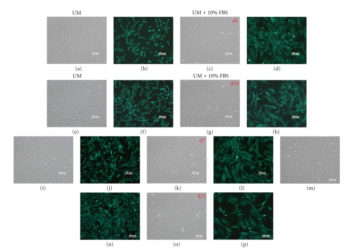
To evaluate the influence of cellular differentiation on transgene expression, transfected cNPCs (at P21) were cultured in UM containing 10% FBS for at least two weeks. Fluorescence microscopy detected eGFP expression in almost every cell, indicating that the transfection procedure was efficient and that the pCAG-PyF101-eGFP plasmids were stably maintained and actively transcribed without significant attenuation due to cell division or growth under in vitro differentiation conditions (Figure 1).
Figure 1.
GFP expression in cNPCs transfected with pCAG-PyF101-eGFP plasmid. Transfected cNPC (passage 21) were maintained under proliferation conditions (UM) or switched to growth factor-free differentiation conditions (UM + 10% FBS) to evaluate potential loss of transgene expression. Cultures were photographed at 6, 7, 10, and 15 days. Sustained expression of green fluorescence protein (GFP) was observed for both conditions at all time points. Paired images are shown for each time point and include phase contrast (a, c, e, g, i, k, m, o) and fluorescence (b, d, f, h, j, l, n, p) micrographs of the same areas in culture.
Flow cytometric analysis was performed to further confirm eGFP expression in transfected cNPCs. Almost all transfected cNPCs cultured in either UM or UM containing 10% FBS (differentiation medium) expressed eGFP (Figure 2). Interestingly, eGFP fluorescence intensity increased in transfected cells cultured in UM containing 10% FBS. We also detected two subpopulations of transfected cells that expressed eGFP at different levels (“high” and “medium high”). This observation may relate to the heterogenous morphology and size distribution of differentiating cells, some of which are larger in size which might serve to dilute the eGFP concentration within the cell.
Figure 2.
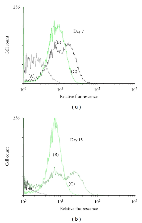
FACS analysis of GFP expression in nontransfected and transfected cNPCs. FACS analysis was performed on nontransfected and transfected cNPCs to show the expression of the GFP reporter gene. The vertical axis shows cell count, and the horizontal axis shows relative fluorescence. (a) Nontransfected cNPCp25 cultured in UM; (b) transfected cNPCp25 cultured in UM; (c) transfected cNPCp25 cultured in UM containing 10% FBS for 7 days (a) and 15 days (b).
3.3. Overexpression of GDNF in cNPCs
To further demonstrate the application of the pCAG vector for efficient transgene delivery beyond an eGFP reporter, we next overexpressed human GDNF in cNPCs. The effect of transduction on cellular proliferation was evaluated using an IncuCyte live cell monitoring system, allowing sequential observation of cell number in undisturbed cultures (Figures 3(a) and 3(b)). Both transfected and nontransfected cNPCs were seeded in 24-well plates in UM. Cell numbers were evaluated in terms of nuclear count and assessment of relative confluence (percentage). The resulting data showed that transfected cNPCs continued to proliferate, albeit at a slower rate than that of nontransfected cNPCs (Figures 3(c) and 3(d)). Interestingly, the monolayer cultures of the GDNF-transfected cNPCs appeared to be healthier, with fewer floating cells, less cell clumps, and minimal evidence of cell death.
Figure 3.
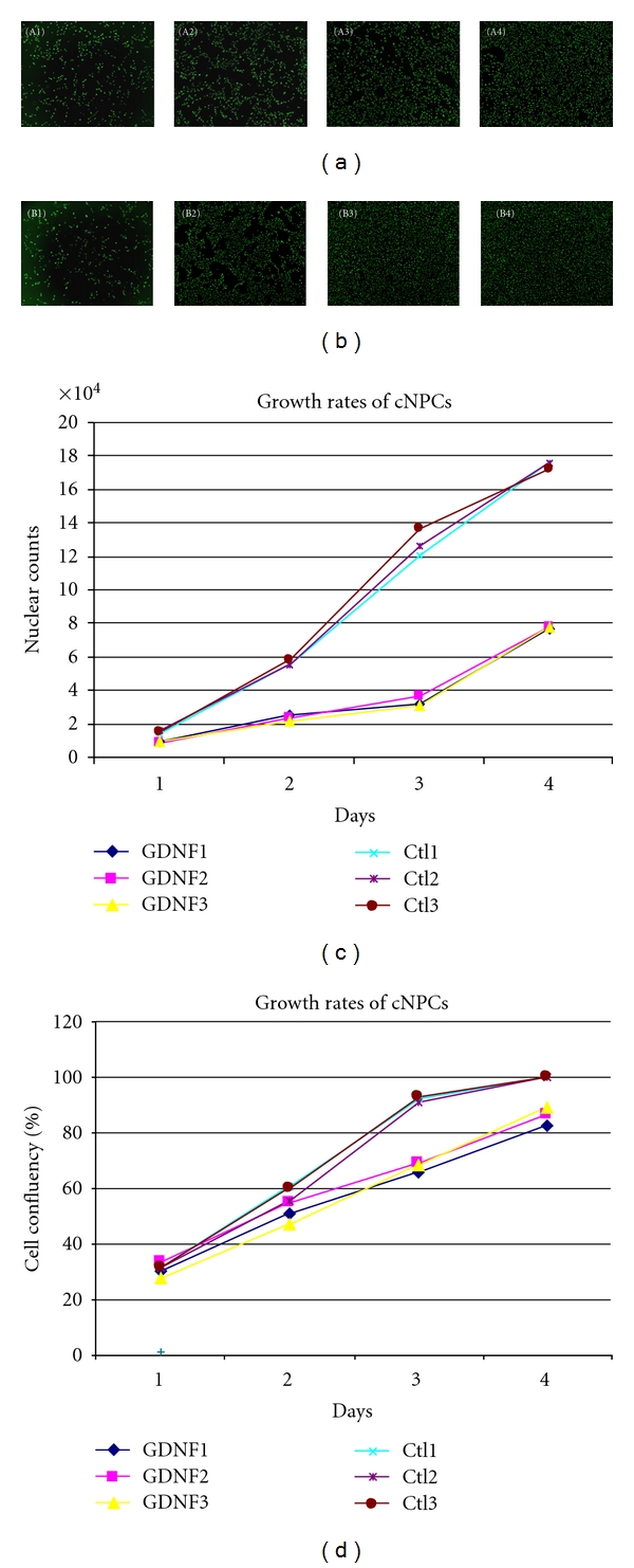
Growth of nontransfected and pCAG-PyF101-eGDNF plasmid transfected cNPCs. The fluorescence stained nuclei of (a) transfected and (b) nontransfected cNPCs. cNPCs were cultured in 24-well plate in UM for 4 days. Cells were labeled with a marker for nuclear DNA, and an IncuCyte live cell monitoring system was used to assess the growth rate of cells on each day. (a) GDNF-transfected cNPCs; A1–4: Day1–4. (b) Nontransfected cNPCs; B1–4: Day1–4. (c, d) Growth curves of GDNF-transfected and nontransfected cNPCs (labeled GDNF and Ctl, resp.) imaged and analyzed using an IncuCyte live cell monitoring system. Cells were stained with Vybrant Dycycle fluorescence nuclear-specific dye daily for 4 consecutive days (c). At the same time, cells in duplicate sets of wells without nuclear stain were measured for percentage cell confluency (d). The tight grouping of control data in (d) makes it difficult to discern Ctl1, which is present in the upper group and reaches confluence rapidly along with the other nontransfected cNPCs. IncuCyte programs were used for both analyses (c, d).
3.4. Confirmation of Increased GDNF Protein Production by Transfected cNPCs
The levels of GDNF produced by GDNF-transfected cNPCs were measured by ELISA. Cells were cultured in UM or UM containing 10% FBS, and fresh media were added 24 hours prior to collection of conditioned media for ELISA analysis. The data showed that GDNF-expressing cNPCs produced large amount of GDNF, even 60 days after initial transfection. In addition, cells cultured in UM as well as those cultured in UM containing 10% FBS produced similar amounts of GDNF (Figure 4), indicating that GDNF expression was maintained under in vitro differentiation conditions. We also determined the level of GDNF present within transfected cells. ELISA indicated that intracellular GDNF level was low (<10 ng/106 cells/day, data not shown) compared to the amount of GDNF present in culture media (227–258 ng/106 cells/day). Therefore the majority of GDNF produced was secreted into the culture media.
Figure 4.
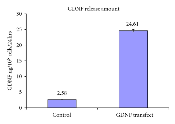
The amount of GDNF produced by nontransfected and transfected cNPCs measured by ELISA. Nontransfected and transfected cNPCs were seeded in UM. Culture media were refreshed 24 hours prior to collection of conditioned media for ELISA. Samples of culture media conditioned by transfected or control cells were analyzed to determine the level of GDNF present. Error bars = SEM.
3.5. Immunocytochemistry Confirms GDNF and Absence of Treatment-Related Changes in cNPCs
Immunocytochemical analysis was performed to confirm elevated GDNF protein expression within treated cNPCs and to evaluate the potential effects of pCAG transduction and GDNF overexpression on the ontogenetic status and lineage potential of the cells. First, an anti-GDNF antibody was used to detect GDNF protein in transfected cNPCs (Figure 5). This showed that GDNF expression was modest in nontransfected cNPCs (Figure 5(a)), but was strongly expressed in the transfected cells (Figure 5(b)). Although GDNF protein was also prominent in FBS-treated cNPCs, the fluorescence level was weaker, implying a more dilute distribution of GDNF, perhaps reflecting the larger size of the differentiating cells (Figure 5(c)).
Figure 5.
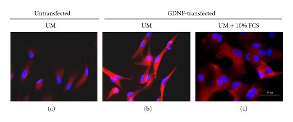
GDNF expression profiles in nontransfected and transfected cNPCp19. GDNF expression profiles were evaluated by immunocytochemistry (ICC) on cNPCs using an anti-human rabbit poly clone antibody. (a) Nontransfected cNPCs in UM; (b) pCAG- PyF101-GDNF plasmid transfected cNPCs in UM; (c) pCAG-PyF101-GDNF plasmid transfected cNPCs in UM containing 10% FBS for 5 days.
Immunocytochemical analysis of a range of neural progenitor markers showed that GDNF overexpression did not significantly affect the expression of neural progenitor and proliferation markers (Figure 6). Thus, GDNF-expressing cNPCs retained their identity as neural progenitors.
Figure 6.
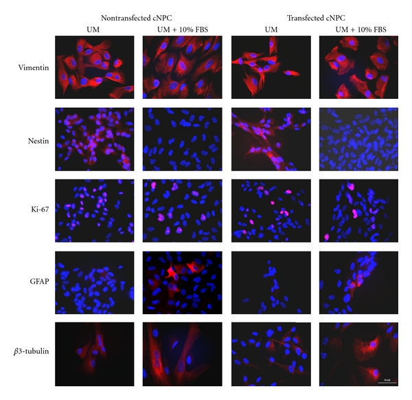
Expression profiles of marker genes in transfected cNPCs by ICC. Gene expression profiles of several neural progenitor, cell proliferation, and differentiation markers were evaluated by immunocytochemistry (ICC). Transfected cNPCs were cultured in UM, or UM containing 10% FBS, for 5 days and then immunolabeled with different epitope-specific antibodies to detect the markers shown.
4. Discussion
Here we report the use of a nonintegrating, plasmid-based vector to effectively transfect neural progenitor cells with an exogenous gene encoding a neuroprotective growth factor. Specifically, this plasmid vector, also containing the CAGG hybrid promoter and polyoma virus mutant enhancer PyF101a, was used to deliver the human GDNF gene to progenitor cells cultured from the fetal cat brain (cNPCs). This is one of the few studies investigating the genetic modification of NPCs derived from nonrodent, nonprimate mammalian species [24] and the first study to demonstrate the applicability of plasmid-based vector technology to feline NPCs.
Gene transfer represents a powerful tool for enhancing the desired characteristics of a therapeutic cell type. Early work exploiting transgenic reporter genes and disease models has been followed by more ambitious strategies, including cytokine delivery, immune modulation, and, more recently, cellular reprogramming [31, 32]. Nevertheless, the enthusiasm surrounding these advances has been tempered by the realization that integration of exogenous transgenes into the recipient genome can result in significant complications, including malignant transformation of cells and death of treated patients. For this reason, the use of a nonintegrating plasmid-based vector is of interest in that it might avoid the potential adverse perturbations of host cell gene regulation associated with uncontrolled alteration of the chromosomal DNA-coding sequence.
One challenge connected with the use of nonintegrating vectors is the transcriptional silencing of nonchromosomal DNA sequences by host cells. Use of the highly transcribed chicken β-actin [33] and its derivative composite promoter CAGG has recently gained popularity as a strategy for countering this phenomenon, providing a robust tool for deriving long-term constitutive transfectants [29, 30, 33, 34]. The findings of the current study demonstrate that, in combination, the plasmid-based vector system and CAGG promoter can effectively transfect feline NPCs. These results suggest that this method could find applicability in the delivery of various other genes of interest to feline NPCs, as well as possibly other feline cells, and immature and differentiated cells from additional mammalian species.
One interesting observation is that the GDNF-transfected cells exhibited a slower growth curve than untransfected controls. The reason for this was not delineated in the current study, but could relate to a number of considerations. One of these is that the cells were genetically manipulated and this could be deleterious in a number of ways. Another is that the cells overexpress the signaling molecule GDNF, which could in turn exert physiological influences on apoptosis or rate of proliferation. In another study with murine retinal progenitor cells [35], we showed that exogenous GDNF was antiapoptotic and did not impede proliferation, suggesting that the slower growth seen in the current study likely results from the genetic modification process or resultant protein overexpression, rather than from subsequent GDNF-induced signaling.
The cells generated and banked during this study provide a uniquely modified cell type with potential scalability. As such, these genetically modified feline NPCs could be of translational interest in the setting of veterinary applications. Further studies involving transplantation will be necessary to explore the safety and therapeutic potential of these cells. In terms of feline retinal degeneration, a suitable recipient exists in the form of the retinal dystrophic Abyssinian cat [8].
Author's Contribution
X. Joann You and J. Yang contributed equally to this work.
Acknowledgments
The authors are grateful to Professor Peter Andrews for developing the pCAG-PyF101-eGFP plasmid and to Steven Menges for assistance with the paper. In addition, the authors would like to thank the Lincy Foundation, the Discovery Eye Foundation, the Andrei Olenicoff Memorial Foundation, and the Polly and Michael Smith Foundation for their generous financial support of this work.
References
- 1.Gage FH. Mammalian neural stem cells. Science. 2000;287(5457):1433–1438. doi: 10.1126/science.287.5457.1433. [DOI] [PubMed] [Google Scholar]
- 2.McKay R. Stem cells in the central nervous system. Science. 1997;276(5309):66–71. doi: 10.1126/science.276.5309.66. [DOI] [PubMed] [Google Scholar]
- 3.van der Kooy D, Weiss S. Why stem cells? Science. 2000;287(5457):1439–1441. doi: 10.1126/science.287.5457.1439. [DOI] [PubMed] [Google Scholar]
- 4.Svendsen CN, Caldwell MA. Neural stem cells in the developing central nervous system: implications for cell therapy through transplantation. Progress in Brain Research. 2000;127:13–34. doi: 10.1016/s0079-6123(00)27003-9. [DOI] [PubMed] [Google Scholar]
- 5.Takahashi M, Palmer TD, Takahashi J, Gage FH. Widespread integration and survival of adult-derived neural progenitor cells in the developing optic retina. Molecular and Cellular Neurosciences. 1998;12(6):340–348. doi: 10.1006/mcne.1998.0721. [DOI] [PubMed] [Google Scholar]
- 6.Young MJ, Ray J, Whiteley SJ, Klassen H, Gage FH. Neuronal differentiation and morphological integration of hippocampal progenitor cells transplanted to the retina of immature and mature dystrophic rats. Molecular and Cellular Neurosciences. 2000;16(3):197–205. doi: 10.1006/mcne.2000.0869. [DOI] [PubMed] [Google Scholar]
- 7.Van Hoffelen SJ, Young MJ, Shatos MA, Sakaguchi DS. Incorporation of murine brain progenitor cells into the developing mammalian retina. Investigative Ophthalmology and Visual Science. 2003;44(1):426–434. doi: 10.1167/iovs.02-0269. [DOI] [PubMed] [Google Scholar]
- 8.Klassen H, Schwartz PH, Ziaeian B, et al. Neural precursors isolated from the developing cat brain show retinal integration following transplantation to the retina of the dystrophic cat. Veterinary Ophthalmology. 2007;10(4):245–253. doi: 10.1111/j.1463-5224.2007.00547.x. [DOI] [PubMed] [Google Scholar]
- 9.Hori J, Ng TF, Shatos M, Klassen H, Streilein JW, Young MJ. Neural progenitor cells lack immunogenicity and resist destruction as allografts. Stem Cells. 2003;21(4):405–416. doi: 10.1634/stemcells.21-4-405. [DOI] [PubMed] [Google Scholar]
- 10.Warfvinge K, Kiilgaard JF, Lavik EB, et al. Retinal progenitor cell xenografts to the pig retina: morphologic integration and cytochemical differentiation. Archives of Ophthalmology. 2005;123(10):1385–1393. doi: 10.1001/archopht.123.10.1385. [DOI] [PubMed] [Google Scholar]
- 11.Warfvinge K, Kiilgaard JF, Klassen H, et al. Retinal progenitor cell xenografts to the pig retina: immunological reactions. Cell Transplantation. 2006;15(7):603–612. doi: 10.3727/000000006783981594. [DOI] [PubMed] [Google Scholar]
- 12.Blesch A, Tuszynski MH. GDNF gene delivery to injured adult CNS motor neurons promotes axonal growth, expression of the trophic neuropeptide CGRP, and cellular protection. Journal of Comparative Neurology. 2001;436(4):399–410. doi: 10.1002/cne.1076. [DOI] [PubMed] [Google Scholar]
- 13.Björklund A, Rosenblad C, Winkler C, Kirik D. Studies on neuroprotective and regenerative effects of GDNF in a partial lesion model of Parkinson’s disease. Neurobiology of Disease. 1997;4(3-4):186–200. doi: 10.1006/nbdi.1997.0151. [DOI] [PubMed] [Google Scholar]
- 14.Behrstock S, Ebert A, McHugh J, et al. Human neural progenitors deliver glial cell line-derived neurotrophic factor to parkinsonian rodents and aged primates. Gene Therapy. 2006;13(5):379–388. doi: 10.1038/sj.gt.3302679. [DOI] [PubMed] [Google Scholar]
- 15.Klein SM, Behrstock S, Mchugh J, et al. GDNF delivery using human neural progenitor cells in a rat model of ALS. Human Gene Therapy. 2005;16(4):509–521. doi: 10.1089/hum.2005.16.509. [DOI] [PubMed] [Google Scholar]
- 16.Frasson M, Picaud S, Léveillard T, et al. Glial cell line-derived neurotrophic factor induces histologic and functional protection of rod photoreceptors in the rd/rd mouse. Investigative Ophthalmology and Visual Science. 1999;40(11):2724–2734. [PubMed] [Google Scholar]
- 17.Sanftner LHM, Abel H, Hauswirth WW, Flannery JG. Glial cell line derived neurotrophic factor delays photoreceptor degeneration in a transgenic rat model of retinitis pigmentosa. Molecular Therapy. 2001;4(6):622–629. doi: 10.1006/mthe.2001.0498. [DOI] [PubMed] [Google Scholar]
- 18.Andrieu-Soler C, Aubert-Pouëssel A, Doat M, et al. Intravitreous injection of PLGA microspheres encapsulating GDNF promotes the survival of photoreceptors in the rd1/rd1 mouse. Molecular Vision. 2005;11:1002–1011. [PubMed] [Google Scholar]
- 19.Hauck SM, Kinkl N, Deeg CA, Swiatek-De Lange M, Schöffmann S, Ueffing M. GDNF family ligands trigger indirect neuroprotective signaling in retinal glial cells. Molecular and Cellular Biology. 2006;26(7):2746–2757. doi: 10.1128/MCB.26.7.2746-2757.2006. [DOI] [PMC free article] [PubMed] [Google Scholar]
- 20.Kretz A, Jacob AM, Tausch S, Straten G, Isenmann S. Regulation of GDNF and its receptor components GFR-α1, -α2 and Ret during development and in the mature retino-collicular pathway. Brain Research. 2006;1090(1):1–14. doi: 10.1016/j.brainres.2006.01.131. [DOI] [PubMed] [Google Scholar]
- 21.Harper MM, Grozdanic SD, Blits B, et al. Transplantation of BDNF-secreting mesenchymal stem cells provides neuroprotection in chronically hypertensive rat eyes. Investigative Ophthalmology & Visual Science. 2011;52(7):4506–4515. doi: 10.1167/iovs.11-7346. [DOI] [PMC free article] [PubMed] [Google Scholar]
- 22.Ostenfeld T, Tai YT, Martin P, Déglon N, Aebischer P, Svendsen CN. Neurospheres modified to produce glial cell line-derived neurotrophic factor increase the survival of transplanted dopamine neurons. Journal of Neuroscience Research. 2002;69(6):955–965. doi: 10.1002/jnr.10396. [DOI] [PubMed] [Google Scholar]
- 23.Gamm DM, Wang S, Lu B, et al. Protection of visual functions by human neural progenitors in a rat model of retinal disease. Plos ONE. 2007;2(3, article e338) doi: 10.1371/journal.pone.0000338. [DOI] [PMC free article] [PubMed] [Google Scholar]
- 24.You XJ, Gu P, Wang J, et al. Efficient transduction of feline neural progenitor cells for delivery of glial cell line-derived neurotrophic factor using a feline immunodeficiency virus-based lentiviral construct. Journal of Ophthalmology. 2011;2011:11 pages. doi: 10.1155/2011/378965. Article ID 378965. [DOI] [PMC free article] [PubMed] [Google Scholar]
- 25.van Gaal EV, Hennink WE, Crommelin DJA, Mastrobattista E. Plasmid engineering for controlled and sustained gene expression for nonviral gene therapy. Pharmaceutical Research. 2006;23(6):1053–1074. doi: 10.1007/s11095-006-0164-2. [DOI] [PubMed] [Google Scholar]
- 26.Black J, Vos JM. Establishment of an oriP/EBNA1-based episomal vector transcribing human genomic β-globin in cultured murine fibroblasts. Gene Therapy. 2002;9(21):1447–1454. doi: 10.1038/sj.gt.3301808. [DOI] [PubMed] [Google Scholar]
- 27.Van Craenenbroeck K, Vanhoenacker P, Haegeman G. Episomal vectors for gene expression in mammalian cells. European Journal of Biochemistry. 2000;267(18):5665–5678. doi: 10.1046/j.1432-1327.2000.01645.x. [DOI] [PubMed] [Google Scholar]
- 28.Niwa H, Yamamura K, Miyazaki J. Efficient selection for high-expression transfectants with a novel eukaryotic vector. Gene. 1991;108(2):193–200. doi: 10.1016/0378-1119(91)90434-d. [DOI] [PubMed] [Google Scholar]
- 29.Liew CG, Draper JS, Walsh J, Moore H, Andrews PW. Transient and stable transgene expression in human embryonic stem cells. Stem Cells. 2007;25(6):1521–1528. doi: 10.1634/stemcells.2006-0634. [DOI] [PubMed] [Google Scholar]
- 30.Alexopoulou AN, Couchman JR, Whiteford JR. The CMV early enhancer/chicken β actin (CAG) promoter can be used to drive transgene expression during the differentiation of murine embryonic stem cells into vascular progenitors. BMC Cell Biology. 2008;9, article 2 doi: 10.1186/1471-2121-9-2. [DOI] [PMC free article] [PubMed] [Google Scholar]
- 31.Takahashi K, Yamanaka S. Induction of pluripotent stem cells from mouse embryonic and adult fibroblast cultures by defined factors. Cell. 2006;126(4):663–676. doi: 10.1016/j.cell.2006.07.024. [DOI] [PubMed] [Google Scholar]
- 32.Yu J, Hu K, Smuga-Otto K, et al. Human induced pluripotent stem cells free of vector and transgene sequences. Science. 2009;324(5928):797–801. doi: 10.1126/science.1172482. [DOI] [PMC free article] [PubMed] [Google Scholar]
- 33.Chung S, Andersson T, Sonntag KC, Björklund L, Isacson O, Kim KS. Analysis of different promoter systems for efficient transgene expression in mouse embryonic stem cell lines. Stem Cells. 2002;20(2):139–145. doi: 10.1634/stemcells.20-2-139. [DOI] [PMC free article] [PubMed] [Google Scholar]
- 34.Kosuga M, Enosawa S, Li XK, et al. Strong, long-term transgene expression in rat liver using chicken β-actin promoter associated with cytomegalovirus immediate-early enhancer (CAG promoter) Cell Transplantation. 2000;9(5):675–680. doi: 10.1177/096368970000900513. [DOI] [PubMed] [Google Scholar]
- 35.Wang J, Yang J, Gu P, Klassen H. Effects of glial cell line-derived neurotrophic factor on cultured murine retinal progenitor cells. Molecular Vision. 2010;16:2850–2866. [PMC free article] [PubMed] [Google Scholar]


