Abstract
The P2X7 receptor gene (P2RX7) is highly polymorphic with five previously described loss-of-function (LOF) single-nucleotide polymorphisms (SNP; c.151+1G>T, c.946G>A, c.1096C>G, c.1513A>C and c.1729T>A) and one gain-of-function SNP (c.489C>T). The purpose of this study was to determine whether the functional P2RX7 SNPs are associated with lumbar spine (LS) bone mineral density (BMD), a key determinant of vertebral fracture risk, in post-menopausal women. We genotyped 506 post-menopausal women from the Aberdeen Prospective Osteoporosis Screening Study (APOSS) for the above SNPs. Lumbar spine BMD was measured at baseline and at 6–7 year follow-up. P2RX7 genotyping was performed by homogeneous mass extension. We found association of c.946A (p.Arg307Gln) with lower LS-BMD at baseline (P=0.004, β=−0.12) and follow-up (P=0.002, β=−0.13). Further analysis showed that a combined group of subjects who had LOF SNPs (n=48) had nearly ninefold greater annualised percent change in LS-BMD than subjects who were wild type at the six SNP positions (n=84; rate of loss=−0.94%/year and −0.11%/year, respectively, P=0.0005, unpaired t-test). This is the first report that describes association of the c.946A (p.Arg307Gln) LOF SNP with low LS-BMD, and that other LOF SNPs, which result in reduced or no function of the P2X7 receptor, may contribute to accelerated bone loss. Certain polymorphic variants of P2RX7 may identify women at greater risk of developing osteoporosis.
Keywords: P2RX7, LS-BMD, single-nucleotide polymorphisms
Introduction
Maintenance of a healthy skeleton to prevent bone disease is dependent on the finely tuned balance between the amount of bone resorption by osteoclasts and bone formation by osteoblasts. Exactly how this is achieved is not fully understood, although several regulatory systems are involved including the RANKL/OPG axis, LRP5/Wnt signaling and more recently purinergic signalling. The latter system involves extracellular nucleotides signalling via specific cell surface P2 purinergic receptors, which consist of two sub-families termed P2X and P2Y receptors. P2Y receptors are metabotropic, heptahelical G protein-coupled receptors of which there are currently eight recognised sub-types, while the P2X are ligand-gated ionotropic channel receptors of which there are currently seven identified sub-types.1 In bone, multiple P2X and P2Y receptors have been demonstrated to be functionally expressed by both osteoblasts and osteoclasts. Activation of these receptors modulates cellular activities, such as proliferation and apoptosis, with subsequent effects on both bone formation and resorption in the bone microenvironment.2, 3, 4, 5, 6, 7, 8, 9, 10, 11
The P2X7 receptor (P2X7R), upon brief activation by ATP at a concentration higher than is required for activation of any of the other P2 receptors, functions as a cation channel. However, prolonged or repeated activation of the P2X7R leads to the formation of a non-selective pore permeable to solutes of up to 900 Da that ultimately leads to cell death.12 Transient activation of the receptor is now known to lead to reversible pseudoapoptosis13 while longer exposure to agonist leads to processing and release of interleukin (IL)-1β14 and IL-18 from monocytes and macrophages.15, 16, 17 Activation of these cell types is known to lead to an upregulation of P2X7R expression.18 This then amplifies the production and release of IL-1β and IL-18 with subsequent induction of IL-6, IL-8 and TNF-α. Given that osteoclasts are derived from the same progenitor cells as macrophages and that these inflammatory cytokines have an important role in regulating bone remodelling19, 20 the P2X7R presents as an ideal target for the regulation of bone remodelling.
The functional expression of P2X7R by osteoclasts has been conclusively demonstrated. We and others have shown that human osteoclasts both in vitro and in vivo express P2X7R protein, and that activation of the P2X7R induced cell death,21, 22 while blockade resulted in reduced multinucleated osteoclast formation and mature osteoclast formation.2, 5 Further studies using rabbit and murine models have highlighted the importance of P2X7R activation in osteoclasts via increased nuclear localisation of the transcription factor NF-κB, an important regulator of osteoclast formation and activity, independently of RANKL.23
We have also shown that a sub-population of human osteoblasts express functional P2X7R and that activation leads to apoptosis in these cells.6 In addition, we and others have shown that P2X7R activation leads to membrane blebbing in osteoblasts,6, 10 a process mediated by stimulation of PLD and PLA2 with subsequent production of the potent lipid mediator lysophosphatidic acid (LPA), which then acts through its G protein-coupled receptor to induce membrane blebbing via a pathway dependent on Rho-associated kinase.10 Both LPA and Rho-associated kinase have important roles in osteoblasts.24, 25, 26
Given the above reported roles of the P2X7R in both osteoclast and osteoblast physiology, profound changes in the bones of P2X7R knock-out (KO) mice would be expected. Analysis of two different P2X7R KO mice models has revealed differences in skeletal phenotypes,27 which may be explained by the retention of a functional splice isoform in the Glaxo mouse model.28 In contrast, the Pfizer P2X7R KO model shows a reduction in total and cortical bone content in the femur, reduced periosteal bone formation, increased trabecular bone resorption in the tibia29 and a reduced sensitivity to mechanical loading.30 We have also demonstrated that osteoblasts constitutively release nucleotides into the bone microenvironment and that this release can be positively modulated by mechanical loading,3, 31 supporting a role for the P2X7R in mechanotransduction and subsequent anabolic responses in bone. If the P2X7R transduces everyday loading into the appropriate responses within bone to help maintain skeletal health then differences in expression and/or activation of P2X7R will result in aberrant responses and possibly predispose people to bone disease.
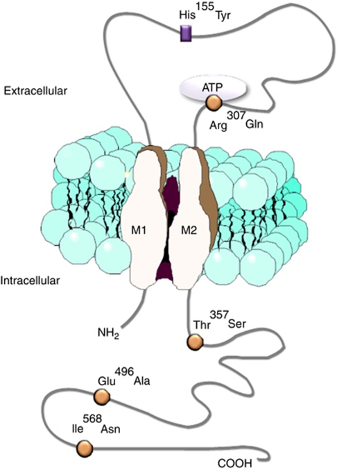
The gene for the P2X7R (P2RX7) is highly polymorphic and at least six non-synonymous single-nucleotide polymorphisms (SNPs: Figure 1) have been previously described as having effects on P2X7R function.32 The most common variant c.1513A>C, produces an amino acid change at position 496 (p.Glu496Ala) in the C terminus, which impairs multiple P2X7R functions, including the ability of the channel to undergo dilation and release of IL-1β, IL-18 and matrix metalloproteinase-9 from macrophages.17, 33, 34 The c.1729T>A variant (p.I568N) abolishes trafficking of the receptor to the cell surface,35 the c.946G>A variant (p.Arg307Gln) abolishes ATP binding to the extracellular domain of P2X7R,36 the c.1096C>G variant (p.Thr357Ser) results in reduced pore formation that is restored with upregulation of P2X7R expression32 and the intronic c.151+1G>T variant results in a null allele.37 The effect of these variants on ATP responsiveness is additive, as heterozygosity for any loss-of-function (LOF) variant leads to a 50% reduction in response whereas homozygosity for a given variant or compound heterozygosity for two LOF variants results in ablated ATP response.37 The c.489C>T variant (p.His155Tyr), located in the extracellular domain of the receptor involved in ATP binding, has been shown to be a weak gain-of-function (GOF) P2RX7 polymorphism evidenced by increased ATP-dependent calcium influx and ethidium uptake.38
Figure 1.
Diagrammatic representation of the protein structure of the P2X7R. The positions of amino acid changes as a result of the five polymorphisms included in this study are shown on the diagram. The three C-terminal and one ATP-binding site polymorphisms confer LOF (circles) while His155Tyr gives a weak GOF (triangle). The sixth polymorphism is located in intron 1.
One LOF P2RX7 polymorphism, the c.1513C allele (p.Glu496Ala) has recently been associated with increased susceptibility to extra pulmonary tuberculosis39 while in the context of bone, the c.1513C allele and the c.1729A allele (p.Ile568Gln) have been shown to be associated with an increased 10-year fracture risk in post-menopausal women.22
Given the above observations, we have investigated whether six P2RX7 SNPs, which have been previously identified and have putative effects on the receptor function, are associated with alterations in bone mineral density (BMD) in post-menopausal women.
Materials and methods
Aberdeen Prospective Osteoporosis Screening Study (APOSS) cohort and BMD measurements
The longitudinal APOSS is a population-based screening programme for osteoporotic fracture risk in females.40 Participants were recruited at random using Community Health Index records from within a 25-mile radius of Aberdeen, a city in the North East of Scotland with a population of ∼250 000.41, 42 BMD measurements were made at the initial baseline visit (V1), which took place between 1990 and 1994 when the women were aged 45–54 years (n=5114), and from a follow-up visit (V2) between 1997 and 1999. BMD measurements of the lumbar spine (LS; L2–L4) were performed by dual-energy X-ray absorptiometry using one of two Norland XR26 or XR36 densitometers (Norland Corp., Fort Atkinson, WI, USA). Annualised percentage change in BMD was calculated after V2. At V2, participants donated blood samples for DNA analysis (n=3266). Information on age at assessment, body mass index (BMI) at assessment, previous contraceptive pill use (V1 only) and hormone replacement therapy (HRT) use were also recorded. This study was approved by the Grampian Research Ethics Committee (97/0106 and 97/0230). For this study, APOSS participants at baseline who were post-menopausal, not on HRT and not taking any other medications influencing bone turnover (calcium supplements, sex hormones, steroid tablets, steroid inhalers, diuretics and tamoxifen) were genotyped (n=506).
DNA SNP analysis
DNA was extracted from peripheral blood obtained during the second visit using standard techniques as described previously.43 Six non-synonomous SNPs in P2RX7 with functional consequences for the receptor were analysed in 506 samples by a homogeneous mass extension assay (HME) at the Australian Genome Research Facility (St Lucia, Queensland, Australia). The samples that failed HME were re-analysed using restriction enzyme digestion of appropriate PCR products or by Taqman assay as described previously.32, 39 Polymorphisms in the coding sequence of the P2RX7 were numbered based on the original mRNA sequence, GenBank accession number Y09561.1.44
Statistical analysis
The statistical package SPSS version 15.0. (SPSS Inc., Chicago, IL, USA) was used for all statistical analysis. SNP genotype categories were recoded as a dummy variable as follows: homozygous wild type (WT)=1, heterozygote=2 and homozygous variant=3. BMD differences between genotype groups were corrected for age, BMI, contraceptive pill use and HRT use (as appropriate for the time point examined) using linear regression analysis, and are reported as P-values. Where P-values were <0.05, these were corrected using the Bonferroni multiple test correction for six SNPs. The threshold for statistical significance was a corrected P-value <0.05. Effect sizes are reported as unstandardised β±SEM.
Subjects who had one or more minor allele for a LOF SNP at either c.151+1G>T, c.946G>A, c.1096C>G, c.1513A>C or c.1729T>A while having major alleles at the other position were categorised as the LOF group (n=48), those who had the c.489T GOF SNP but major alleles at all the other positions were categorised as the GOF group (n=144) and those subjects who had the major alleles at all six SNPs positions were categorised as the WT group (n=84). Differences in annualised percentage change in BMD between two individual groups were examined using the unpaired t-test with Welch's correction due to unequal variances between the groups.
Results
Characteristics of genotyped subjects
Summary values for age, height, BMI, LS-BMD, contraceptive pill use, HRT use and annualised % change in LS-BMD for the 506 genotyped subjects are shown in Table 1. These women were slightly older than the rest of APOSS, had lower LS-BMD at both visits and a lower rate of bone loss.
Table 1. Descriptive statistics for the genotyped APOSS subjects.
| Subject characteristic | V1 | V2 |
|---|---|---|
| Age (years) | 49.7 (0.1) | 56.0 (0.1) |
| Height (cm) | 160.3 (0.5) | 160.0 (0.3) |
| BMI (kg/m) | 25.29 (0.2) | 26.5 (0.2) |
| LS-BMD (g/cm) | 1.00 (0.01) | 0.97 (0.01) |
| Annualised change in LS-BMD (%) | −0.39 (0.06) | |
| Contraceptive pill use (no use ever, previous; %) | 45.3, 54.7 | — |
| HRT status a V2 (never, previous, present; %) | — | 47.6, 18.8, 33.5 |
Abbreviations: APOSS, Aberdeen Prospective Osteoporosis Screening Study; BMI, body mass index; HRT, hormone replacement therapy; LS-BMD, lumbar spine bone mineral density.
Numbers are mean values with standard error in brackets. V1 is the baseline measurement, and V2 is at the follow-up visit. NB All women were post-menopausal and not on HRT or other medication at baseline.
Genotype data
Overall, SNP call rates were 97% for c.151+1G>T (rs35933842), c.946G>A (rs28360457), c.1096C>G (rs2230911) and c.1729T>A (rs1653624) and 95% for c.489C>T (rs208294) and c.1513A>C (rs3751143). Table 2 shows the predicted amino-acid change for each SNP. All six SNPs were consistent with Hardy–Weinberg equilibrium (all P-values >0.2).
Table 2. Results from linear regression analysis of individual P2RX7 SNPs and V1 LS BMD.
| LS-BMD V1 (±SEM) g/cm2 | |||||||
|---|---|---|---|---|---|---|---|
| rs # | Base (amino-acid) change | MAF | WT | HET | HOMO | P-value (corrected) | β-value |
| rs35933842 | c.151+1G>T | 0.01 | 1.00 (0.01) | 0.98 (0.04) | * | 0.4 | −0.003 |
| rs208294 | c.489C>T (p.H155Y) | 0.43 | 0.99 (0.01) | 1.01 (0.01) | 1.00 (0.02) | 0.3 | 0.042 |
| rs28360457 | c.946G>A (p.R307Q) | 0.02 | 1.00 (0.01) | 0.88 (0.03) | * | 0.004 (0.024) | −0.122 |
| rs2230911 | c.1096C>G (p.T357S) | 0.07 | 1.00 (0.01) | 1.03 (0.02) | 1.09 (0.12) | 0.08 | 0.074 |
| rs3751143 | c.1513A>C (p.E496A) | 0.17 | 1.00 (0.01) | 1.00 (0.02) | 1.01 (0.06) | 0.8 | 0.010 |
| rs1653624 | c.1729T>A (p.I568N) | 0.02 | 1.00 (0.01) | 0.99 (0.03) | 1.25** | 0.9 | 0.006 |
Abbreviations: LS-BMD, lumbar spine bone mineral density; MAF, minor allele frequency; SNPs, single-nucleotide polymorphisms; WT, wild type.
Where P<0.05 (bold), the Bonferonni's correction was applied for six SNPs and the corrected P-value is in brackets.
*n=0.
**n=1.
P2RX7 c.946G>A (p.Arg307Gln) SNP is associated with lower LS BMD in post-menopausal women
Analysis of the six previously published P2RX7 SNPs revealed that c.946G>A (p.Arg307Gln) was significantly associated with lower LS BMD both at study enrolment (V1) and at the 6-year follow-up visit (V2). Linear regression analysis of the individual SNP data (correcting for age, BMI, previous contraceptive pill use (for V2 only) and HRT status) showed that V1 LS BMD was significantly lower in heterozygous individuals (GA, n=18) compared with WT (GG, n=474; Pcorrected=0.024, β=−0.122 (Table 2)), and that this effect was maintained for V2 LS BMD (Pcorrected=0.012, β=−0.130 (Table 3)). No individuals were homozygous for the A allele at this SNP. The average annualised percentage change in LS-BMD did not differ significantly by c.946G>A genotype (−0.39%/year (SEM 0.06) for GG subjects and −0.57%/year (SEM 0.43) for GA subjects (P=0.7)), suggesting that the c.946G >A may be exerting effects on bone mass at an earlier age.
Table 3. Results from linear regression analysis of individual P2RX7 SNPs and V2 LS BMD.
| LS-BMD V2 (±SEM) g/cm2 | |||||||
|---|---|---|---|---|---|---|---|
| rs # | Base (amino-acid) change | MAF | WT | HET | HOMO | P-value (corrected) | β-value |
| rs35933842 | c.151+1G>T | 0.01 | 0.97 (0.01) | 0.93 (0.03) | * | 0.3 | −0.041 |
| rs208294 | c.489C>T (p.H155Y) | 0.43 | 0.96 (0.01) | 0.98 (0.01) | 0.97 (0.02) | 0.3 | 0.044 |
| rs28360457 | c.946G>A (p.R307Q) | 0.02 | 0.97 (0.01) | 0.84 (0.04) | * | 0.002 (0.012) | −0.130 |
| rs2230911 | c.1096C>G (p.T357S) | 0.07 | 0.97 (0.01) | 0.98 (0.02) | 1.00 (0.16) | 0.38 | 0.037 |
| rs3751143 | c.1513A>C (p.E496A) | 0.17 | 0.97 (0.01) | 0.97 (0.01) | 0.99 (0.06) | 0.37 | 0.038 |
| rs1653624 | c.1729T>A (p.I568N) | 0.02 | 0.97 (0.01) | 0.93 (0.02) | 1.22** | 0.63 | −0.020 |
Abbreviations: LS-BMD, lumbar spine bone mineral density; MAF, minor allele frequency; SNPs, single-nucleotide polymorphisms; WT, wild type.
Where P<0.05 (bold), the Bonferonni's correction was applied for six SNPs and the corrected P-value is in brackets.
*n=0.
**n=1.
LOF P2RX7 SNPs are associated with greater rate of bone loss at the LS in post-menopausal women
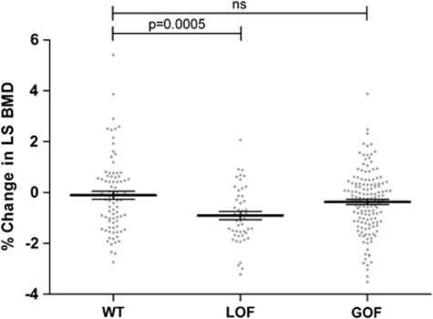
Further analysis showed that compared with subjects who were WT at all six SNP positions (n=84), subjects who had a LOF SNP at either c.151+1G>T, c.G946A, c.1096C>G, c.1513A>C or c.1729T>A (n=48) had almost ninefold greater annualised percent change from baseline in LS BMD (−0.9354%/year for the LOF group and −0.1057%/year for the WT group, P=0.0005 (Figure 2)). The percentage change in LS BMD for a group of subjects who had the c.489T GOF SNP but were WT at the other five LOF SNPs (n=144) was not statistically significantly different from subjects who were WT at all six SNP positions (−0.3676%/year for GOF group, P=0.1072 (Figure 2)).
Figure 2.
Difference in annualised percentage change in LS-BMD. WT subjects (n=84); LOF, subjects who are have any LOF SNP but are WT at the c.489T GOF position (n=47); GOF, subjects who have a c.489T GOF SNP but WT at the LOF SNP positions (n=144). Individual values plotted with bars being the mean±SEM.
Discussion
Previous in vitro studies from our group and others have revealed that functional P2X7Rs have profound effects on bone cells, regulating both formation and survival of osteoclasts,2, 5, 22 as well as enhancing bone formation through an osteoblast autonomous mechanism9 and inducing apoptosis of a sub-population of osteoblasts.6 A fine balance between the activities of these cells is required for the maintenance of a healthy skeleton. Any perturbations of this balance in the favour of osteoclasts would result in increased bone resorption/bone loss and an increased risk of developing osteoporosis. In humans, the P2RX7 is highly polymorphic with 26 non-synonymous SNPs listed on the NCBI database (Build 131), of which six have been functionally characterised.32, 34, 35, 36, 37, 38 A recent report by Ohlendorff et al22 demonstrated that two P2RX7 SNPs, c.1513A>C (p.Glu496Ala) and c.1729T>A (p.I568N), are associated with an increased 10-year fracture risk in post-menopausal women.
In this study, we have found an association of a major LOF SNP in P2RX7, the c.946G>A (p.Arg307Gln), with low BMD in the LS in post-menopausal females, both at the initial and at the 6 year follow-up visit. As only women who were post-menopausal at baseline, not on HRT and not taking any other medications influencing bone turnover (calcium supplements, sex hormones, steroid tablets, steroid inhalers, diuretics and tamoxifen) were selected for this study, the genotyped subset are more homogenous than the entire APOSS cohort and are free from any confounding effects on bone loss or baseline BMD. The c.946G>A polymorphism changes arginine to glutamine at residue 307 and abolishes binding of ATP to the receptor.36 The functional effect of this amino acid change is likely to be magnified because of the trimeric nature of the receptor and the need for three molecules of ATP to bind for the receptor to become functional. Permeability studies of subjects heterozygous for c.946G>A (Figure 1 in Gu et al, 200436) show complete absence of ATP-mediated responses, which supports a dominant-negative nature of this variant on function even in heterozygous dosage. Thus, c.946G>A may be classified as a dominant-negative polymorphism and this may explain its profound effects on BMD and bone turnover. Indeed, the profound effects of the c.946G>A SNP on bone are further highlighted and replicated in the Danish Osteoporosis Prevention Study, which found that subjects who were heterozygous for the c.946G>A (Arg307Gln variant) had >40% greater bone loss at the hip over the 0- to10-year interval than subjects who were WT at this position (Jørgensen et al45). Furthermore, this hypothesis is supported by a recent study showing that rare variants causing complete loss of P2X7R function were overrepresented among patients with total hip replacement revision and that the c.946G>A allele increased cumulative hazard of total hip replacement revision.46 In our study, heterozygosity for c.151+1G>T that leads to one null allele37 had no impact on LS-BMD, while neither of the most prevalent variants, c.1513A>C, nor c.1729T>A polymorphisms alone showed any significant decrease in LS BMD at either the first or the follow-up visit in our cohort, consistent with the previous report of Ohlendorff et al22 (Tables 2 and 3).
Owing to the highly polymorphic nature of the P2RX7 and previous studies describing the effect of compound heterozygosity on function,32 we performed further analysis by grouping the subjects based on their status at all six functional SNP positions. We defined a WT group that consisted of subjects who had the major allele at all six SNP positions, a LOF group that consisted of subjects who had a minor allele at any one of the LOF SNP positions while having the major alleles at the other positions and a gain GOF group that had the c.489T GOF SNP while having the major alleles at the other LOF SNPs. Although this reduced the size of the groups, identification of the subjects who had none of the functional P2RX7 SNP alleles enabled us to identify a significant, almost ninefold increase in the rate of bone loss at the LS in the group of individuals who carried a LOF SNP allele in the P2RX7. Rate of LS-BMD in the GOF group was not significantly different to WT, perhaps reflecting the weak functional effect of this polymorphism. Interestingly, the WT group of individuals had an almost fourfold lower average annualised percentage change in LS-BMD than the average for the whole cohort, although this was not statistically significant (P=0.10, average values=−0.11%/year, SEM 0.16 and −0.39%/year, SEM 0.06, respectively). We believe that this further highlights the importance of a fully functional P2X7R to ensure the effective mechanotransduction of everyday load bearing over a lifetime, which is essential to the form and function of the skeleton.
We do not currently know precisely how the functional activity of osteoblasts and osteoclasts are affected by either LOF or GOF P2RX7 SNPs. However, given that the P2X7R is known to mediate osteoclast apoptosis and osteoblast bone formation any genetic changes, which affect P2X7R function would presumably affect the fine balance of bone loss and bone formation needed to maintain a healthy skeleton. In addition, previous studies have demonstrated that the P2X7R forms complexes with proteins of the cytoskeleton known to be involved in mechanotransduction,47, 48 and the P2X7R KO mouse has a disuse phenotype29 as well as a reduced response to mechanical loading.30 Given that mechanical loading is the most anabolic stimulus known to the skeleton and exercise is less effective after attaining peak bone mass49 early identification of individuals with polymorphisms conferring a major decrease in P2X7R function would help to target alternative therapies to build and maintain bone mass.
Recent studies have suggested that the P2X7R and P2X4 receptors may form heterotrimers when overexpressed by transfection,50 however, further data on possible heteromeric associations in native cells are needed before the possible implications of mutations in the P2X4 receptor gene (P2XR4) on P2X7R-mediated effects can be determined.
This study is the first to demonstrate an association of LOF polymorphisms in P2RX7 and LS-BMD, a key determinant of vertebral fracture risk. As the observed effect size of the c.946G>A is quiet large (β=0.12) compared with previously known SNPs, one might expect a locus of such a large effect size to be have been previously detected in the published genome-wide association studies (GWAS) in osteoporosis. Although the initial GWAS for osteoporosis have confirmed the roles for many previously suspected candidate genes such as RANK (TNFRSF11A), RANKL (TNFSF11) and LRP551 the results to date account for only a small amount of the genetic component of traits such as BMD. Most GWAS studies focus on genes/markers with top-ranking statistical significance and the current GWAS platforms do not examine rare genetic variants, including the P2RX7 c.946G>A, which has a population frequency of around 1.0% in Caucasians. Moreover, this SNP is outside the haplotype block encompassing exons 11–13 of P2RX752 thus reducing the probability that this SNP is in linkage disequilibrium with a more common variant.
In conclusion, the result of this study, in addition to the previously published data and that of Jørgensen et al,45 provides evidence that P2RX7 is involved in the regulation of LS-BMD and may, in the future, represent an early diagnostic tool for the management of osteoporosis.
Acknowledgments
We thank the APOSS participants for their support; Dr Teare (Mathematical Modelling and Genetic Epidemiology, UoS) and Dr Walters (School of Mathematics and Statistics, UoS) for help and advice on statistical analysis and preparation of this manuscript. We also acknowledge funding support from: Arthritis Research UK (AG, WDF and JAG) and the European Commission under the 7th Framework Programme (proposal #202231) performed as a collaborative project among the members of the ATPBone Consortium (Copenhagen University, University College London, University of Maastricht, University of Ferrara, University of Liverpool, University of Sheffield and Université Libre de Bruxelles), and is a sub study under the main study ‘Fighting osteoporosis by blocking nucleotides: purinergic signalling in bone formation and homeostasis; (AG, WDF and JAG) the National Health and Medical Research Council of Australia and the Leukemia Foundation of Australia (JW) and a Scottish Funding Council Strategic Research Development Grant, ‘Generation Scotland: Genetic Health in the 21st Century' (LJH).
The authors declare no conflict of interest.
References
- Burnstock G, Williams M. P2 purinergic receptors: modulation of cell function and therapeutic potential. J Pharmacol Exp Ther. 2000;295:862–869. [PubMed] [Google Scholar]
- Agrawal A, Buckley KA, Bowers K, et al. The effects of P2X7 receptor antagonists on the formation and function of human osteoclasts in vitro. Purinergic Signal. 2010;6:307–315. doi: 10.1007/s11302-010-9181-z. [DOI] [PMC free article] [PubMed] [Google Scholar]
- Bowler WB, Buckley KA, Gartland A, et al. Extracellular nucleotide signaling: a mechanism for integrating local and systemic responses in the activation of bone remodeling. Bone. 2001;28:507–512. doi: 10.1016/s8756-3282(01)00430-6. [DOI] [PubMed] [Google Scholar]
- Buckley KA, Hipskind RA, Gartland A, et al. Adenosine triphosphate stimulates human osteoclast activity via upregulation of osteoblast-expressed receptor activator of nuclear factor-kappa B ligand. Bone. 2002;31:582–590. doi: 10.1016/s8756-3282(02)00877-3. [DOI] [PubMed] [Google Scholar]
- Gartland A, Buckley KA, Bowler WB, et al. Blockade of the pore-forming P2X7 receptor inhibits formation of multinucleated human osteoclasts in vitro. Calcif Tissue Int. 2003;73:361–369. doi: 10.1007/s00223-002-2098-y. [DOI] [PubMed] [Google Scholar]
- Gartland A, Hipskind RA, Gallagher JA, et al. Expression of a P2X7 receptor by a subpopulation of human osteoblasts. J Bone Miner Res. 2001;16:846–856. doi: 10.1359/jbmr.2001.16.5.846. [DOI] [PubMed] [Google Scholar]
- Jorgensen NR, Henriksen Z, Sorensen OH, et al. Intercellular calcium signaling occurs between human osteoblasts and osteoclasts and requires activation of osteoclast P2X7 receptors. J Biol Chem. 2002;277:7574–7580. doi: 10.1074/jbc.M104608200. [DOI] [PubMed] [Google Scholar]
- Orriss IR, Utting JC, Brandao-Burch A, et al. Extracellular nucleotides block bone mineralization in vitro: evidence for dual inhibitory mechanisms involving both P2Y2 receptors and pyrophosphate. Endocrinology. 2007;148:4208–4216. doi: 10.1210/en.2007-0066. [DOI] [PubMed] [Google Scholar]
- Panupinthu N, Rogers JT, Zhao L, et al. P2X7 receptors on osteoblasts couple to production of lysophosphatidic acid: a signaling axis promoting osteogenesis. J Cell Biol. 2008;181:859–871. doi: 10.1083/jcb.200708037. [DOI] [PMC free article] [PubMed] [Google Scholar]
- Panupinthu N, Zhao L, Possmayer F, et al. P2X7 nucleotide receptors mediate blebbing in osteoblasts through a pathway involving lysophosphatidic acid. J Biol Chem. 2007;282:3403–3412. doi: 10.1074/jbc.M605620200. [DOI] [PubMed] [Google Scholar]
- Gallagher JA. ATP P2 receptors and regulation of bone effector cells. J Musculoskelet Neuronal Interact. 2004;4:125–127. [PubMed] [Google Scholar]
- Di Virgilio F. The P2Z purinoceptor: an intriguing role in immunity, inflammation and cell death. Immunol Today. 1995;16:524–528. doi: 10.1016/0167-5699(95)80045-X. [DOI] [PubMed] [Google Scholar]
- Mackenzie AB, Young MT, Adinolfi E, et al. Pseudoapoptosis induced by brief activation of ATP-gated P2X7 receptors. J Biol Chem. 2005;280:33968–33976. doi: 10.1074/jbc.M502705200. [DOI] [PubMed] [Google Scholar]
- Ferrari D, Pizzirani C, Adinolfi E, et al. The P2X7 receptor: a key player in IL-1 processing and release. J Immunol. 2006;176:3877–3883. doi: 10.4049/jimmunol.176.7.3877. [DOI] [PubMed] [Google Scholar]
- Mehta VB, Hart J, Wewers MD. ATP-stimulated release of interleukin (IL)-1beta and IL-18 requires priming by lipopolysaccharide and is independent of caspase-1 cleavage. J Biol Chem. 2001;276:3820–3826. doi: 10.1074/jbc.M006814200. [DOI] [PubMed] [Google Scholar]
- Rampe D, Wang L, Ringheim GE. P2X7 receptor modulation of beta-amyloid- and LPS-induced cytokine secretion from human macrophages and microglia. J Neuroimmunol. 2004;147:56–61. doi: 10.1016/j.jneuroim.2003.10.014. [DOI] [PubMed] [Google Scholar]
- Sluyter R, Dalitz JG, Wiley JS. P2X7 receptor polymorphism impairs extracellular adenosine 5′-triphosphate-induced interleukin-18 release from human monocytes. Genes Immun. 2004;5:588–591. doi: 10.1038/sj.gene.6364127. [DOI] [PubMed] [Google Scholar]
- Humphreys BD, Dubyak GR. Modulation of P2X7 nucleotide receptor expression by pro- and anti-inflammatory stimuli in THP-1 monocytes. J Leukoc Biol. 1998;64:265–273. doi: 10.1002/jlb.64.2.265. [DOI] [PubMed] [Google Scholar]
- Mundy GR. Osteoporosis and inflammation. Nutr Rev. 2007;65:S147–S151. doi: 10.1111/j.1753-4887.2007.tb00353.x. [DOI] [PubMed] [Google Scholar]
- Rifas L. Bone and cytokines: beyond IL-1, IL-6 and TNF-alpha. Calcif Tissue Int. 1999;64:1–7. doi: 10.1007/s002239900570. [DOI] [PubMed] [Google Scholar]
- Naemsch LN, Dixon SJ, Sims SM. Activity-dependent development of P2X7 current and Ca2+ entry in rabbit osteoclasts. J Biol Chem. 2001;276:39107–39114. doi: 10.1074/jbc.M105881200. [DOI] [PubMed] [Google Scholar]
- Ohlendorff SD, Tofteng CL, Jensen JE, et al. Single nucleotide polymorphisms in the P2X7 gene are associated to fracture risk and to effect of estrogen treatment. Pharmacogenet Genomics. 2007;17:555–567. doi: 10.1097/FPC.0b013e3280951625. [DOI] [PubMed] [Google Scholar]
- Korcok J, Raimundo LN, Ke HZ, et al. Extracellular nucleotides act through P2X7 receptors to activate NF-kappaB in osteoclasts. J Bone Miner Res. 2004;19:642–651. doi: 10.1359/JBMR.040108. [DOI] [PubMed] [Google Scholar]
- Grey A, Banovic T, Naot D, et al. Lysophosphatidic acid is an osteoblast mitogen whose proliferative actions involve G(i) proteins and protein kinase C, but not P42/44 mitogen-activated protein kinases. Endocrinology. 2001;142:1098–1106. doi: 10.1210/endo.142.3.8011. [DOI] [PubMed] [Google Scholar]
- Masiello LM, Fotos JS, Galileo DS, et al. Lysophosphatidic acid induces chemotaxis in MC3T3-E1 osteoblastic cells. Bone. 2006;39:72–82. doi: 10.1016/j.bone.2005.12.013. [DOI] [PubMed] [Google Scholar]
- Radeff JM, Nagy Z, Stern PH. Rho and Rho kinase are involved in parathyroid hormone-stimulated protein kinase C alpha translocation and IL-6 promoter activity in osteoblastic cells. J Bone Miner Res. 2004;19:1882–1891. doi: 10.1359/JBMR.040806. [DOI] [PubMed] [Google Scholar]
- Gartland A, Buckley KA, Hipskind RA, et al. Multinucleated osteoclast formation in vivo and in vitro by P2X7 receptor-deficient mice. Crit Rev Eukaryot Gene Expr. 2003;13:243–253. doi: 10.1615/critreveukaryotgeneexpr.v13.i24.150. [DOI] [PubMed] [Google Scholar]
- Nicke A, Kuan YH, Masin M, et al. A functional P2X7 splice variant with an alternative transmembrane domain 1 escapes gene inactivation in P2X7 knock-out mice. J Biol Chem. 2009;284:25813–25822. doi: 10.1074/jbc.M109.033134. [DOI] [PMC free article] [PubMed] [Google Scholar]
- Ke HZ, Qi H, Weidema AF, et al. Deletion of the P2X7 nucleotide receptor reveals its regulatory roles in bone formation and resorption. Mol Endocrinol. 2003;17:1356–1367. doi: 10.1210/me.2003-0021. [DOI] [PubMed] [Google Scholar]
- Li J, Liu D, Ke HZ, et al. The P2X7 nucleotide receptor mediates skeletal mechanotransduction. J Biol Chem. 2005;280:42952–42959. doi: 10.1074/jbc.M506415200. [DOI] [PubMed] [Google Scholar]
- Buckley KA, Golding SL, Rice JM, et al. Release and interconversion of P2 receptor agonists by human osteoblast-like cells. Faseb J. 2003;17:1401–1410. doi: 10.1096/fj.02-0940com. [DOI] [PubMed] [Google Scholar]
- Shemon AN, Sluyter R, Fernando SL, et al. A Thr357 to Ser polymorphism in homozygous and compound heterozygous subjects causes absent or reduced P2X7 function and impairs ATP-induced mycobacterial killing by macrophages. J Biol Chem. 2006;281:2079–2086. doi: 10.1074/jbc.M507816200. [DOI] [PubMed] [Google Scholar]
- Gu BJ, Wiley JS. Rapid ATP-induced release of matrix metalloproteinase 9 is mediated by the P2X7 receptor. Blood. 2006;107:4946–4953. doi: 10.1182/blood-2005-07-2994. [DOI] [PubMed] [Google Scholar]
- Gu BJ, Zhang W, Worthington RA, et al. A Glu-496 to Ala polymorphism leads to loss of function of the human P2X7 receptor. J Biol Chem. 2001;276:11135–11142. doi: 10.1074/jbc.M010353200. [DOI] [PubMed] [Google Scholar]
- Wiley JS, Dao-Ung LP, Li C, et al. An Ile-568 to Asn polymorphism prevents normal trafficking and function of the human P2X7 receptor. J Biol Chem. 2003;278:17108–17113. doi: 10.1074/jbc.M212759200. [DOI] [PubMed] [Google Scholar]
- Gu BJ, Sluyter R, Skarratt KK, et al. An Arg307 to Gln polymorphism within the ATP-binding site causes loss of function of the human P2X7 receptor. J Biol Chem. 2004;279:31287–31295. doi: 10.1074/jbc.M313902200. [DOI] [PubMed] [Google Scholar]
- Skarratt KK, Fuller SJ, Sluyter R, et al. A 5′ intronic splice site polymorphism leads to a null allele of the P2X7 gene in 1–2% of the Caucasian population. FEBS Lett. 2005;579:2675–2678. doi: 10.1016/j.febslet.2005.03.091. [DOI] [PubMed] [Google Scholar]
- Cabrini G, Falzoni S, Forchap SL, et al. A His-155 to Tyr polymorphism confers gain-of-function to the human P2X7 receptor of human leukemic lymphocytes. J Immunol. 2005;175:82–89. doi: 10.4049/jimmunol.175.1.82. [DOI] [PubMed] [Google Scholar]
- Fernando SL, Saunders BM, Sluyter R, et al. A polymorphism in the P2X7 gene increases susceptibility to extrapulmonary tuberculosis. Am J Respir Crit Care Med. 2007;175:360–366. doi: 10.1164/rccm.200607-970OC. [DOI] [PubMed] [Google Scholar]
- MacDonald HM, McGuigan FA, New SA, et al. COL1A1 Sp1 polymorphism predicts perimenopausal and early postmenopausal spinal bone loss. J Bone Miner Res. 2001;16:1634–1641. doi: 10.1359/jbmr.2001.16.9.1634. [DOI] [PubMed] [Google Scholar]
- Garton MJ, Torgerson DJ, Donaldson C, et al. Recruitment methods for screening programmes: trial of a new method within a regional osteoporosis study. BMJ. 1992;305:82–84. doi: 10.1136/bmj.305.6845.82. [DOI] [PMC free article] [PubMed] [Google Scholar]
- Torgerson DJ, Garton MJ, Donaldson C, et al. Recruitment methods for screening programmes: trial of an improved method within a regional osteoporosis study. BMJ. 1993;307:99. doi: 10.1136/bmj.307.6896.99. [DOI] [PMC free article] [PubMed] [Google Scholar]
- Grant SF, Reid DM, Blake G, et al. Reduced bone density and osteoporosis associated with a polymorphic Sp1 binding site in the collagen type I alpha 1 gene. Nat Genet. 1996;14:203–205. doi: 10.1038/ng1096-203. [DOI] [PubMed] [Google Scholar]
- Rassendren F, Buell GN, Virginio C, et al. The permeabilizing ATP receptor, P2X7. Cloning and expression of a human cDNA. J Biol Chem. 1997;272:5482–5486. doi: 10.1074/jbc.272.9.5482. [DOI] [PubMed] [Google Scholar]
- Jørgensen NR, Husted LB, Skarratt KK, et al. Single nucleotide polymorphisms in the P2X7 receptor gene are associated with post-menopausal bone loss and vertebral fractures Eur J Hum Genet 2011. epub ahead of print, doi: 10.1038/ejhg.2011.253 [DOI] [PMC free article] [PubMed]
- Mrazek F, Gallo J, Stahelova A, et al. Functional variants of the P2RX7 gene, aseptic osteolysis, and revision of the total hip arthroplasty: a preliminary study. Hum Immunol. 2010;71:201–205. doi: 10.1016/j.humimm.2009.10.013. [DOI] [PubMed] [Google Scholar]
- Gu BJ, Rathsam C, Stokes L, et al. Extracellular ATP dissociates nonmuscle myosin from P2X(7) complex: this dissociation regulates P2X(7) pore formation. Am J Physiol Cell Physiol. 2009;297:C430–C439. doi: 10.1152/ajpcell.00079.2009. [DOI] [PubMed] [Google Scholar]
- Kim M, Jiang LH, Wilson HL, et al. Proteomic and functional evidence for a P2X7 receptor signalling complex. EMBO J. 2001;20:6347–6358. doi: 10.1093/emboj/20.22.6347. [DOI] [PMC free article] [PubMed] [Google Scholar]
- Turner CH, Robling AG. Mechanisms by which exercise improves bone strength. J Bone Miner Metab. 2005;23 (Suppl:16–22. doi: 10.1007/BF03026318. [DOI] [PubMed] [Google Scholar]
- Guo C, Masin M, Qureshi OS, et al. Evidence for functional P2X4/P2X7 heteromeric receptors. Mol Pharmacol. 2007;72:1447–1456. doi: 10.1124/mol.107.035980. [DOI] [PubMed] [Google Scholar]
- Styrkarsdottir U, Halldorsson BV, Gretarsdottir S, et al. Multiple genetic loci for bone mineral density and fractures. N Engl J Med. 2008;358:2355–2365. doi: 10.1056/NEJMoa0801197. [DOI] [PubMed] [Google Scholar]
- Fuller SJ, Stokes L, Skarratt KK, et al. Genetics of the P2X7 receptor and human disease. Purinergic Signal. 2009;5:257–262. doi: 10.1007/s11302-009-9136-4. [DOI] [PMC free article] [PubMed] [Google Scholar]


