Abstract
Excessive secretion of triglyceride-rich very low-density lipoproteins (VLDL-TG) contributes to diabetic dyslipidemia. Earlier studies have indicated a possible role for the hypothalamus and autonomic nervous system in the regulation of VLDL-TG. In the current study, we investigated whether the autonomic nervous system and hypothalamic neuropeptide Y (NPY) release during fasting regulates hepatic VLDL-TG secretion. We report that, in fasted rats, an intact hypothalamic arcuate nucleus and hepatic sympathetic innervation are necessary to maintain VLDL-TG secretion. Furthermore, the hepatic sympathetic innervation is necessary to mediate the stimulatory effect of intracerebroventricular administration of NPY on VLDL-TG secretion. Since the intracerebroventricular administration of NPY increases VLDL-TG secretion by the liver without affecting lipolysis, its effect on lipid metabolism appears to be selective to the liver. Together, our findings indicate that the increased release of NPY during fasting stimulates the sympathetic nervous system to maintain VLDL-TG secretion at a postprandial level.
The secretion of triglyceride-rich very low-density lipoproteins (VLDL-TG) is increased in type 2 diabetic patients (1). Licht et al. (2) showed that hypertriglyceridemia in patients with the metabolic syndrome strongly correlates with changes in activity of the autonomic nervous system (ANS). This indicates that besides the availability of free fatty acids (FFAs) and hormones, such as insulin, the ANS might be involved in the regulation of VLDL-TG (3,4).
Recently, Stafford et al. (5) reported that central infusion of neuropeptide Y (NPY) increases VLDL-TG. The mechanism of this effect remains unclear. We hypothesized that the ANS mediates the effect of NPY on VLDL-TG secretion and that this mechanism is part of the physiological response during fasting, when lipids become the main energy source. First, NPY neurons in the arcuate nucleus (ARC) of the hypothalamus are activated in response to fasting, and the extracellular availability of NPY in the paraventricular nucleus (PVN) is increased (6–8). Second, Viñuela and Larsen (9) showed that intracerebroventricular administration of NPY activates neurons in the PVN projecting to the sympathetic preganglionic neurons. Third, we and others showed that preautonomic neurons in the PVN are anatomically connected to the liver (10,11). These pharmacological and anatomical data support the concept that NPY neurons in the ARC communicate with peripheral metabolic organs via the ANS. Along this line, the intracerebroventricular administration of NPY induces insulin resistance and prevents the inhibitory effect of hyperinsulinemia on hepatic glucose production via activation of the sympathetic nervous system (SNS) (12–14).
In this study, we tested the hypothesis that during fasting, elevated hypothalamic NPY release regulates hepatic VLDL-TG secretion via autonomic inputs to the liver. We first investigated the importance of the ANS in VLDL-TG secretion during fasting by transecting either the sympathetic or parasympathetic nerves to the liver and measuring VLDL-TG secretion after fasting. Since NPY neurons in the ARC are activated during fasting, we then investigated whether a central NPY infusion alters VLDL-TG metabolism via the SNS. Subsequently, we investigated VLDL-TG secretion after fasting in rats with a chemical lesion of the ARC, a component of the sympathetic outflow circuit to the liver (10). It was shown previously in Siberian hamsters that these ARC-lesioned animals do not show increased NPY immunoreactivity after fasting (15). In the final experiment, we investigated the effect of intracerebroventricular NPY on the availability of substrate for VLDL-TG secretion through lipolysis.
RESEARCH DESIGN AND METHODS
Male Wistar rats weighing 280–310 g (Harlan Nederland, Horst, the Netherlands) were ordered and housed in individual cages with a 12/12 light/dark schedule (lights on at 7:00 a.m.). Standard rodent chow and water were available ad libitum, unless stated otherwise. All procedures were approved by the animal care committee of the Royal Netherlands Academy of Arts and Sciences.
Surgery.
After 1 week in the facility, rats underwent surgeries according to the different experimental designs. All rats were fitted with an intra-atrial silicone cannula into the right jugular vein and a second silicone cannula into the left carotid artery (16,17). For experiments involving denervation of the liver, hepatic sympathetic or parasympathetic branches were denervated according to the methodology of previous reports (18). A total liver denervation was achieved by cutting the sympathetic and parasympathetic branches to the liver. The effectiveness of the hepatic sympathetic denervation was checked by measurement of norepinephrine content (17). We previously validated our method for selective hepatic parasympathectomy by using retrograde viral tracing (18). For experiments involving acute intracerebroventricular NPY treatment, a stainless steel guide cannula (Plastics One, Roanoke, VA) was implanted into the third ventricle (19). After recovery to presurgery body weight for at least 10 days, rats were connected to an infusion swivel (Instech Laboratories, Plymouth Meeting, PA) 1 day before the experiment for adaption.
Fasting experiments.
To investigate whether the ANS regulates VLDL-TG secretion in the fasted state, rats received a sham, selective sympathetic, selective parasympathetic, or total liver denervation. The fasted condition in our experiments was administered after recovery to presurgery body weight. Rats were placed in a clean cage without food from 5:00 p.m. onwards. The next day at 12:00 p.m., an arterial baseline blood sample was taken and VLDL-TG clearance was subsequently blocked by an intravenous dosage of 300 mg/kg tyloxapol (Sigma-Aldrich, Seelze, Germany) (5,20). At 20-min intervals, blood samples were drawn from the carotid catheter.
NPY and sympathetic denervation experiments.
For determination that NPY stimulates VLDL-TG secretion via the SNS, rats were implanted with a third ventricle cannula and received a sham or sympathetic denervation of the liver. After recovery to presurgery body weight, rats were placed in clean solid-bottom cages with a measured amount of chow. The next morning at 8:00 a.m., food was removed from the cage and weighed. This represents the PP condition in our experiments. A baseline arterial blood sample was drawn at 12:00 p.m., and VLDL-TG clearance was subsequently blocked by an intravenous dosage of 300 mg/kg tyloxapol, and at 20-min intervals arterial blood samples were taken. Forty minutes after the tyloxapol injection, we started an intracerebroventricular infusion of NPY (1 μg/μL) or vehicle (purified water; Milli-Q) for 2 h (bolus 5 μL/5 min, followed by 5 μL/h). In vitro control experiments showed that addition of 15 μL NPY or vehicle to 300 μL artificial cerebrospinal fluid (pH = 7.5), the reported total volume of rat cerebrospinal fluid (21), did not affect the pH of the artificial cerebrospinal fluid. At the end of the experiment, 5 μL colored dye was injected via the intracerebroventricular guiding probe to confirm the probe placement.
MSG experiments.
To show that the ARC is necessary for regulating VLDL-TG secretion after fasting, we used monosodium glutamate (MSG) to chemically ablate the ARC. The offspring of pregnant Wistar dams (Harlan Nederland) was treated with MSG (4 mg/g s.c.; Sigma, St. Louis, MO) or saline on days 1, 3, 5, 7, and 9 postnatally (22). After reaching a minimum body weight of 300 g, MSG-treated rats were subjected to a sham, hepatic sympathetic, or parasympathetic denervation as described above. After recovery, these rats underwent the same overnight fasting experiment as described above. One group of sham MSG rats was tested postprandially. MSG lesions were checked by NPY immunohistochemistry (Supplementary Data 1).
Stable isotope experiments.
For establishment of whether central NPY affects lipolysis, rats received a cannula into the third ventricle. After recovery to their presurgery body weight, rats were fasted from 8:00 a.m. onward and at 11:00 a.m. a blood sample from the carotid catheter was drawn for background isotope enrichment. To study lipolysis (glycerol appearance) and endogenous glucose production, a solution of 1.69 mg/mL [1,1,2,3,3-d5]glycerol and 4.66 mg/mL [6,6-2H2]glucose was infused (as a primed 243 μL in 5 min: continuous 500 μL/h infusion). After 90 min, a steady state was established and three baseline blood samples were taken at 5-min intervals. Subsequently, the intracerebroventricular NPY or vehicle infusion was started (i.e., under the same conditions as the previous experiment). Blood samples were taken every 20 min for 2 h during NPY infusion.
Analytical methods.
Glucose concentrations were determined during the experiment in blood spots using a glucose meter (Freestyle, Abbott, Hoofddorp, the Netherlands). Triglycerides were assayed using a kit from Roche (Mannheim, Germany). The VLDL-TG secretion was determined by the slope of the rise in triglycerides over time by linear regression analysis. The WAKO NEFA HR kit (Wako Chemicals, Neuss,Germany) was used to measure FFAs in plasma. Through use of radioimmunoassay kits, plasma insulin (LINCO Research, St. Charles, MO) and corticosterone (ICN Biomedicals, Costa Mesa, CA) were measured. Isotope enrichments were measured using gas chromatography–mass spectrometry. Plasma glucose and [6,6-2H2]glucose enrichment were measured as previously described (23). Endogenous glucose production (EGP) was calculated by the methods of Steele (24). [1,1,2,3,3-d5]glycerol was measured as described by Patterson et al. (25). Glycerol concentration and [1,1,2,3,3-d5]glycerol enrichment were used to calculate glycerol kinetics with Steele’s equation for steady-state conditions. Triglycerides in liver were measured after a single-step lipid extraction with methanol and chloroform (26). The pellets were finally dissolved in 2% Triton X-100 (Sigma-Aldrich), and triglycerides were measured using the “Trig/GB” kit (Roche). Expression and phosphorylation of acetyl-CoA carboxylase (ACC) protein from a liver homogenate were determined by SDS-PAGE Western blotting (Supplementary Data 2).
RNA isolation and real-time PCR.
After the experiments, liver tissue was removed after an overdose of pentobarbital administered intravenously. Liver mRNA was isolated using the Magna Pure LC HS mRNA kit (Roche Molecular Biochemicals, Mannheim, Germany). Approximately 10 mg tissue was homogenized in the lysis buffer supplied with the kit using the Magna Lyser according to the kit protocol (Roche Molecular Biochemicals). The cDNA synthesis was performed using the Transcriptor First Strand cDNA Synthesis kit for RT-PCR with oligo d(T) primers (Roche Molecular Biochemicals). All primers (Supplementary Table 1) were either intron spanning or checked for DNA contamination using a reaction without reverse transcriptase. Real-time PCR was performed using the Lightcycler 480 (Roche Molecular Biochemicals) and the Lightcycler 480 Sybr Green I Master kit (Roche Molecular Biochemicals). Samples were corrected for their mRNA content using HPRT as a reference gene. The reference gene was not altered by the different interventions used in the NPY and denervation experiment (NPY P = 0.51; denervation P = 0.37) and fasting and denervation experiment (fasting P = 0.16; denervation P = 0.71). Samples were baseline corrected and individually checked for their PCR efficiency (27) using LC480 Conversion and LC9beta software, provided by Dr. J. M. Ruijter (Academic Medical Center (AMC), Amsterdam, the Netherlands). The median of the efficiency was calculated for each assay, and samples that had a greater difference than 0.05 of the efficiency median value were not taken into account (0–5%). The original amounts of target cDNA were calculated by LC480 Conversion and LC9beta software; calculation was based on the mean efficiency of the amplicon (28).
Statistical analysis.
Data are presented as means ± SEM. Data were first analyzed by one-way ANOVA. For the experiment combining denervation and NPY infusion and the comparison between the sham and sympathetic denervation in the fasted and PP state, a two-way ANOVA was used. A significant (P ≤ 0.05) global effect of ANOVA was followed by post hoc tests of individual group differences (Fisher protected least significant difference). For continuous measurements during this study, a general linear model analysis with repeated measurements was used, with “Treatment” as between-animal factor and “Time” as within-animal factor. Significance was defined at P < 0.05.
RESULTS
The sympathetic nervous system is necessary to regulate VLDL-TG secretion during fasting.
We hypothesized that in the fasted state, the ANS is necessary to regulate VLDL-TG secretion. To test this hypothesis, we combined overnight fasting with a selective hepatic denervation (i.e., sham denervation, sympathetic denervation, parasympathetic denervation, or a total denervation). We measured hepatic VLDL-TG secretion after injection of tyloxapol. Tyloxapol inhibits lipoprotein lipase, thereby blocking the uptake of triglycerides by the peripheral tissues. In the absence of chylomicrons carrying triglycerides from the gut, the increase in plasma triglycerides reflects VLDL-TG secretion (20). In the fasted state, VLDL-TG secretion in the selective sympathetically (Sx) denervated rats was significantly lower than in sham and selective parasympathetically (Px) denervated rats (Fig. 1A and B). Importantly, VLDL-TG secretion in Px rats was not significantly different from the sham controls. Moreover, a total hepatic (Tx) denervation did not add to the effect of sympathetic denervation alone (Fig. 1B). The effectiveness of the sympathetic denervation was confirmed by markedly reduced levels of norepinephrine in liver tissue (Fig. 1C). The decrease in VLDL-TG secretion in Sx rats did not result in an increase of liver triglyceride content (P = 0.54).
FIG. 1.
Sympathetic denervation decreases VLDL-TG secretion in the fasted state. A: Plasma triglyceride levels after intravenous tyloxapol injection in sham and Sx liver-denervated rats. B: Calculated VLDL-TG secretion in sham, Sx, Px, or Tx liver-denervated rats during fasting (one-way ANOVA P = 0.003). C: Norepinephrine values in the liver (one-way ANOVA P < 0.001). Values are means ± SEM of six to nine rats per group. *P < 0.05, **P < 0.01.
None of the denervation protocols affected body weight (P = 0.21) or food intake before fasting (P = 0.90) or baseline plasma corticosterone (P = 0.25), insulin (P = 0.27), FFA (P = 0.71), or glucose (P = 0.61) concentrations. These experiments show that an intact hepatic sympathetic innervation is necessary to maintain VLDL-TG secretion during fasting.
Sympathetic liver denervation prevents the stimulatory effect of NPY on VLDL-TG secretion.
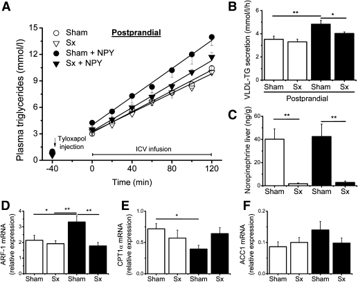
To test our hypothesis that during fasting the increased release of hypothalamic NPY is responsible for stimulating hepatic VLDL-TG secretion via the SNS, we combined the intracerebroventricular infusion of NPY with a selective sympathetic liver denervation. In contrast to the fasted condition in the previous experiment, the rats were instead subjected to the PP condition (4-h fast) to have a low endogenous NPY tone and nearly undetectable chylomicrons in plasma (5). In the PP condition, the sympathetic denervation itself did not change VLDL-TG secretion compared with the sham control (3.30 ± 0.22 vs. 3.52 ± 0.28 mmol/L/h). Infusion of NPY in the third ventricle of the brain in sham-operated rats strongly increased VLDL-TG secretion compared with the vehicle control (4.82 ± 0.35 vs. 3.52 ± 0.28 mmol/L/h) (Figs. 2A and B). However, intracerebroventricular NPY administration in Sx rats no longer resulted in a significant increase of VLDL-TG secretion compared with the vehicle control (4.02 ± 0.14 vs. 3.30 ± 0.22 mmol/L/h). Finally, the NPY-induced VLDL-TG secretion was significantly lower in Sx compared with sham-denervated NPY-infused rats (4.02 ± 0.14 vs. 4.82 ± 0.35 mmol/L/h) (Fig. 2B). The marked decrease in liver norepinephrine levels confirmed a successful selective sympathetic liver denervation (Fig. 2C). There were no significant differences in body weight (P = 0.28) or food intake (P = 0.45) in the night before the experiment. During the experiment, we observed no differences in plasma corticosterone (Time × Treatment P = 0.85) or insulin (Time × Treatment P = 0.24) between the four groups. To further dissect the metabolic pathways in the liver by which central NPY controls VLDL-TG secretion, we analyzed gene expression of key hepatic enzymes. NPY infusion increased mRNA levels of ADP-ribosylation factor (ARF-1) only in sham-operated rats (Fig. 2D). The mRNA levels of other genes involved in VLDL secretion, including apolipoprotein B (ApoB) and microsomal triglyceride transfer protein (MTTP), were not modified by NPY treatment. NPY infusion decreased carnitine palmitoyltransferase 1 α (CPT1α) mRNA levels in the sham-denervated rats but not in the Sx groups (Fig. 2E). We found no differences in expression of genes promoting lipogenesis, including acetyl-coenzyme A carboxylase alpha (ACC1) (Fig. 2F), acetyl-coenzyme A carboxylase beta (ACC2), fatty acid synthase (FAS), stearoyl-CoA desaturase 1 (SCD1), peroxisome proliferator–activated receptor γ (PPARγ), and sterol regulatory element–binding transcription factor 1c (SREBP1c). Western blot analysis revealed no changes in total ACC protein or phosphorylated ACC–to–ACC protein ratio after NPY infusion (data not shown).
FIG. 2.
Hepatic Sx prevents the stimulatory effect of the intracerebroventricular (ICV) administration of NPY on VLDL-TG secrection compared with hepatic sham-denervated animals. A: Plasma triglyceride levels after tyloxapol injection and intracerebroventricular infusion of NPY (1 μg/μL) in sham and Sx liver-denervated rats compared with intracerebroventricular vehicle infusion in sham and Sx liver-denervated rats. B: Calculated VLDL-TG secretion rates in the sham and Sx groups after intracerebroventricular infusion of vehicle (□) or NPY (■) (two-way ANOVA NPY P = 0.001). C: Liver norepinephrine levels in the different groups (two-way ANOVA Denervation P < 0.001). D–F: Relative gene expression in the liver is shown for ARF-1 (two-way ANOVA NPY × Denervation P = 0.046), CPT1α (two-way ANOVA NPY × Denervation P = 0.046), and ACC1 (two-way ANOVA NPY × Denervation P = 0.216). Values are means ± SEM of seven to nine rats per group. *P < 0.05, **P < 0.01.
Nutritional status has clear effects on genes regulating hepatic lipid metabolism.
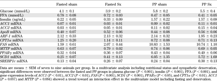
With regard to nutritional status, the previous experiments show the following: 1) during fasting conditions, a sympathetic liver denervation lowers VLDL-TG secretion compared with sham-denervated animals; 2) in the PP condition, Sx denervated rats do not show a lower VLDL-TG secretion compared with sham-denervated animals; and 3) comparing experiments 1 and 2 shows that in intact animals, VLDL-TG secretion is not significantly different between PP and fasted sham animals. Together, these results clearly indicate that only during fasting conditions is the SNS necessary to stimulate VLDL-TG secretion in order to maintain VLDL-TG secretion at a PP level. We therefore compared expression levels of key genes involved in lipid metabolism, between the sham and Sx denervated rats in the fasted and the PP experiments, to determine which pathways are regulated by the SNS after fasting (Table 1). Lower baseline concentrations of plasma glucose and insulin and higher baseline plasma concentrations of FFA illustrate the clear metabolic differences between the fasted and PP rats (Table 1). Fasted rats show decreased mRNA expression of genes involved in lipogenesis, including ACC1, ACC2, FAS, SCD1, and PPARγ, and increased mRNA expression of a gene promoting oxidation, CPT1α (Table 1). ACC1 (P = 0.057) and MTTP (P = 0.084) showed a trend toward an interaction effect in the multivariate model including fasting and denervation. Western blot analysis revealed no changes in total ACC protein or the phosphorylated ACC–to–ACC protein ratio between the Sx and sham groups (data not shown). Thus, although several genes involved in hepatic lipid metabolism are clearly affected by the nutritional status, analysis of mRNA expression of the fasted and PP denervated rats did not reveal the molecular pathways involved in lower VLDL-TG secretion in the Sx rats during fasting.
TABLE 1.
Comparison of baseline plasma parameters and gene expression in liver tissue between fasted and PP rats with a sham or Sx denervation
Rats with a lesioned arcuate nucleus cannot maintain VLDL-TG secretion during fasting.
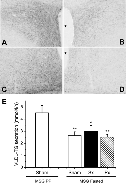
We subsequently investigated whether rats with a lesion of the ARC, an important component of the sympathetic outflow circuit to the liver (10), can maintain VLDL-TG secretion at a PP level during fasting. Adequate MSG treatment was shown by a pronounced decrease of NPY immunoreactive fibers in the ARC and PVN (Fig. 3A–D) in adult rats. First, comparing MSG-treated sham liver-denervated rats in the PP and fasted state revealed a significant decrease in baseline plasma triglyceride concentration (Table 2) and a 42% decrease in VLDL-TG secretion (Fig. 3E). But in the fasted MSG-treated rats, a sympathetic liver denervation did not result in an additional significant decrease in VLDL-TG secretion as shown in the first experiment in rats with an intact ARC. The MSG-treated rats in the different groups did not differ in body weight on the day before the experiment or in baseline plasma corticosterone levels (Table 2). Norepinephrine values were significantly lower in the MSG Sx group (P < 0.05). The fasted condition was clearly reflected in the significantly decreased plasma glucose concentrations compared with the PP rats (Table 2). With our sample size, we observed no significant effect of fasting on plasma insulin concentrations as seen in nontreated animals. However, MSG-treated animals were clearly hyperinsulinemic and showed larger variance in plasma insulin levels compared with nontreated animals (Tables 1 and 2). These data show that fasted MSG rats cannot maintain VLDL-TG secretion to a PP level. Furthermore, in fasted MSG-treated rats, sympathetic denervation does not affect VLDL-TG secretion compared with fasted sham denervated rats.
FIG. 3.
MSG rats cannot maintain VLDL-TG secretion during fasting. A–D: MSG treatment resulted in decreased NPY immunoreactivity in the PVN (B) and ARC (D) compared with saline-administered rats (A and C). The asterisk indicates the third ventricle. E: Calculated VLDL-TG secretion in PP and fasted sham, Sx, and Px liver-denervated MSG rats (one-way ANOVA P = 0.008). Values are means ± SEM of six to eight rats per group. *P < 0.05, **P < 0.01 compared with the PP sham MSG group.
TABLE 2.
Comparison of baseline parameters between the MSG groups PP sham and fasted sham, Sx and Px denervated rats
Central NPY does not affect lipolysis.
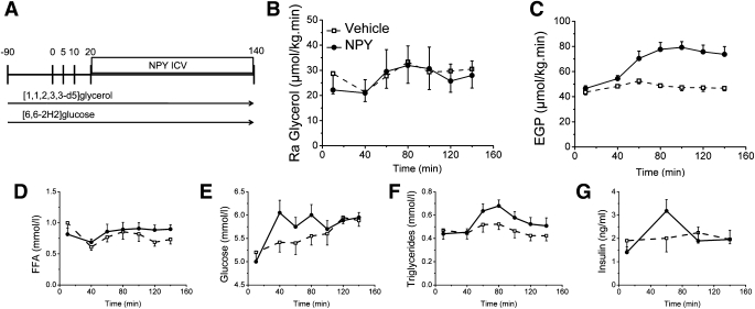
Finally, we investigated whether the increased VLDL-TG secretion after central NPY infusion could be due to increased substrate availability of FFAs through lipolysis. We investigated the effect of intracerebroventricularly administered NPY on lipolysis with a double stable isotope technique measuring endogenous glucose production and lipolysis simultaneously. The stable isotopes [1,1,2,3,3-d5]glycerol and [6,6-2H2]glucose were used (Fig. 4A). No significant effects of central NPY on lipolysis, assessed either by glycerol appearance or plasma FFA levels, could be observed (Figs. 4B and D). On the other hand, a clear effect of central NPY on endogenous glucose production was observed (Fig. 4C). Plasma triglyceride and glucose concentrations showed a trend toward an increase (Fig. 4E and F). We observed no significant differences in plasma corticosterone (Time × Treatment P = 0.13), glucagon (Time × Treatment P = 0.81), or insulin (Fig. 4G) concentrations between the groups.
FIG. 4.
Intracerebroventricular NPY does not affect lipolysis. A: Experimental protocol of the double stable isotope study to measure lipolysis and EGP during NPY or vehicle intracerebroventricular treatment. B–G: Acute effect of intracerebroventricular NPY or vehicle on rate of appearance (Ra) of glycerol (Time × Treatment P = 0.90), EGP (Time × Treatment P < 0.01), plasma FFA (Time × Treatment P = 0.10), plasma glucose (Time × Treatment P = 0.06), plasma triglycerides (Time × Treatment P = 0.10), and plasma insulin (Time × Treatment P = 0.12) concentrations. Values are means ± SEM of six to seven rats per group.
DISCUSSION
In recent years, the important role of the central nervous system in controlling liver metabolism has become more and more evident. A number of insightful experiments have considerably increased our understanding of the mechanisms of the hypothalamus to control glucose metabolism through the ANS (12–14,17,18). Our current experiments show that an intact hepatic sympathetic innervation and arcuate nucleus are also necessary to maintain VLDL-TG secretion during fasting. In agreement, a central infusion of NPY cannot increase VLDL-TG secretion in Sx liver-denervated rats. Together, these data indicate that the increased release of hypothalamic NPY during fasting maintains hepatic VLDL-TG secretion via the sympathetic input to the liver. This mechanism could be of physiological importance during fasting, when lipids are the main energy source. However, this mechanism could play a pathophysiological role in conditions characterized by a constant high activity of NPY, as found in animal models of obesity and hypertriglyceridemia (6,8,29). Recent data in humans support this hypothesis, as high sympathetic activity and low parasympathetic activity significantly correlate with components of the metabolic syndrome, including hypertriglyceridemia (2). Additionally, genetic screening revealed a novel polymorphism in the NPY1-5 gene to be associated with reduced serum triglyceride levels in a severely obese cohort (30).
The notion that the autonomic innervation of the liver is not only important for the control of carbohydrate metabolism but also for hepatic lipid metabolism is supported by the findings of a previous study applying phenol to the liver (31). In our study, we used microsurgery to selectively denervate the sympathetic and parasympathetic inputs to the liver and dissect the separate roles of these antagonistic branches of the ANS. We conclude that during fasting specifically, the SNS is important in the regulation of VLDL-TG metabolism. Our results also extend those of the study of Stafford et al. (5) showing that intracerebroventricular NPY increases VLDL-TG secretion in the PP condition and intracerebroventricular infusion of the Y1 antagonist decreases VLDL-TG secretion during fasting.
The MSG model induces a destruction of 80–90% of the neurons in the ARC, while sparing glial cells or axons passing through the nucleus (22,32). It was shown by others that during fasting the increased NPY immunoreactivity in the ARC and PVN does not occur in the MSG model (15). We are, however, aware that the neurotoxic lesion is not restricted to NPY neurons but affects other neurons in the ARC as well (22). In spite of the caveats of the MSG model, the data from our experiments in MSG animals support a role for the arcuate nucleus and possibly NPY in activating the SNS to control hepatic lipid metabolism during fasting, as the experiments were performed in rats with similar body weights. Furthermore, MSG animals were still responsive to exogenous NPY (data not shown).
The strength of this study is that by a selective hepatic denervation we were able to show the neural pathway by which hypothalamic NPY increases VLDL-TG secretion. This is important, as NPY is also known to exhibit endocrine effects (33). We propose that NPY neurons in the ARC activate second-order neurons in the PVN, which in turn activate the preganglionic sympathetic neurons in the intermediolateral column of the spinal cord. This notion is supported by the study by Viñuela and Larsen (9), who combined retrograde tracing from the intermediolateral column with intracerebroventricular administration of NPY and clearly demonstrated that sympathetic preautonomic hypothalamic neurons are activated by intracerebroventricular NPY. However, the lack of infected NPY neurons in the ARC after viral tracing from the liver (11) indicates that the NPY projection from the ARC to the preautonomic neurons might involve an interneuron. Alternatively, we cannot exclude from our experiments that NPY activates the SNS at the level of the brain stem. Although an inhibition of the melanocortin system could also be important during fasting, a previous study (5) did not find an acute effect of central melanocortin stimulation or blockade on VLDL-TG secretion. Moreover, contrary to the effect of chronic intracerebroventricular NPY administration, chronic blockade of the central melanocortin system does not change plasma levels of triglycerides independently of increased food intake, although it does result in an obese phenotype (34–36). Therefore, in acute and chronic conditions hypothalamic NPY seems more important than the melanocortin system in regulating hepatic VLDL-TG secretion. Conversely, the effects of the melanocortin system on white adipose tissue are well established (36).
The molecular mechanism through which the activation of the SNS after intracerebroventricular NPY regulates VLDL-TG secretion probably includes an increased VLDL-TG assembly and decreased β-oxidation. The second step in VLDL assembly is dependent of ARF-1, which was increased in our NPY-infused rats. This may reflect an increased production of mature VLDL particles (37). We also observed decreased mRNA levels of CPT1α, indicating decreased oxidation of FFAs in the liver. When oxidation is inhibited, more substrate can be guided to the alternative route, i.e., resulting in a higher VLDL-TG secretion. Therefore, both of the above-mentioned mechanisms may contribute to increased VLDL-TG secretion during the intracerebroventricular administration of NPY. Surprisingly, the effects of exogenous NPY on oxidation were contrary to changes occurring during fasting (when endogenous NPY levels are high), when increased oxidation was observed. Our results point to the possibility that the central nervous system stimulates the liver to maintain VLDL-TG secretion in competition with the peripheral effects of fasting. This is in accordance with our observation that VLDL-TG secretion does not increase after fasting in spite of the fact that NPY levels are physiologically high.
We show that central NPY has no effect on lipolysis and therefore does not contribute to increased VLDL-TG secretion. Others have shown that NPY inhibits lipolysis in vitro and after systemic administration, which possibly reflects a peripheral effect of NPY derived from the autonomic nerve endings on adipocytes (38–40).
In summary, we provide evidence that the activation of NPY neurons in the hypothalamus has a stimulatory role on hepatic VLDL-TG secretion through the SNS. We believe our data are of importance in understanding the physiological and pathophysiological role of the central nervous system in controlling lipid metabolism.
Supplementary Material
ACKNOWLEDGMENTS
This study was supported by NWO-ZonMw (TOP 91207036).
No potential conflicts of interest relevant to this article were reported.
E.B. researched data, contributed to discussion, wrote the manuscript, and reviewed and edited the manuscript. L.P. researched data and reviewed and edited the manuscript. M.T.A., E.Fo., A.J.B., J.K., and A.A. contributed to discussion and reviewed and edited the manuscript. E.Fl. and A.K. contributed to discussion, wrote the manuscript, and reviewed and edited the manuscript. E.B. and A.K. are the guarantors of this work and, as such, had full access to all the data in the study and take responsibility for the integrity of the data and the accuracy of the data analysis.
The authors thank Cindy Kunne from the Tytgat Institute for Liver and Intestinal Research, Academic Medical Center (AMC), University of Amsterdam, Amsterdam, the Netherlands, for the liver triglycerides measurement and Yvette Kettelarij and Marja Neeleman from the Department of Clinical Chemistry, Laboratory of Endocrinology, AMC, University of Amsterdam, for technical assistance.
Footnotes
This article contains Supplementary Data online at http://diabetes.diabetesjournals.org/lookup/suppl/doi:10.2337/db11-1142/-/DC1.
REFERENCES
- 1.Sørensen LP, Andersen IR, Søndergaard E, et al. Basal and insulin mediated VLDL-triglyceride kinetics in type 2 diabetic men. Diabetes 2011;60:88–96 [DOI] [PMC free article] [PubMed] [Google Scholar]
- 2.Licht CM, Vreeburg SA, van Reedt Dortland AK, et al. Increased sympathetic and decreased parasympathetic activity rather than changes in hypothalamic-pituitary-adrenal axis activity is associated with metabolic abnormalities. J Clin Endocrinol Metab 2010;95:2458–2466 [DOI] [PubMed] [Google Scholar]
- 3.Gibbons GF. Assembly and secretion of hepatic very-low-density lipoprotein. Biochem J 1990;268:1–13 [DOI] [PMC free article] [PubMed] [Google Scholar]
- 4.Chirieac DV, Chirieac LR, Corsetti JP, Cianci J, Sparks CE, Sparks JD. Glucose-stimulated insulin secretion suppresses hepatic triglyceride-rich lipoprotein and apoB production. Am J Physiol Endocrinol Metab 2000;279:E1003–E1011 [DOI] [PubMed] [Google Scholar]
- 5.Stafford JM, Yu F, Printz R, Hasty AH, Swift LL, Niswender KD. Central nervous system neuropeptide Y signaling modulates VLDL triglyceride secretion. Diabetes 2008;57:1482–1490 [DOI] [PMC free article] [PubMed] [Google Scholar]
- 6.Takahashi KA, Cone RD. Fasting induces a large, leptin-dependent increase in the intrinsic action potential frequency of orexigenic arcuate nucleus neuropeptide Y/Agouti-related protein neurons. Endocrinology 2005;146:1043–1047 [DOI] [PubMed] [Google Scholar]
- 7.Bai FL, Yamano M, Shiotani Y, et al. An arcuato-paraventricular and -dorsomedial hypothalamic neuropeptide Y-containing system which lacks noradrenaline in the rat. Brain Res 1985;331:172–175 [DOI] [PubMed] [Google Scholar]
- 8.Lambert PD, Wilding JP, Turton MD, Ghatei MA, Bloom SR. Effect of food deprivation and streptozotocin-induced diabetes on hypothalamic neuropeptide Y release as measured by a radioimmunoassay-linked microdialysis procedure. Brain Res 1994;656:135–140 [DOI] [PubMed] [Google Scholar]
- 9.Viñuela MC, Larsen PJ. Identification of NPY-induced c-Fos expression in hypothalamic neurones projecting to the dorsal vagal complex and the lower thoracic spinal cord. J Comp Neurol 2001;438:286–299 [DOI] [PubMed] [Google Scholar]
- 10.la Fleur SE, Kalsbeek A, Wortel J, Buijs RM. Polysynaptic neural pathways between the hypothalamus, including the suprachiasmatic nucleus, and the liver. Brain Res 2000;871:50–56 [DOI] [PubMed] [Google Scholar]
- 11.Stanley S, Pinto S, Segal J, et al. Identification of neuronal subpopulations that project from hypothalamus to both liver and adipose tissue polysynaptically. Proc Natl Acad Sci USA 2010;107:7024–7029 [DOI] [PMC free article] [PubMed] [Google Scholar]
- 12.Marks JL, Waite K. Intracerebroventricular neuropeptide Y acutely influences glucose metabolism and insulin sensitivity in the rat. J Neuroendocrinol 1997;9:99–103 [DOI] [PubMed] [Google Scholar]
- 13.van den Hoek AM, Voshol PJ, Karnekamp BN, et al. Intracerebroventricular neuropeptide Y infusion precludes inhibition of glucose and VLDL production by insulin. Diabetes 2004;53:2529–2534 [DOI] [PubMed] [Google Scholar]
- 14.van den Hoek AM, van Heijningen C, Schröder-van der Elst JP, et al. Intracerebroventricular administration of neuropeptide Y induces hepatic insulin resistance via sympathetic innervation. Diabetes 2008;57:2304–2310 [DOI] [PMC free article] [PubMed] [Google Scholar]
- 15.Leitner C, Bartness TJ. Food deprivation-induced changes in body fat mobilization after neonatal monosodium glutamate treatment. Am J Physiol Regul Integr Comp Physiol 2008;294:R775–R783 [DOI] [PubMed] [Google Scholar]
- 16.Steffens AB. A method for frequent sampling blood and continuous infusion of fluids in the rat without disturbing the animal. Physiol Behav 1969;4:833–836 [Google Scholar]
- 17.Klieverik LP, Sauerwein HP, Ackermans MT, Boelen A, Kalsbeek A, Fliers E. Effects of thyrotoxicosis and selective hepatic autonomic denervation on hepatic glucose metabolism in rats. Am J Physiol Endocrinol Metab 2008;294:E513–E520 [DOI] [PubMed] [Google Scholar]
- 18.Kalsbeek A, La Fleur S, Van Heijningen C, Buijs RM. Suprachiasmatic GABAergic inputs to the paraventricular nucleus control plasma glucose concentrations in the rat via sympathetic innervation of the liver. J Neurosci 2004;24:7604–7613 [DOI] [PMC free article] [PubMed] [Google Scholar]
- 19.Obici S, Feng Z, Tan J, Liu L, Karkanias G, Rossetti L. Central melanocortin receptors regulate insulin action. J Clin Invest 2001;108:1079–1085 [DOI] [PMC free article] [PubMed] [Google Scholar]
- 20.Otway S, Robinson DS. The use of a non-ionic detergent (Triton WR 1339) to determine rates of triglyceride entry into the circulation of the rat under different physiological conditions. J Physiol 1967;190:321–332 [DOI] [PMC free article] [PubMed] [Google Scholar]
- 21.van den Berg MP, Romeijn SG, Verhoef JC, Merkus FW. Serial cerebrospinal fluid sampling in a rat model to study drug uptake from the nasal cavity. J Neurosci Methods 2002;116:99–107 [DOI] [PubMed] [Google Scholar]
- 22.Meister B, Ceccatelli S, Hökfelt T, Andén NE, Andén M, Theodorsson E. Neurotransmitters, neuropeptides and binding sites in the rat mediobasal hypothalamus: effects of monosodium glutamate (MSG) lesions. Exp Brain Res 1989;76:343–368 [DOI] [PubMed] [Google Scholar]
- 23.Ackermans MT, Pereira Arias AM, Bisschop PH, Endert E, Sauerwein HP, Romijn JA. The quantification of gluconeogenesis in healthy men by (2)H2O and [2-(13)C]glycerol yields different results: rates of gluconeogenesis in healthy men measured with (2)H2O are higher than those measured with [2-(13)C]glycerol. J Clin Endocrinol Metab 2001;86:2220–2226 [DOI] [PubMed] [Google Scholar]
- 24.Steele R. Influences of glucose loading and of injected insulin on hepatic glucose output. Ann N Y Acad Sci 1959;82:420–430 [DOI] [PubMed] [Google Scholar]
- 25.Patterson BW, Mittendorfer B, Elias N, Satyanarayana R, Klein S. Use of stable isotopically labeled tracers to measure very low density lipoprotein-triglyceride turnover. J Lipid Res 2002;43:223–233 [PubMed] [Google Scholar]
- 26.Srivastava NK, Pradhan S, Mittal B, Kumar R, Gowda GN. An improved, single step standardized method of lipid extraction from human skeletal muscle tissue. Anal Lett 2006;39:297–315 [Google Scholar]
- 27.Ramakers C, Ruijter JM, Deprez RH, Moorman AF. Assumption-free analysis of quantitative real-time polymerase chain reaction (PCR) data. Neurosci Lett 2003;339:62–66 [DOI] [PubMed] [Google Scholar]
- 28.Ruijter JM, Ramakers C, Hoogaars WM, et al. Amplification efficiency: linking baseline and bias in the analysis of quantitative PCR data. Nucleic Acids Res 2009;37:e45. [DOI] [PMC free article] [PubMed] [Google Scholar]
- 29.McKibbin PE, Cotton SJ, McMillan S, et al. Altered neuropeptide Y concentrations in specific hypothalamic regions of obese (fa/fa) Zucker rats. Possible relationship to obesity and neuroendocrine disturbances. Diabetes 1991;40:1423–1429 [DOI] [PubMed] [Google Scholar]
- 30.Blumenthal JB, Andersen RE, Mitchell BD, et al. Novel neuropeptide Y1 and Y5 receptor gene variants: associations with serum triglyceride and high-density lipoprotein cholesterol levels. Clin Genet 2002;62:196–202 [DOI] [PubMed] [Google Scholar]
- 31.Tavares FL, Seelaender MC. Hepatic denervation impairs the assembly and secretion of VLDL-TAG. Cell Biochem Funct 2008;26:557–565 [DOI] [PubMed] [Google Scholar]
- 32.Simson EL, Gold RM, Standish LJ, Pellett PL. Axon-sparing brain lesioning technique: the use of monosodium-L-glutamate and other amino acids. Science 1977;198:515–517 [DOI] [PubMed] [Google Scholar]
- 33.Härfstrand A, Eneroth P, Agnati L, Fuxe K. Further studies on the effects of central administration of neuropeptide Y on neuroendocrine function in the male rat: relationship to hypothalamic catecholamines. Regul Pept 1987;17:167–179 [DOI] [PubMed] [Google Scholar]
- 34.Zarjevski N, Cusin I, Vettor R, Rohner-Jeanrenaud F, Jeanrenaud B. Chronic intracerebroventricular neuropeptide-Y administration to normal rats mimics hormonal and metabolic changes of obesity. Endocrinology 1993;133:1753–1758 [DOI] [PubMed] [Google Scholar]
- 35.Baran K, Preston E, Wilks D, Cooney GJ, Kraegen EW, Sainsbury A. Chronic central melanocortin-4 receptor antagonism and central neuropeptide-Y infusion in rats produce increased adiposity by divergent pathways. Diabetes 2002;51:152–158 [DOI] [PubMed] [Google Scholar]
- 36.Nogueiras R, Wiedmer P, Perez-Tilve D, et al. The central melanocortin system directly controls peripheral lipid metabolism. J Clin Invest 2007;117:3475–3488 [DOI] [PMC free article] [PubMed] [Google Scholar]
- 37.Asp L, Claesson C, Boren J, Olofsson SO. ADP-ribosylation factor 1 and its activation of phospholipase D are important for the assembly of very low density lipoproteins. J Biol Chem 2000;275:26285–26292 [DOI] [PubMed] [Google Scholar]
- 38.Labelle M, Boulanger Y, Fournier A, St Pierre S, Savard R. Tissue-specific regulation of fat cell lipolysis by NPY in 6-OHDA-treated rats. Peptides 1997;18:801–808 [DOI] [PubMed] [Google Scholar]
- 39.Margareto J, Aguado M, Osés-Prieto JA, et al. A new NPY-antagonist strongly stimulates apoptosis and lipolysis on white adipocytes in an obesity model. Life Sci 2000;68:99–107 [DOI] [PubMed] [Google Scholar]
- 40.Kreier F, Fliers E, Voshol PJ, et al. Selective parasympathetic innervation of subcutaneous and intra-abdominal fat—functional implications. J Clin Invest 2002;110:1243–1250 [DOI] [PMC free article] [PubMed] [Google Scholar]
Associated Data
This section collects any data citations, data availability statements, or supplementary materials included in this article.


