Abstract
Background
Dictyostelium cells exhibit an unusual response to hyperosmolarity that is distinct from the response in other organisms investigated: instead of accumulating compatible osmolytes as it has been described for a wide range of organisms, Dictyostelium cells rearrange their cytoskeleton and thereby build up a rigid network which is believed to constitute the major osmoprotective mechanism in this organism. To gain more insight into the osmoregulation of this amoeba, we investigated physiological processes affected under hyperosmotic conditions in Dictyostelium.
Results
We determined pH changes in response to hyperosmotic stress using FACS or 31P-NMR. Hyperosmolarity was found to acidify the cytosol from pH 7.5 to 6.8 within 5 minutes, whereas the pH of the endo-lysosomal compartment remained constant. Fluid-phase endocytosis was identified as a possible target of cytosolic acidification, as the inhibition of endocytosis observed under hypertonic conditions can be fully attributed to cytosolic acidification. In addition, a deceleration of vesicle mobility and a decrease in the NTP pool was observed.
Conclusion
Together, these results indicate that hyperosmotic stress triggers pleiotropic effects, which are partially mediated by a pH signal and which all contribute to the downregulation of cellular activity. The comparison of our results with the effect of hyperosmolarity and intracellular acidification on receptor-mediated endocytosis in mammalian cells reveals striking similarities, suggesting the hypothesis of the same mechanism of inhibition by low internal pH.
Background
Cells steadily face fluctuations of the external osmolarity due to dehydration. Occasionally, dramatic changes in osmolarity can occur, resulting in a stress condition [1]. Hyperosmolarity of the external medium leads to the extrusion of water and the concomitant shrinkage of cells [2]. Within a few minutes, the cells activate mechanisms, termed "regulatory volume increase" (RVI), to regain their volume [3]. Under prolonged hyperosmotic conditions, compatible osmolytes, e.g. polyols or amines are accumulated inside the cells [3]. These osmolytes exhibit a stabilizing effect on proteins and thereby avoid the deleterious effect of protein aggregation. In addition, the expression of stress proteins, as chaperones and DNA repair proteins was observed in various organisms in response to hypertonicity [4,5,6].
Recently it could be shown, that the amoeba Dictyostelium discoideum exhibits an unusual response to hypertonic stress which is distinct from the response observed in other organisms [7]: the cells remain shrunken after water extrusion and they neither accumulate compatible osmolytes nor express stress proteins. Instead, Dictyostelium largely rearranges cellular proteins between compartments [7]. Especially the enhanced recruitment of major cytoskeletal proteins to the cell cortex, i.e. the layer beneath the plasma membrane plays a pivotal role, as increased amounts of actin and myosin II co-localize at the cell cortex of hyperosmotically shocked cells and form a rigid network [8]. The formation of this shell-like structure is believed to be the major osmoprotective mechanism in Dictyostelium [9]. The osmoprotective role of the cytoskeletal reinforcement is also supported by the fact that several cytoskeletal proteins are essential for viability under hypertonic conditions [10, 11].
The knowledge about the signalling pathways involved in osmoregulation is scarce. In response to hyperosmotic stress, guanylyl cyclase is activated, resulting in an increase in cGMP concentration. This induces the phosphorylation of the myosin II heavy chain kinase, which in turn results in the phosphorylation of myosin II heavy chain [8, 12, 13]. Thereby, the myosin II filaments are disassembled as a prerequesite for their rearrangement to the cell cortex [14]. In addition, DokA, a homologue of bacterial histidine kinases, was shown to be essential under hypertonic conditions, suggesting a role for 2-component regulatory elements in osmoregulation [15].
Furthermore, cAMP is believed to play an essential role during the spore state, which represents a naturally occurring hyperosmotic condition induced by the developmental program of the amoeba. The adenylyl cyclase G was shown to be an osmosensor, which suppresses germination under hyperosmotic conditions in spore cells [16]. This correlates with the observation that high cAMP concentrations are maintained during spore dormancy [17].
In addition, acidification of the external medium has been observed in response to hypertonicity, which was attributed to the secretion of protons [18]. We therefore addressed the question, whether this increase in external proton concentration correlates with a change in pH in an intracellular compartment. Cytosolic pH changes have been shown to act as signals in regulating proteins and cellular processes in various organisms [19]. The processes regulated by cytosolic pH changes include e.g. glycolysis, protein synthesis, DNA synthesis, motility and activation from dormant states [19, 20]. Generally, the acidification of the cytosol correlated with a decrease, whereas the alkalinization correlated with an increase in activity or efficiency of cellular processes [19]. In addition to cytosolic pH, homeostasis in the endo-lysosomal pH has been shown to be essential for cellular processes: perturbations of the pH affect endocytosis as well as the post-translational modification and sorting of proteins in Dictyostelium cells [21,24].
In this paper we present evidence that high osmolarity results in a significant acidification and a concomitant decrease in endocytic activity, as well as a depletion of the NTP pool, indicating a general decrease in cellular activity.
Results
Hyperosmotic stress leads to intracellular acidification
It was shown recently, that the external medium was acidified, when Dictyostelium cells were exposed to high tonicity [18]. Therefore we tested, whether this external acidification is paralleled by a change in intracellular pH. To monitor pH changes, we used BCECF as a pH-dependent fluorescent dye. After incubation with the non- fluorescent, cell-permeable derivative BCECF-AM, efficient cleavage of the AM ester by cell esterases as well as homogeneous labeling of the cells was confirmed by fluorescence microscopy (data not shown). As considerable leakage of the dye out of the cells and a concomitant increase in fluorescence in the buffer was observed within minutes, flow cytometry was chosen to measure intracellular fluorescence.
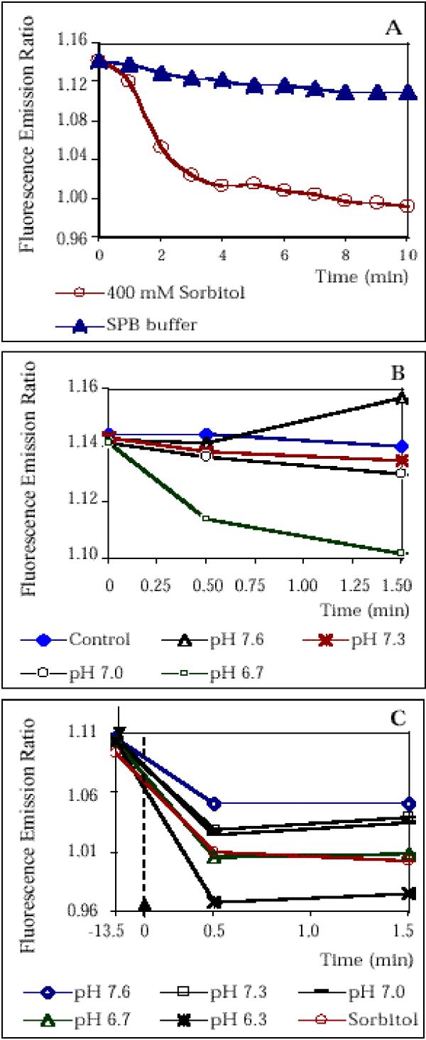
Cells labeled with BCECF were washed and resuspended in SPB buffer. To study pH changes under hypertonic conditions, the cells were first analyzed recording at the emission wavelengths FL1 (530/30 nm) and FL2 (585/42 nm). Then, SPB buffer/2 M sorbitol was added to a final concentration of 400 mM sorbitol and flow cytometry measurements were performed at intervals of 30 sec. The average emission ratio FL1/FL2 was calculated for each time point as a relative measure for the intracellular pH. BCECF-labeled cells, which were exposed to hyperosmotic conditions, exhibited a strong decrease in the emission ratio, indicating a decrease in pH, whereas little changes of the emission ratio of control cells suspended in SPB buffer were observed (Fig. 1A). Apparently, the major changes in emission ratio occurred during the first 1-4 min of hyperosmotic shock, whereas the decrease in the emission ratio was slowed down 5-10 min after onset of the shock. However, the observed decrease was still 4-fold bigger in shocked cells than in control cells in this time slot. This suggests, that the process of pH drop in shocked cells persists at least to T = 10 min.
Figure 1.
FACS measurements using BCECF as fluorescent pH indicator show that hyperosmotic stress results in intracellular acidification (A) AX-2 cells were labeled with BCECF and were subsequently exposed to hyperosmotic stress. Fluorescence emission was determined at FL1 and FL2 by flow cytometry and the fluorescence emission ratio FL1/FL2 was calculated. The decrease in FL1/FL2 of cells exposed to hyperosmotic shock ("400 mM Sorbitol") reflects a decrease in pH. As control, the measurements were performed with cells suspended in SPB buffer. (B) Calibration of intracellular pH of cells in low osmolarity buffer. BCECF-labeled cells were suspended in calibration solutions with different Pseudo-Null pH values [25] and FL1/FL2 was determined by flow cytometry at intervals. The solution with Pseudo-Null pH 7.3 slightly decreased FL1/FL2 compared to the control cells suspended in SPB buffer and the solution with Pseudo-Null pH 7.6 increases FL1/FL2 compared to the control. Hence, the actual intracellular pH is estimated to be pH 7.3 to pH 7.4. (C) Calibration of the pH of hyperosmotically shocked cells. After BCECF labeling, Dictyostelium cells were suspended SPB buffer and FL1/FL2 was determined at T=-13.5 min. Sorbitol solution was added (indicated by the upper arrow) and the cells were incubated for 10 min. The cells were centrifuged and resuspended in high osmolarity calibration solutions with different Pseudo-Null pH values at T = 0 min (indicated by the lower arrow). FL1/FL2 was determined by flow cytometry at intervals. The solution with Pseudo-Null pH 6.7 slightly decreased FL1/FL2 compared to the control ("Sorbitol"; cells suspended in SPB buffer/400 mM sorbitol) at T = 0.5 min, before intracellular pH starts to recover. This suggests, that the actual intracellular pH is 6.7-6.8. The points of time specified in Fig. 1C are not drawn to scale.
To determine the absolute changes in intracellular pH, calibration of the emission ratio was performed largely according to the Pseudo-Null calibration method for human cells [25] with modifications accounting for Dictyostelium cells and for the hyperosmotic shock condition. Calibration was first performed with cells in low osmolarity buffer to determine pre-shock pH. Suspending the cells in the calibration solution with Pseudo-Null pH 7.3 caused a slight decrease in the emission ratio, whereas the calibration solution with Pseudo-Null pH 7.6 caused a distinct increase in the emission ratio, indicating that the actual intracellular pH is 7.4-7.5 (Fig. 1B). In order to determine the intracellular pH of hyperosmotically shocked cells, the cells were first shaken in high osmolarity buffer (430 mOsm) for 10 min, before the cells were centrifuged and suspended in high osmolarity calibration solutions at T = 13.5 min. The calibration solution with Pseudo-Null pH 6.7 caused a very slight decrease in the emission ratio after 0.5 min, whereas the calibration solution with Pseudo-Null pH value 7.0 caused a marked increase in the emission ratio, indicating that the actual intracellular pH is 6.7-6.8 (Fig. 1C). The intracellular pH is calibrated at T = 15 min as hyperosmotic conditions prevail during calibration. Hence, the results indicate, that the intracellular pH drops from 7.4-7.5 to 6.7-6.8 in cells exposed to hypertonic stress for 15 min.
Hyperosmotic stress leads to cytosolic acidification, but leaves the pH of the endo-lysosomal system unaffected
In parallel to the flow cytometry measurements, the intracellular pH drop under hypertonic conditions was investigated by 31P-NMR. This technique has the advantage, that the measurements are conducted directly on aerobic cell suspensions allowing detection of the pH with an excellent time resolution [26]. The strong broadening of intracellular resonances induced by sorbitol addition precluded the use of intracellular Pi to measure cytosolic pH. To determine the effect of hyperosmotic shock on cytosolic and lysosomal pH, methyl phosphonate (MEP) and 2-aminoethyl phosphonate (AEP) were used as pH probes in P-NMR experiments. Previous 31P-NMR studies have established that cytosolic pH is 7.3-7.5 [26], and that Dictyostelium cells possess an acidic lysosomal compartment at pH 4.3-4.5 [21, 27]. The phosphonates MEP and AEP are internalized by macropinocytosis and MEP is transferred to the cytosol much more efficiently than AEP.
The 31P-NMR chemical shifts (ppm) of these phosphonates are sensitive to pH and are therefore directly related to the pH of the compartments in which they are located. AEP has a pKa of 6.3 and its chemical shift decreased from 19.1 to 16.5 ppm when the pH increased from 4.0 to 9.0. MEP has a higher pKa (7.6) and its chemical shift decreased from 24.5 to 20.6 ppm when the pH increased as above. AEP allows simultaneous measurement of cytosolic and lysosomal pH, while MEP allows more precise determination of cytosolic pH. In the case of the NMR experiments, a final theoretical osmolarity of 340 mOsm was chosen, as a higher osmolarity was not feasible for technical reasons.
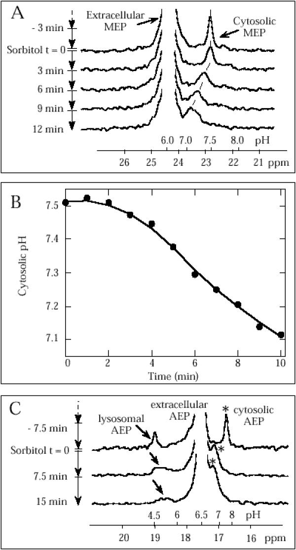
As shown in Fig. 2B (see also Fig. 2A), the cytosol progressively acidified from pH 7.5 to 7.1 following addition of sorbitol to a final concentration of 300 mOsm. This cytosolic acidification was confirmed with the AEP probe (Fig. 2C). Lysosomal pH (about pH 4.5) remained relatively stable during 7.5 min, then pH increased (Fig. 2C). Hence, the experiments show, that the acidification under hypertonic conditions can be confined to the cytosolic compartment. The decrease in pH is steady, suggesting a further decrease after 10 min of hyperosmotic shock.
Figure 2.
In vivo31 P-NMR analysis of the effect of hyperosmotic shock on cytosolic pH in Dictyostelium cells (A) The sequence of 31P-NMR spectra represents a typical experiment conducted with the probe MEP. After addition of MEP and preincubation during 60 min, a control spectrum was recorded for 3 min immediately before sorbitol addition (upper trace). Sorbitol (0.3 M) was then added at T = 0 and data were collected during successive 3-min intervals up to 12 min. NMR spectra shown are the sum of 180 free induction decays with 1-s interpulse delays and correspond to a 3-min accumulation. Only the spectral region around +24 ppm corresponding to MEP probe is shown. The pH scale shown on abcissa was calculated from the in vivo calibration curve pH = 7.6 + log(24.5 - δ)/(δ - 20.6), 8 being the chemical shift of MEP resonance. (B) Temporal evolution of cytosolic pH after onset of hyperosmotic shock in Dictyostelium cells calculated from the position of cytosolic MEP resonance (see A). (C) In vivo31P-NMR analysis of the effect of hyperosmotic shock on lysosomal pH in Dictyostelium cells. The sequence of successive 31P-NMR spectra represents a typical experiment conducted with the probe AEP. After addition of AEP and preincubation for 60 min, a control spectrum was recorded for 7.5 min immediately before sorbitol addition (upper trace). Sorbitol (0.3 M) was then added at T = 0 min and data were collected during two consecutive 7.5-min intervals. Only the spectral region around +18 ppm corresponding to AEP probe is shown. The pH scale indicated on the graph was calculated from the in vivo AEP calibration curve pH = 6.3 + log(19.1 - δ)/(δ - 16.5), δ being the chemical shift of AEP resonance. Lysosomal and cytosolic AEP satellite peaks, lowfield and upfield of extracellular AEP, respectively, are indicated by arrow and star symbols, respectively.
Hyperosmolarity results in deenergetization of Dictyostelium cells
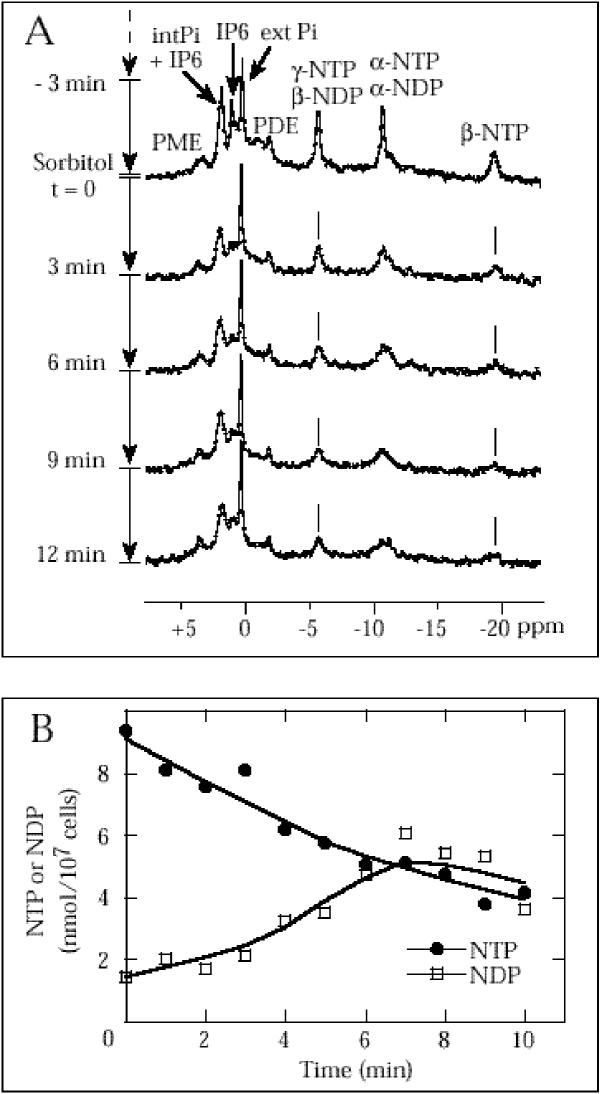
As cytosolic acidification is often accompanied by a down-regulation and inactivation of cellular activity [19, 20], we investigated, whether the bioenergetic status, characterized by the phosphorylated metabolite content, is affected by hypertonicity. In vivo31P-NMR was used to assess the phosphorylated metabolite content in Dictyostelium cells (Fig. 3A). An effect of sorbitol addition is a strong broadening of all intracellular signals which is a likely consequence of cellular shrinkage (compare T = 0 min and T = 6 min for example). Nevertheless, except for intracellular Pi (see arrow in Fig. 3A), major resonances remained easily identified. As shown in Fig. 3B, calculation of the NTP and NDP levels from the successive 31P-NMR spectra showed that the nucleoside triphosphate level decreased progressively after addition of 0.3 M sorbitol with T1/2 = 8 min. In parallel, NDP levels increased so that the total nucleotide content (NTP+NDP) remained constant. Other phosphorylated metabolites were not affected appreciably. The results therefore indicate, that the NTP pool of Dictyostelium amoebae is progressively depleted under hypertonic conditions.
Figure 3.
In vivo31 P-NMR analysis of the effect of osmotic shock on phosphorylated metabolites in Dictyostelium cells (A) The experiments were performed as described for the determination of cytosolic pH by 31P-NMR using MEP as probe. Only the spectral region (+5 to -20 ppm) corresponding to phosphorylated metabolites is shown. (B) Temporal evolution of intracellular nucleoside di- and triphosphate content during hyperosmotic shock in Dictyostelium cells. The relative intracellular NTP content was calculated from the area of the P-NTP peak at about -20 ppm and the NDP content was deduced from the area of the (β-NDP + γNTP) peak at about -5 ppm. The NTP and NDP content (nmol/107 cells) before hyperosmotic shock (values at T = 0) were measured on perchloric acid extracts. Abb.: PME, phosphomonoesters; PDE, phosphodiesters; cytPi, extPi, cytosolic and extracellular Pi; NTP, NDP, nucleoside tri- and diphosphate; IP6, inositol hexakisphosphate.
Hyperosmotic stress inhibits fluid-phase endo- and exocytosis
As shown above, the NTP content declines and the cytosol is acidified, indicating a general downregulation of cellular activity. We therefore addressed the question, whether also fluid-phase endocytosis, the only mechanism for nutrient uptake in axenic medium, is inhibited under hypertonic conditions.
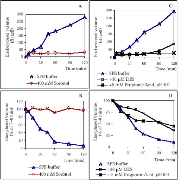
To examine fluid-phase endocytosis, we measured the ability of hyperosmotically shocked cells to internalize the fluorescent fluid-phase marker FITC-Dextran. The cells were incubated with 2 mg/ml FITC-Dextran for 2 h. Samples were withdrawn at various intervals and the fluorescence of cell lysates was determined by fluorescence spectroscopy. Control cells, which were suspended in SPB buffer avidly accumulated FITC-Dextran (Fig. 4A). In contrast, endocytosis was inhibited in cells, which were suspended in hypertonic sorbitol buffer containing FITC-Dextran: very little fluorescence was associated with cells exposed to hypertonicity and no increase in fluorescence was observed during 2 h of shock, indicating a total inhibition of endocytosis.
Figure 4.
Hyperosmotic stress results in the abolishment of endo- and exocytosis partially due to cytosolic acidification (A) Cells which were resuspended in SPB buffer avidly endocytose the fluorescent fluid-phase marker FITC-Dextran, whereas no fluid uptake was observed with cells exposed to 400 mM sorbitol in SPB buffer. (B) Correspondingly, cells in SPB buffer rapidly exocytosed ingested FITC-Dextran, whereas no loss of fluorescence was measured with cell suspended in 400 mM sorbitol, indicating that exocytosis is blocked. (C) Cells, which were artificially acidified by suspending the cells in SPB buffer/5 mM propionic acid (pH 6.0)/FITC-Dextran did not accumulate any FITC-Dextran, indicating that endocytosis was blocked. Also suspending the cells in SPB buffer/30 μM DES/FITC-Dextran resulted in the inhibition of endocytosis. However, a resumption of endocytic activity was observed after 1 h of hyperosmotic shock, presumably due to adaptation. As control, cells were suspended in SPB buffer/FITC-Dextran. (D) Artificial acidification of cells partially inhibits exocytosis. Control cells labeled with FITC-Dextran exocytosed about 90% of the dye within 2 h, whereas a loss of 55%-60% of cell-associated fluorescence was measured with cells exposed to 5 mM propionic acid or 30 μM DES.
To study the effect of hyperosmotic shock on exocytosis, we labeled Dictyostelium cells with FITC-Dextran for 2 h, washed the cells and resuspended them either in hyperosmolar buffer or in SPB buffer as control. The control cells steadily exocytosed the fluorescent dye, resulting in a 90% reduction of cell-associated fluorescence after 2 h (Fig. 4B). No reduction in cell-associated fluorescence was measured with cells, which were resuspended in high osmolarity sorbitol buffer, which clearly shows, that also fluid-phase exocytosis was blocked. Hence, hyperosmotic stress blocks the endo-lysosomal pathway.
Inhibition of fluid-phase endocytosis can be attributed to cytosolic acidification
We wanted to further elucidate the question, whether the inhibition of both fluid-phase endo- and exocytosis under hypertonic conditions could be mediated by cytosolic acidification. Therefore, we measured the endo- and exocytic activity of cells which were artificially acidified. This was on the one hand achieved by suspending the cells in SPB buffer containing 5 mM propionic acid (pH 6.0), which results in general intracellular acidification. On the other hand, diethylstilbestrol (DES) was added, which inhibits the plasma membrane proton ATPase and thereby acidifies the cytosol [28].
Fluid-phase endocytosis was found to be blocked in cells acidified by propionic acid (Fig. 4C), whereas avid endocytosis was measured in the control cell suspension. Also, in the presence of DES a strong inhibition of endocytosis could be observed. During the first 60 min after onset of hyperosmotic stress, no accumulation of fluorescent dye was measured, suggesting that cytosolic acidification can account for the inhibition of endocytosis under hyperosmotic conditions. During 60-120 min of hyperosmotic stress, a slight increase in intracellular fluorescence was observed. This effect is presumably due to adaptation of the cells to DES, as a similarly quick adaptation to Concanamycin A was observed, which is an inhibitor of the vacuolar proton- translocating ATPase [29].
Artificial acidification also affected exocytosis, however, inhibition was significantly weaker than under hyperosmotic shock conditions. Both the exposure of the cells to 5 mM propionic acid and to DES resulted in a decrease in cell-associated fluorescence to about 40% of the fluorescence measured at T = 0 after 2 h (Fig. 4D). Again, a strong inhibitory effect of DES was observed during the first 60 min of hyperosmotic stress, whereas a loss of the inhibitory effect of DES could be determined 60-120 min after onset of the experiment. Hence, exocytic activity is only reduced to about 50% after intracellular and cytosolic acidification for 2 h, whereas total inhibition is observed under hypertonic stress conditions. Again, the effect of intracellular acidification could be confined to the cytosol, as cytosolic acidification and intracellular acidification have the same effect.
The inhibition of fluid-phase endocytosis under hypertonicity can be mimicked by artificial cytosolic acidification, whereas the artificial acidification only partially inhibited exocytosis. This suggests the hypothesis that the inhibition of endocytosis under hypertonic conditions is mediated by the cytosolic acidification whereas the inhibition of exocytosis requires additional factors.
Membrane flow is slowed down under hyperosmotic conditions
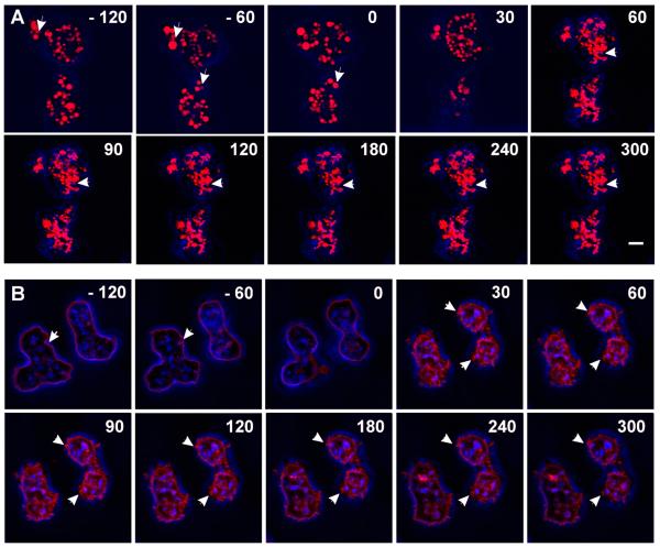
We then investigated, whether the cessation of fluid-phase endocytosis under hyperosmotic conditions correlates with general deceleration of vesicle mobility. We therefore incubated Dictyostelium cells with TRITC-Dextran, which specifically labels the vesicles of the endo-lysosomal system. Subsequently, the cells were washed, suspended in SPB buffer and analyzed by confocal fluorescence microscopy. As shown in Fig. 5A, the vesicles appeared distinct and highly mobile in these cells, as the vesicles rapidly changed their position within 60 sec (indicated by arrows in frames -120 and -60). Upon onset of hyperosmotic stress, cell shrinkage was induced, leading to progressive perturbation of the vesicular system (frames 30, 60). In addition to the structural change, also an altered vesicle mobility could be observed. The vesicles (for example those indicated by arrows in frames 60-300) were stalled at the same place over a time range of 4 minutes, suggesting that the transport of the vesicles was severely impaired under hypertonic conditions.
Figure 5.
Vesicle mobility and membrane flow are downregulated under hypertonic conditions. Cells were either labeled with TRITC-Dextran (A) or with Cy3.5 (B). Points of time at which confocal images were scanned are indicated in seconds referring to the onset of hyperosmotic shock as T = 0. (A) The fluorescent label of TRITC-Dextran in red is superimposed to phase contrast images in blue. TRITC-Dextran labeled vesicles of cells suspended in SPB buffer are mobile (indicated by arrows in frames -120, -60). Upon sorbitol shock, perturbation and immobilization of vesicles was observed. As an example, a region with stalled and distorted vesicles is indicated by arrows in frames 60-300. (B) The fluorescent label of Cy3.5 in red is superimposed to phase contrast images in blue. Cells suspended in SPB buffer occassionally endocytose membrane (arrows in frames -120, -60). Upon hyperosmotic shock, the plasma membrane thickens and forms spikes after cell shrinkage. The spikes are not endocytosed but remain fixed. Typical spikes are indicated with arrows in frames 30-300. Bar = 2 μm.
The down-regulation of membrane-flow under hypertonic conditions was investigated by labeling plasma membrane proteins with Cy3.5 [30]. Subsequently, the cells were washed, suspended in SPB buffer and were analyzed by confocal fluorescence microscopy (Fig. 5B). Cells which were suspended in SPB buffer occasionally endocytosed plasma membrane (indicated by arrows in frames -120, -60). Upon addition of 2 M sorbitol to a final concentration of 400 mM sorbitol, the plasma membrane became highly folded and invaginated, which is indicated by the formation of a thickened fluorescent layer at the plasma membrane and numerous spikes prominent from the cell membrane (indicated by arrows in frame 30). These spikes did not eventually invaginate to form vesicles, but appeared to be stalled, as the spikes were found to be located at the same place for more than 4 min (frames 30-300). Thus, the invagination of plasma membrane as a prerequisite for constant membrane flow appears to be downregulated in hyperosmotically shocked cells. In summary, hyperosmotic stress leads to the cessation not only of endocytosis but also of the transport of endocytosed vesicles in Dictyostelium cells.
Discussion
Dictyostelium cells exhibit unusual stress responses under hypertonic conditions, which are mediated by cGMP and the histidine kinase homologue DokA [8, 15]. In this work, we identified pH changes as a novel element in osmoregulation: a intracellular pH drop induced by the hypertonic stress could be determined using two independent methods. An intracellular pH change from pH 7.4-7.5 to 6.7-6.8 was measured after exposure of the cells to 430 mOsm for 15 min by investigating Dictyostelium cells by flow cytometry. In parallel, intracellular acidification upon hyperosmotic stress could be shown by 31P-NMR, a method that allows both kinetic and compartmental analysis of the pH drop. The results demonstrate, that hyperosmolarity (330 mOsm) causes the cytosolic pH to drop progressively from pH 7.5 to 7.1 during 2-10 min after onset of the shock condition (Fig. 2B). Extrapolation of the pH drop determined by 31P-NMR to T = 15 min results in a pH of 6.8, which is in excellent agreement with the measurements performed by flow cytometry. Thus, an identical response was observed, imposing a ΔOsm of 0.3 Osm (31P-NMR) or of 0.4 Osm (flow cytometry). This suggests, that Dictyostelium does not activate a gradual osmo-response but induces a general osmo-response when a certain threshold of ΔOsm is exceeded. This coincides with the results of recent studies, where it could be shown that the final cell volume of shrunken Dictyostelium NC-4 strain cells is identical for ΔOsm = 0.1, 0.2 and 0.3 Osm [18] and ΔOsm = 0.4 Osm in AX-2 strain cells [7]. In addition to cytosolic acidification, a progressive depletion of the NTP pool and a concomitant increase in NDP content was observed by 31P-NMR. Strikingly similar effects have been described recently with Dictyostelium cells facing hypoxia, suggesting a similar mechanism: withdrawal of oxygen from Dictyostelium cells resulted both in the depletion of the NTP pool to undetectable levels at T = 15 min and in cytosolic acidification, reaching pH 6.7 at T = 15 min [26]. This correlates with the cytosolic pH of cells exposed to hyperosmotic stress for 15 min (6.7-6.8; Fig. 1C) and with T1/2 = 8 min for the decrease in NTP content (Fig. 3B). Possibly, both hypoxia and hypertonicity lead to the downregulation of oxidative phosphorylation and a concomitant accumulation of an acidic fermentation product. In addition, the decrease in the ATP/ADP ratio might as an additional factor contribute to the cytosolic acidification as the conversion of ATP to ADP and Pi results in the release of a proton. Also, the plasma membrane proton ATPase, which is responsible for pH homeostasis in Dictyostelium, is a conceivable target of hypertonicity action, as inhibition of this enzyme causes cytosolic acidification [28]. In contrast, enhanced proton secretion was observed upon exposure of Dictyostelium cells to hyperosmotic stress [18], indicating that the extrusion of protons out of the cytosol still operates. Alternatively, proton secretion could be achieved by the exocytosis of acidic vesicles. However, we could show that exocytosis was totally blocked under hypertonic conditions (Fig. 4B). In addition, endo-lysosomal pH was shown to be largely unaffected by hypertonicity (Fig. 2C). This indicates, that the pH change is confined to the cytosolic compartment and is not perturbing endo-lysosomal pH. Changes in proton concentration can affect the biochemical properties of proteins. In some cases, a regulatory function could be attributed to cytosolic pH changes, indicating that a pH change can act as a signal mediating downstream responses [19]. Generally, cytosolic acidification correlated with a decrease in activity or efficiency of cellular processes, like protein and DNA biosynthesis, glycolysis, motility and activation from dormant states [19]. This raises the question, which might be the targets mediated by cytosolic acidification. In this work we could show, that cytosolic acidification can fully mimick the block of endocytosis observed under hypertonic conditions (Fig. 4C). In addition, cytosolic acidification partially inhibits exocytosis, which indicates, that other factors have to co-operate to achieve the total inhibition of exocytosis of cells exposed to hyperosmolar conditions. This is consistent with the concept of a pH change as a "synergistic" messenger [20], which acts together with other messengers to elicit the physiological responses observed. Cytoskeletal rearrangements and the subsequent inactivation of the dynamic actin system as well as energy depletion might contribute to the total inhibition of the endo-lysosomal pathway, as both bioenergetic status and the integrity of the cytoskeleton have been shown to be essential for a functional endo-lysosomal pathway (see [31]. The enrichment of the elongation factor eEF lα in the actin-rich cytoskeletal fraction of hyperosmotically stressed cells is another osmo-response which can be attributed to cytosolic acidification [7]. It was shown recently, that eEF lα exhibits increased binding to actin when pH is lowered to 6.5. This sequesters the protein from interaction with aminoacyl-tRNA which in turn affects the rate of protein biosynthesis [32]. Dictyostelium cells endocytose fluid by macropinocytosis [33].
At crown-shaped protrusions, extracellular medium is taken up into vesicles with an average diameter of 1.6 μm. This process is dependent on coronin and F-actin, indicating that an intact cytoskeleton is a prerequisite for functional endocytosis [33]. Clathrin-coated pits have been rarely observed in Dictyostelium cells and cannot account for the fluid-uptake rate observed [34]. However, clathrin is essential for functional endocytosis, as fluid-phase endocytosis is reduced by 80% in cells in which clathrin heavy chain expression is impaired [34, 35]. It is assumed, that clathrin plays an important role in later steps of the vesicle pathway as well as for general vesicle trafficking and organization in Dictyostelium [34, 35]. Thus, the mechanism of vesicle budding during fluid uptake in Dictyostelium cells appears to be largely different from the mechanism of clathrin-dependent endocytosis in mammalian cells. The latter accounts for both the internalization of receptors and presumably the uptake of fluid ([36] and references therein). Several treatments including hypertonicity [37] and cytosolic acidification [38] have been shown to "selectively" inhibit receptor-mediated endocytosis. However, the mechanism of action of these treatments, as well as possible pleiotropic effects of hypertonicity and cytosolic acidification are largely unknown. Both treatments cause empty microcages to bud from the plasma membrane [39, 40]. It is believed that this effect accounts for the inhibition of endocytosis, however, it has been reported that also various exocytic steps are affected by hypertonicity and cytosolic acidification [41, 42]. Hence, fluid-phase endocytosis in Dictyostelium and receptor-mediated, clathrin-dependent endocytosis in mammalian cells exhibit striking similarities, as both seemingly unrelated pathways of endocytosis are inhibited by both hypertonicity and cytosolic acidification. This raises the question whether a common mechanism underlies the inhibitory effect. Assuming that such a common mechanism exists, it can be deduced in analogy to our data, that cytosolic acidification also mediates the inhibition of endocytosis by hypertonicity in mammalian cells. In fact, evidence exists, that hypertonicity does cause cytosolic acidification in mammalian cells [43]. A candidate target protein, which is inhibited by a lowered cytosolic pH is the uncoating ATPase, which is responsible for the dissociation of clathrin from coated vesicles [44]. Actually, hyperosmolarity and cytosolic acidification were found to result in an enhanced cytosolic accumulation of coated vesicles presumably near the Golgi apparatus in mammalian cells, whereas the distribution of coated vesicles at the plasma membrane was not appreciably affected [39, 40]. This suggests, that later clathrin-dependent steps in vesicle trafficking [45, 46] are inhibited in mammalian cells, as would be expected for Dictyostelium cells. Our results together with previously published results demonstrate that hyperosmotic stress causes pleiotropic effects in Dictyostelium cells: hyperosmolarity results in progressive cytosolic acidification (Fig. 1C, 2B), depletion of the NTP pool (Fig. 3B), a deceleration of membrane intemalization (Fig. 5), stalling of vesicle movement (Fig. 5), relocation of cytoskeletal proteins [7, 8, 11] and inactivation of the dynamic actin system [7]. This indicates that the pleiotropic effects of hyperosmolarity severely perturb cellular physiology and lead to the establishment of a dormant state resembling the spore state; i.e., elevation of the internal osmolyte concentration [47], inactivation of the dynamic actin system [7], down- regulation of the metabolism and adoption of a compact cell shape have also been observed in the dormant spore state of Dictyostelium cells, revealing striking similarities between sporulation and the hyperosmotic stress response [48]. Hyperosmolarity serves as a signal for spore state maintenance [16, 49]. We therefore suggest, that Dictyostelium cells activate a subset of spore-specific responses upon exposure to hyperosmotic stress, which are sufficient to protect themselves against prolonged hypertonic conditions. Hence, osmo-specific stress responses like compatible osmolyte accumulation or "regulatory volume increase" are not required and are therefore absent in this organism.
Conclusion
We could demonstrate that Dictyostelium cells which are exposed to hypertonic conditions exhibit a significant internal acidification, a depletion of the internal NTP pool as well as a downregulation of vesicular mobility and a total inhibition of fluid-phase endocytosis and exocytosis. In addition, our results show that the cytosolic acidification can be responsible for the block of fluid-phase endocytosis, thereby acting as signal mediator. Together these results suggest, that hyperosmotic stress elicits pleiotropic effects in Dictyostelium cells resulting in a general downregulation of cellular activity.
Materials and methods
Growth of Dictyostelium cells
Dictyostelium strain AX2-214, referred to as wild type, was cultivated axenically in shaken suspension in AX medium at 21°C [50] and harvested during exponential growth.
Hyperosmotic shock in liquid culture
Hyperosmotic shock experiments were performed according to Schuster et al. (1996). Axenically grown AX-2 cells were harvested, washed twice in Soerensen Phosphate Buffer (SPB: 2.0 mM Na2HPO4, 14.6 mM KH2PO4, pH 6.0) and were then adjusted to 2 *107 cells/ml with SPB buffer (osmolarity of SPB buffer: 34 mOsm). The cell suspension was shaken for 1 h. Thereupon, SPB buffer/2 M sorbitol was added to a final concentration of 400 mM sorbitol (osmolarity of the buffer: 430 mOsm). The cell suspension was incubated ≤ 2 hours according to the experiment.
For 31P-NMR experiments, cells were washed twice in 20 mM sodium (2- [N-morpholino] ethanesulfonate) (Mes-Na), pH 6.5, suspended in the same buffer and a final sorbitol concentration of 300 mM was chosen to reduce signal broadening.
Artificial acidification of cells
To acidify all cellular compartments, axenically grown AX-2 cells were washed twice in SPB buffer and were resuspended in SPB buffer/5 mM propionic acid, pH 6.0. The procedure causes the cytosolic pH to drop to pH 6.4 [26]. To specifically acidify the cytosol, axenically grown AX-2 cells were washed twice in SPB buffer and were resuspended in SPB buffer/30 μM diethylstilbestrol (DES; Sigma). DES inhibits the plasma proton ATPase [51] and thereby perturbes cytosolic pH. DES causes a ΔpH of -0.8 [52].
BCECF labeling
Axenically grown AX-2 cells were harvested, washed twice in SPB buffer and adjusted to 2 * 107 cells/ml. Subsequently, BCECF-AM (Molecular Probes) was added to a final concentration of 1 μM and the cell suspension was shaken for 30 min at room temperature. The cells were washed twice in ice-cold SPB buffer and resuspended in SPB buffer to a density of 1 * 107 cells/ml. The cells were kept on ice until use and were readjusted to 21°C for 10 min before measurements were performed.
The activity of the cell esterases, which hydrolyze the AM ester and thereby liberate the fluorescent BCECF, was controlled by fluorescence microscopy. The equipment consisted in a Leitz Aristoplan fluorescence microscope, set at 450-490 nm for excitation and 510-520 nm for emission.
Flow cytometry
The flow cytometry measurements were performed at a FACSCalibur™ flow cytometer equipped with an argon-ion laser producing 15 mW of 488 nm light. CellQuest™ Software was used for analysis (Becton Dickinson). For the determination of the intracellular pH value, Forward Scatter (FSC), Sideward Scatter (SSC), FL1 (530/30 nm) and FL2 (585/42 nm) were monitored. 10000 cells were analyzed during each measurement. FSC was used as trigger and BCECF retention was used to set the region of live cells during pH measurement. The determination of relative changes of intracellular pH values after onset of hyperosmotic shock was performed with AX-2 cells labeled with BCECF. A sample of 1 ml suspended in SPB buffer was analyzed three times at intervals of 30 sec by flow cytometry. Subsequently, 250 μl SPB buffer/2 M sorbitol was added and the suspension was mixed thoroughly. FACS measurements were initiated after 20 sec and then at intervals of 30 sec. After data acquisition, a region including the main cell population was selected in the FSC-SSC diagram and the average value of FL1/FL2 was determined as a relative measure for pH: fluorescence emission of BCECF at FL1 is strongly dependent on pH as a strong decrease in fluorescence is observed when pH is lowered. In contrast, fluorescence emission at FL2 is weakly affected by pH, which makes FL2 suitable as pseudo-isosbestic point. In addition, BCECF has a pKa of 7.0, which makes the dye suited for measurements of the cytosolic pH. As control, the BCECF-labeled cells were suspended in 5 mM propionic acid in SPB buffer, pH 6.0, resulting in a decrease in FL1/FL2, or in 10 mM NH4Cl in SPB-buffer, pH 8.0, which resulted in an increase in FL1/FL2. It has to be pointed out that measurements by flow cytometry are unapplicable to interpret the changes in FL1/FL2 with respect to kinetics due to non-linear pH-dependence of FL1/FL2.
Calibration of BCECF fluorescence emission ratios
Calibration was performed largely according to the Pseudo-Null calibration method for human cell lines [25], which was modified for Dictyostelium cells. Calibration solutions with different Pseudo-Null pH values were prepared as described in Table 1. The Pseudo-Null pH values were calculated as follows [25, 53]: pHPseudo-Null = pHbuffer *log (c(Base)/c(Acid)). To determine the pH value of cells in unshocked cells, AX-2 cells were labeled with BCECF. A sample of 1 ml was analyzed by flow cytometry and was subsequently centrifuged and resuspended in 1 ml calibration solution S1. FACS measurements were initiated after 20 sec and after 80 sec. The same procedure was performed with calibration solutions S2-S6. After data acquisition, a region including the main cell population was selected in the FSC-SSC diagram and the average value of FL1/FL2 was calculated. In theory, the calibration solution has to be identified, that causes no change in FL1/FL2 with respect to the control [25]. The corresponding Pseudo-Null Point pH would be identical to the intracellular pH. In practice, deviations from the control values are common. A calibration solution with a Pseudo-Null Point pH value lower than the actual intracellular pH will transiently lower the intracellular pH, which is reflected by a smaller value of FL1/FL2. Correspondingly, a calibration solution with a Pseudo-Null Point pH value higher than the actual intracellular pH will cause an increase in intracellular pH and in FL1/FL2. By comparing the effects of the various calibration solutions on FL1/FL2, the actual intracellular pH can be extrapolated.
Table 1.
Buffers used in the Pseudo-Null Point pH determination experiment.
| Buffer | Composition | Pseudo-Null pH |
| (BA=butyric acid; TA=trimethyl amine) | ||
| S1 | 3 mM BA/48 mM TA in SPB buffer, pH 7.0 | 7.6 |
| S2 | 3 mM BA/12 mM TA in SPB buffer, pH 7.0 | 7.3 |
| S3 | 3 mM BA/3 mM TA in SPB buffer, pH 7.0 | 7.0 |
| S4 | 12 mM BA/3 mM TA in SPB buffer, pH 7.0 | 6.7 |
| S5 | 48 mM BA/3 mM TA in SPB buffer, pH 7.0 | 6.3 |
| S6 | SPB buffer, pH 7.0 | low osmolarity control |
| S1S | S 1/400 mM sorbitol | 7.6 |
| S2S | S2/400 mM sorbitol | 7.3 |
| S3S | S3/400 mM sorbitol | 7.0 |
| S4S | S4/400 mM sorbitol | 6.7 |
| S5S | S5/400 mM sorbitol | 6.3 |
| S6S | S6/400 mM sorbitol | high osmolarity control |
Calibration of BCECF fluorescence emission ratios was performed according to the Pseudo-Null calibration method for human cell lines [25], with modifications for Dictyostelium cells. The Pseudo-Null pH values were calculated by the formula: pH Pseudo-Null = pHbuffer* log (c(Base)/c(Acid)).
To determine the pH value of cells exposed to hyperosmotic shock for 15 min, AX-2 cells were labeled with BCECF. A sample of 1 ml was analyzed by flow cytometry to control cell integrity. The cells were subsequently exposed to hyperosmotic stress by the addition of 250 μl SPB buffer/2 M sorbitol. After incubation for 10 min, the cells were centrifuged and resuspended in 1 ml S1S. FACS measurements were initiated after 20 sec and 80 sec. The same procedure was performed with calibration solutions S2S-S6S. The analysis was performed as described for the pH value determination for unshocked cells. The control solution for hyperosmotically shocked cells is S6S.
31P- NMR spectroscopy
In vivo31P-NMR spectra of Dictyostelium cells were obtained as described previously, at 22°C on a Bruker AMX400 spectrometer operating at a phosphorus frequency of 162 MHz with 60° pulse width, 0.2 s acquisition time, 0.8 s repetition delay, and gated Waltz proton decoupling [26]. Chemical shifts are given with respect to orthophosphoric acid as an external probe at 0 ppm. Dictyostelium cells (4 * 108 cells/ml) were suspended in 20 mM Mes-Na, 6% D2O (v/v), and 5 μl of antifoam agent 286 (Sigma) at pH 6.5 in a final volume of 20 ml and bubbled with oxygen. Methyl phosphonate (MEP) at 10 mM or 2-aminoethyl phosphonate (AEP) at 7.5 mM was added to the suspension, and data were collected. After 60 min loading, sorbitol was added to a final concentration of 0.3 M, and data were further collected.
In vivo31P-NMR calibration curves were obtained as described previously [26]. In experiments conducted with the addition of MEP, all spectra were plotted with an artificial line broadening of 10 Hz. In the case of experiments with AEP, application of a Gaussian line broadening (GB = 0.05 and LB = -5 Hz) preceded Fourier transformation.
Fluid-phase endocytosis
Fluid-phase endocytosis was measured largely according to [21]. Axenically grown AX-2 cells were washed twice with ice-cold SPB buffer and were resuspended in SPB buffer containing FITC-Dextran (Mw 70 kD; Sigma) at a concentration of 2 mg/ml. The cell density was adjusted to 2 * 107 cells/ml. Samples of 1 ml were withdrawn at intervals and were washed twice with ice-cold SPB buffer. The cells were resuspended in 500 μl lysis buffer (200 mM Tris/HCl, pH 9.0; 1% Triton X-100). To study the influence of hyperosmotic stress, the cells were incubated and washed SPB buffer/400 mM sorbitol. The influence of intracellular or cytosolic acidification on endocytosis were investigated by incubating and washing the cells in SPB buffer/5 mM propionic Acid, pH 6.0 or in SPB buffer/30 μM diethylstilbestrol (DES; Sigma). For the analysis of the probes, a Fluorescence Spectrophotometer F2000 (Hitachi) was used. 50 μl probe was mixed with 1950 μl H2O and fluorescence emission was determined at 520 nm ± 10 nm (Excitation: 495 nm). Calibration of the endocytosed volume was performed in vitro by determining the fluorescence of specific amounts of FITC-Dextran solubilized in lysis buffer.
Fluid-phase exocytosis
Axenically grown AX-2 cells were centrifuged and resuspended in fresh AX medium to a cell density of 2 * 107 cells/ml. FITC-Dextran was added to a final concentration of 2 mg/ml and the cell suspension was shaken for 2-3 h. The cells were washed twice with ice-cold SPB buffer and were resuspended in SPB-buffer to a cell density of 2 * 107 cells/ml. Samples of 1 ml were withdrawn at intervals and were washed twice in ice-cold SPB buffer. The cells were lysed in 500 μl lysis buffer and fluorescence was determined as described in "Fluid-phase endocytosis". To study the influence of hyperosmotic stress, the cells were incubated and washed SPB buffer/400 mM sorbitol. The influence of intracellular or cytosolic acidification on endocytosis were investigated by incubating and washing the cells in SPB buffer/5 mM propionic acid, pH 6.0 or in SPB buffer/30 μM diethylstilbestrol (DES; Sigma).
Confocal fluorescence microscopy experiments
Labeling of the plasma membrane proteins was performed as described previously [30], using a monofunctional NHS ester of the fluorescent dye Cy3.5 (50 μM; Amersham Pharmacia Biotech). After labeling and washing, the cells were allowed to adhere to glass coverslips and were kept on ice. Cells were adjusted to room temperature 10 minutes before the onset of the experiment. Labeling of the endocytic vesicles was performed largely according to [30]. After incubation of the cells with TRITC-Dextran (2 mg/ml) for 2 h, the cells were prepared as described above.
Hyperosmotic stress was initiated by carefully dropping SPB-buffer/2 M sorbitol onto the cell suspension to a final concentration of 0.4 M sorbitol. Microscopic analysis was resumed 30 sec after the addition of the sorbitol buffer. For confocal imaging, a LSM 410 laser scanning microscope (Zeiss) equipped with a X100/1.3 Plan-Neofluor objective was used.
Acknowledgments
Acknowledgments
We would like to thank Günther Gerisch and Jana Kohler for help and discussion with the fluorescence microscopy. This work was supported by grants of the Boehringer Ingelheim Fonds to T.P., of the Deutsche Forschungsgemeinschaft to S.C.S. (Schu778/3-l, Schu778/3-2, Schu778/5-l) and of the University Joseph Fourier of Grenoble, the Centre National de la Recherche Scientifique, and the Commissariat à l'Energie Atomique to M.S., G.K. and J.-B.M..
Contributor Information
Tanja Pintsch, Email: pintsch@switch-biotech.com.
Michel Satre, Email: msatre@cea.fr.
Gérard Klein, Email: gklein@cea.fr.
Jean-Baptiste Martin, Email: jbmartin@cea.fr.
Stephan C Schuster, Email: stephan.schuster@tuebingen.mpg.de.
References
- Yancey HP, Dark ME, Hand SC, Bowlus RD, Somero GN. Living with Water Stress: Evolution of Osmolyte Systems. Science. 1982;217:1214–1222. doi: 10.1126/science.7112124. [DOI] [PubMed] [Google Scholar]
- Baumgarten CM, Feher JJ. Osmosis and the regulation of cell volume. In Cell Physiology Source Book Edited by Sperelakis N pp 180-211 San Diego, CA: Academic Press; 1995.
- Kwon HM, Handler JS. Cell volume regulated transporters of compatible osmolytes. Curr Opin Cell Biol. 1995;7:465–471. doi: 10.1016/0955-0674(95)80002-6. [DOI] [PubMed] [Google Scholar]
- Ruis H, Schuller C. Stress signaling in yeast. BioEssays. 1995;17:959–965. doi: 10.1002/bies.950171109. [DOI] [PubMed] [Google Scholar]
- Cohen D, Wassermann J, Gullans S. Immediate early gene and HSP70 expression in hyperosmotic stress in MDCK cells. American Journal of Physiology. 1991;261:C594–C601. doi: 10.1152/ajpcell.1991.261.4.C594. [DOI] [PubMed] [Google Scholar]
- Dasgupta S, Hohmann TC, Carper D. Hypertonic stress induces α-B-crystallin expression. Exp Eye Res. 1992;54:461–470. doi: 10.1016/0014-4835(92)90058-Z. [DOI] [PubMed] [Google Scholar]
- Zischka H, Oehme F, Pintsch T, Ott A, Keller H, Kellermann J, Schuster SC. Rearrangement of cortex proteins constitutes an osmoprotective mechanism in Dictyostelium. EMBO Journal. 1999;18:4241–4249. doi: 10.1093/emboj/18.15.4241. [DOI] [PMC free article] [PubMed] [Google Scholar]
- Kuwayama H, Ecke M, Gerisch G, Van Haastert PJ. Protection against osmotic stress by cGMP- mediated myosin phosphorylation. Science. 1996;271:207–209. doi: 10.1126/science.271.5246.207. [DOI] [PubMed] [Google Scholar]
- Insall RH. Cyclic GMP and the big squeeze. Osmoregulation. Current Biology. 1996;6:516–518. doi: 10.1016/S0960-9822(09)00453-9. [DOI] [PubMed] [Google Scholar]
- Rivero F, Koppel B, Peracino B, Bozzaro S, Siegert F, Weijer CJ, Schleicher M, Albrecht R, Noegel AA. The role of the cortical cytoskeleton: F-actin crosslinking proteins protect against osmotic stress, ensure cell size, cell shape and motility, and contribute to phagocytosis and development. Journal of Cell Science. 1996;109:2679–2691. doi: 10.1242/jcs.109.11.2679. [DOI] [PubMed] [Google Scholar]
- Aizawa H, Katadae M, Maruya M, Sameshima M, Murakami-Murofushi K, Yahara I. Hyperosmotic stress-induced reorganization of actin bundles in Dictyostelium cells over-expressing cofilin. Genes to Cells. 1999;4:311–324. doi: 10.1046/j.1365-2443.1999.00262.x. [DOI] [PubMed] [Google Scholar]
- Kuwayama H, Van Haastert PJ. Chemotactic and osmotic signals share a cGMP transduction pathway in Dictyostelium discoideum. FEBS Letters. 1998;424:248–252. doi: 10.1016/S0014-5793(98)00183-5. [DOI] [PubMed] [Google Scholar]
- Oyama M. cGMP accumulation induced by hypertonic stress in Dictyostelium discoideum. Journal of Biological Chemistry. 1996;271:5574–5579. [PubMed] [Google Scholar]
- Egelhoff TT, Lee RJ, Spudich JA. Dictyostelium myosin heavy chain phosphorylation sites regulate myosin filament assembly and localization in vivo. Cell. 1993;75:363–371. doi: 10.1016/0092-8674(93)80077-r. [DOI] [PubMed] [Google Scholar]
- Schuster SC, Noegel AA, Oehme F, Gerisch G, Simon MI. The hybrid histidine kinase DokA is part of the osmotic response system of Dictyostelium. EMBO Journal. 1996;15:3880–3889. [PMC free article] [PubMed] [Google Scholar]
- van Es S, Virdy KJ, Pitt GS, Meima M, Sands TW, Devreotes PN, Cotter DA, Schaap P. Adenylyl cyclase G, an osmosensor controlling germination of Dictyostelium spores. Journal of Biological Chemistry. 1996;271:23623–23625. doi: 10.1074/jbc.271.39.23623. [DOI] [PubMed] [Google Scholar]
- Virdy KJ, Sands TW, Kopko SH, van Es S, Meima M, Schaap P, Cotter DA. High cAMP in spores of Dictyostelium discoideum: association with spore dormancy and inhibition of germination. Microbiology. 1999;145:1883–1890. doi: 10.1099/13500872-145-8-1883. [DOI] [PubMed] [Google Scholar]
- Oyama M, Kubota K. H+ secretion induced by hypertonic stress in the cellular slime mold Dictyostelium discoideum. Journal of Biochemistry. 1997;122:64–70. doi: 10.1093/oxfordjournals.jbchem.a021741. [DOI] [PubMed] [Google Scholar]
- Nuccitelli R, Heiple JM. Summary of the Evidence and Discussion Concerning the Involvement of pH in the Control of Cellular Functions. In intracellular pH: Its measurement, regulation, and utilization in cellular functions Edited by Nuccitelli R, Deamer DW pp 567-586 New York: Alan R Liss Inc; 1982.
- Busa WE, Nuccitelli R. Metabolic Regulation via intracellular pH. American Journal of Physiology. 1984;246:R405–R438. doi: 10.1152/ajpregu.1984.246.4.R409. [DOI] [PubMed] [Google Scholar]
- Aubry L, Klein G, Martiel JL, Satre M. Kinetics of endosomal pH evolution in Dictyostelium discoideum amoebae. Study by fluorescence spectroscopy. Journal of Cell Science. 1993;105:861–866. doi: 10.1242/jcs.105.3.861. [DOI] [PubMed] [Google Scholar]
- Bof M, Brenot F, Gonzalez C, Klein G, Martin JB, Satre M. Dictyostelium discoideum mutants resistant to the toxic action of methylene diphosphonate are defective in endocytosis. Journal of CellScience. 1992;101:139–144. [Google Scholar]
- Cardelli JA, Richardson J, Miears D. Role of acidic intracellular compartments in the biosynthesis of Dictyostelium lysosomal enzymes. The weak bases ammonium chloride and chloroquine differentially affect proteolytic processing and sorting. Journal of Biological Chemistry. 1989;264:3454–3463. [PubMed] [Google Scholar]
- Cardelli JA. Regulation of lysosomal trafficking and function during growth and development of Dictyostelium discoideum. In Advances in Cell Molecular Biology of Membranes Edited by Storrie B, Murphy R, vol 1 pp 341-390: JAI Press, Inc; 1993.
- Chow S, Hedley D, Tannock I. Flow cytometric calibration of intracellular pH measurements in viable cells using mixtures of weak acids and bases. Cytometry. 1996;24:360–367. doi: 10.1002/(SICI)1097-0320(19960801)24:4<360::AID-CYTO7>3.3.CO;2-H. [DOI] [PubMed] [Google Scholar]
- Satre M, Martin JB, Klein G. Methyl phosphonate as a 31P-NMR probe for intracellular pH measurements in Dictyostelium amoebae. Biochimie. 1989;71:941–948. doi: 10.1016/0300-9084(89)90076-X. [DOI] [PubMed] [Google Scholar]
- Padh H, Ha J, Lavasa M, Steck TL. A post-lysosomal compartment in Dictyostelium discoideum. Journal of Biological Chemistry. 1993;268:6742–6747. [PubMed] [Google Scholar]
- Furukawa R, Wampler JE, Fechheimer M. Measurement of the cytoplasmic pH of Dictyostelium discoideum using a low light level microspectrofluorometer. Journal of Cell Biology. 1988;107:2541–2549. doi: 10.1083/jcb.107.6.2541. [DOI] [PMC free article] [PubMed] [Google Scholar]
- Temesvari LA, Rodriguez-Paris JM, Bush JM, Zhang L, Cardelli JA. Involvement of the vacuolar proton-translocating ATPase in multiple steps of the endo-lysosomal system and in the contractile vacuole system of Dictyostelium discoideum. Journal of Cell Science. 1996;109:1479–1495. doi: 10.1242/jcs.109.6.1479. [DOI] [PubMed] [Google Scholar]
- Gabriel D, Hacker U, Kohler J, Müller-Taubenberger A, Schwartz J-M, Westphal M, Gerisch G. The contractile vacuole network of Dictyostelium as a distinct organelle: its dynamics visualized by a GFP marker protein. Journal of Cell Biology. 1999;112:3995–4005. doi: 10.1242/jcs.112.22.3995. [DOI] [PubMed] [Google Scholar]
- Aubry L, Klein G, Satre M. Cytoskeletal dependence and modulation of endocytosis in Dictyostelium discoideum amoebae. In Dictyostelium-A Model System for Cell and Developmental Biology Edited by Maeda Y, Inouye K, Takeuchi I pp 65-74 Tokyo: Universal Academy Press Inc. 1997.
- Liu G, Tang J, Edmonds BT, Murray J, Levin S, Condeelis J. F-actin sequesters elongation factor lalpha from interaction with aminoacyl-tRNA in a pH-dependent reaction. Journal of Cell Biology. 1996;135:953–963. doi: 10.1083/jcb.135.4.953. [DOI] [PMC free article] [PubMed] [Google Scholar]
- Hacker U, Albrecht R, Maniak M. Fluid-phase uptake by macropinocytosis in Dictyostelium. Journal of Cell Science. 1997;110:105–112. doi: 10.1242/jcs.110.2.105. [DOI] [PubMed] [Google Scholar]
- O'Halloran TJ, Anderson RG. Clathrin heavy chain is required for pinocytosis, the presence of large vacuoles, and development in Dictyostelium. Journal of Cell Biology. 1992;118:1371–1377. doi: 10.1083/jcb.118.6.1371. [DOI] [PMC free article] [PubMed] [Google Scholar]
- Ruscetti T, Cardelli JA, Niswonger ML, O'Halloran TJ. Clathrin heavy chain functions in sorting and secretion of lysosomal enzymes. Journal of Cell Biology. 1994;126:343–352. doi: 10.1083/jcb.126.2.343. [DOI] [PMC free article] [PubMed] [Google Scholar]
- Lamaze C, Schmid SL. The emergence of clathrin-independent pinocytic pathways. Current Opinion in Cell Biology. 1995;7:573–580. doi: 10.1016/0955-0674(95)80015-8. [DOI] [PubMed] [Google Scholar]
- Daukas G, Zigmond SH. Inhibition of receptor-mediated but not fluid-phase endocytosis in polymorphonuclear leukocytes. Journal of Cell Biology. 1985;101:1673–1679. doi: 10.1083/jcb.101.5.1673. [DOI] [PMC free article] [PubMed] [Google Scholar]
- Sandvig K, Olsnes S, Petersen OW, Van Deurs B. Acidification of the cytosol inhibits endocytosis from coated pits. Journal of Cell Biology. 1987;105:679–689. doi: 10.1083/jcb.105.2.679. [DOI] [PMC free article] [PubMed] [Google Scholar]
- Heuser JE, Anderson RG. Hypertonic media inhibit receptor-mediated endocytosis by blocking clathrin-coated pit formation. Journal of Cell Biology. 1989;108:389–400. doi: 10.1083/jcb.108.2.389. [DOI] [PMC free article] [PubMed] [Google Scholar]
- Heuser J. Effects of cytoplasmic acidification on clathrin lattice morphology. Journal of Cell Biology. 1989;108:401–411. doi: 10.1083/jcb.108.2.401. [DOI] [PMC free article] [PubMed] [Google Scholar]
- Docherty PA, Snider MD. Effects of hypertonic and sodium-free medium on transport of a membrane glycoprotein along the secretory pathway in cultured mammalian cells. Journal of Cellular Physiology. 1991;146:34–42. doi: 10.1002/jcp.1041460106. [DOI] [PubMed] [Google Scholar]
- Cosson P, de Curtis I, Pouyssegur J, Griffiths G, Davoust J. Low cytoplasmic pH inhibits endocytosis and transport from the trans-Golgi network to the cell surface. Journal of Cell Biology. 1989;108:377–387. doi: 10.1083/jcb.108.2.377. [DOI] [PMC free article] [PubMed] [Google Scholar]
- Heuser J, Schlesinger PH, Roos A. pH control of clathrin-mediated endocytosis. Journal of Cell Biology. 1987;105:91a. [Google Scholar]
- Schlossman DM, Schmid SL, Braell WA, Rothman JE. An enzyme that removes Clathrin coats: purification of an Uncoating ATPase. Journal of Cell Biology. 1984;99:723–733. doi: 10.1083/jcb.99.2.723. [DOI] [PMC free article] [PubMed] [Google Scholar]
- Robinson MS. The role of clathrin, adapters and dynamin in endocytosis. Current Opinion in Cell Biology. 1994;6:538–544. doi: 10.1016/0955-0674(94)90074-4. [DOI] [PubMed] [Google Scholar]
- Stoorvogel W, Oorschot V, Geuze HJ. A novel class of clathrin-coated vesicles budding from endosomes. Journal of Cell Biology. 1996;132:21–33. doi: 10.1083/jcb.132.1.21. [DOI] [PMC free article] [PubMed] [Google Scholar]
- Klein G, Cotter DA, Martin JB, Satre M. A natural-abundance 13C-NMR study of Dictyostelium discoideum metabolism. Monitoring of the spore germination process. European Journal of Biochemistry. 1990;193:A135–142. doi: 10.1111/j.1432-1033.1990.tb19314.x. [DOI] [PubMed] [Google Scholar]
- Pintsch T, Schuster SC. Hyperosmolarity: stress or signal. Bioforum Int. 2000;1:25–26. [Google Scholar]
- Cotter DA. The effects of osmotic pressure changes on the germination of Dictyostelium discoideum spores. Canadian Journal of Microbiology. 1977;23:1170–1177. doi: 10.1139/m77-176. [DOI] [PubMed] [Google Scholar]
- Ashworth JM, Watts DJ. Metabolism of the cellular slime mould Dictyostelium discoideum grown in axenic culture. Biochem J. 1970;119:175–182. doi: 10.1042/bj1190175. [DOI] [PMC free article] [PubMed] [Google Scholar]
- Pogge-von Strandmann R, Kay RR, Dufour J-P. An electrogenic proton pump in plasma membranes from the cellular slime mould Dictyostelium discoideum. FEBS Letters. 1984;175:422–427. doi: 10.1016/0014-5793(84)80781-4. [DOI] [Google Scholar]
- Furukawa R, Wampler JE, Fechheimer M. Measurement of the cytoplasmic pH of Dictyostelium-discoideum using a low light level microspectrofluorometer. Journal of Cell Biology. 1988;107:2541–2550. doi: 10.1083/jcb.107.6.2541. [DOI] [PMC free article] [PubMed] [Google Scholar]
- Eisner DA, Kenning NA, O'Neill SC, Pocock G, Richards CD, Valdeolmillos M. A novel method for absolute calibration of intracellular pH indicators. Pflügers Archiv. 1989;413:553–558. doi: 10.1007/BF00594188. [DOI] [PubMed] [Google Scholar]


