Abstract
Collagen fiber re-alignment is one postulated mechanism of tendon structural response to load. While collagen fiber distribution has been shown to vary by tendon location in the supraspinatus tendon, changes in local re-alignment behavior have not been examined throughout postnatal development. Postnatal tendons, with immature collagen fibrils, may respond to load in a much different manner than collagen fibers with mature fiber-fiber and fiber-matrix connections. Local collagen fiber re-alignment is quantified throughout tensile mechanical testing in a developmental mouse supraspinatus tendon model and corresponding mechanical properties measured. Collagen fiber re-alignment occurred during preconditioning for 28 day old tendons, at the toe-region for 10 day tendons and at the linear-region for 4 day tendon midsubstance. Mechanical properties increased with developmental age. Linear modulus was lower at the insertion site compared to the midsubstance location at all time points. Local differences in collagen fiber distributions were found at 10 and 28 days for all mechanical testing points (except the 10 day transition point). This study found that collagen fiber re-alignment depends on developmental age and suggests that collagen fibrillogenesis may influence the tendon’s ability to structurally respond to load. Additionally, results indicate that the insertion site and tendon midsubstance locations develop differently.
Keywords: Collagen fiber distributions, tendon post-natal development, collagen fibrillogenesis, preconditioning, polarized light, structural changes
3. Introduction
One proposed mechanism of tendon structural response to mechanical load is collagen fiber re-alignment. Changes in collagen fiber re-alignment have been shown to vary by tendon location in both the human and rat supraspinatus tendon (SST). 13, 16 In a recent study, the tendon-to-bone insertion site in the rat SST demonstrated a more disorganized collagen distribution than the midsubstance throughout the entire mechanical testing protocol. 16 Neonatal tendons, with immature collagen fibrils, may respond to load in a much different manner and at a different time scale than collagen fibers with mature fiber-fiber and fiber-matrix connections. However, collagen fiber re-alignment has not yet been examined throughout neonatal development.
The development of tendon hierarchical structure during fibrillogenesis can be marked by at least three stages, 5, 6, 31 which have been previously documented in the mouse model. 2, 31 Initially during development, collagen molecules assemble to form immature fibril intermediates (4 days old) which assemble end-to-end during linear fibril growth (10 days old). In addition, the distribution of collagen fibril diameter size changes throughout development. 17–20 During early development, the distribution is tight consisting mainly of small diameter fibrils. Throughout development, the distribution increases as fibrils coalesce laterally to form large diameter fibers. At four days old, a normal distribution of small diameter fibrils was observed and fibril intermediates had begun their initial assembly. By ten days old, the transition from fibril assembly to growth begins and fibril diameters are heterogeneous; large diameter fibrils are present at this stage in addition to smaller diameter fibrils. By thirty days old, a broad distribution of fibril diameters is present with large diameter fibrils being characteristic. While changes in tendon structure throughout development are thought to correlate with increasing stiffness of tendon during maturation, the ability of collagen fibers to re-align in the direction of loading throughout mechanical testing has not been examined in developmental tendon. 27 It is currently unknown if the changing structure seen throughout development affects where collagen fiber re-alignment will occur during the mechanical testing process.
Additionally, the mature SST has demonstrated local differences between the tendon insertion site and midsubstance locations. The tendon insertion site is characterized by a more disorganized collagen fiber distribution and lower linear modulus than the tendon midsubstance. 14, 16, 30 Mature tendon-to-bone insertion sites are marked by 4 distinct zones. 30 It has been suggested that the continuous gradation in tissue composition and structure found at the insertion site is necessary to aid in efficient load transfer between tendon and bone as well as to minimize stress concentrations. 3, 4, 30 A mature fibrocartilaginous insertion site is not formed until 21 days post-natal in the mouse SST, therefore local changes between the insertion site and tendon midsubstance are only expected after 21 days postnatal development. 12 Additionally, it is unknown if tendon will structurally respond to load as a continuous unit before the insertion site is fully developed or if the tendon will be able to successfully transmit loads between the tendon and bone before the insertion site is fully developed. Further, changes in collagen fiber re-alignment and a shift in the mean angle of collagen fibers have been identified in mature rat SST during preconditioning. 16 Collagen fiber realignment is postulated to be a mechanism of preconditioning,16, 23, 28 however, the effect of the number of preconditioning cycles on collagen fiber re-alignment has not yet been examined.
While there is currently a basic knowledge of compositional and structural changes during development, the mechanical properties of tendon throughout development are not usually measured due to their fragile size and nature. 31 Recent work in our laboratory demonstrated the ability to mechanically test developmental mouse Achilles tendon utilizing custom-made fixtures that protect the tendon prior to tensile loading. 1 However, tendon properties are known to vary by location and function and the mechanical properties of developmental supraspinatus tendon have not yet been examined.
Therefore, the objective of this study was to determine developmental changes in local mechanical and structural behavior in order to address how tendon structurally responds to load as well as to examine tendon inhomogeneity throughout development in a mouse SST model. Additionally, this study will evaluate if collagen fiber re-alignment is dependent on the specific preconditioning protocol (5, 10 or 20 cycles). We hypothesized that: toe- and linear-region moduli will increase throughout development; local differences between midsubstance and insertion site moduli will be detectable at late neonatal development; during late neonatal development, tendons will demonstrate the largest shift in fiber re-alignment during preconditioning while early neonatal development tendons will experience fiber re-alignment during the toe region and with 20 cycles of preconditioning; local differences between midsubstance and insertion site fiber alignment will be detectable in late neonatal development tendon; and that a significant correlation exists between mechanical and organizational properties.
4. Materials and Methods
4. 1 Sample Preparation
This study was approved by the University of Pennsylvania IACUC. Postnatal mice in a C57/BL/6 (Jackson Laboratory) background were bred in house. All litters were reduced to 6 pups within one day of birth to reduce variance from litter size. 10 Pups were weaned at 21 days after birth and separated by sex. Supraspinatus tendons were harvested from male and female postnatal mice at 4, 10, and 28 days old. Three different mechanical testing preconditioning protocols (5, 10, 20 cycles) were randomly assigned (n=9–11 per age per protocol). Tendons from the same mouse were not used for the same mechanical testing protocol. Supraspinatus tendons were carefully dissected out under a dissection microscope for mechanical testing. Excess tissue was removed with the tendon still attached to the humeral head. Tendon cross-sectional area was measured using a custom built device consisting of LVDTs, a CCD laser, and translation stages. 9 Stain lines were placed on the tendons to denote the tendon insertion site and midsubstance to track strain optically7 and denote areas for alignment analysis. The humeral head was trimmed to a small bone chip and then both ends of the tendon were secured between pieces of sandpaper with a cyanoacrylate adhesive. Verhoeff’s stain was used to mark the tendons as previously described in many studies (e. g. Ansorge 2011). 1 The stain was gently applied using 100 μm thread saturated with Verhoeff’s stain to mark the top layer of collagen fibers. Pilot studies performed with and without stain measured similar grip-to-grip mechanical properties indicating that the staining did not damage the neonatal tendons. Grip to grip and stain line to stain line gauge lengths were as follows (grip:stain): 4 and 10 days old (1. 5 mm: 0. 5 mm), 28 days old (2. 5 mm:1 mm). Tendons were secured in the grips and a grip holder was used to ensure that the tendons remained unloaded during handling and mounting for mechanical testing. 1
4. 2 Mechanical Testing
Samples were placed in a room temperature PBS bath and loaded in a tensile testing system (Instron, Norwood, MA) integrated with a polarized light setup, consisting of a linear backlight (Dolan-Jenner, Boxborough, MA), 90°-offset rotating polarizer sheets (Edmund Optics, Barrington, NJ) on either side of the test sample, and a digital camera (Basler, Exton, PA) (Fig. 1). 14 Prior to testing, the stepper motor encoder (Lin Engineering, Santa Clara, CA) was initialized by resetting the encoder value with the polarizer sheets set at a position corresponding to 0° of angular rotation. A 10N load cell was used for all tests. Each supraspinatus tendon underwent the following mechanical testing protocol: preloaded to a nominal load (0. 005N for 4 and 10 day tendons, 0. 02N for 28 day tendons), preconditioned for either 5, 10 or 20 cycles (0. 005–0. 008N for 4–10 days old, 0. 02–0. 04N for 28 days old) at a rate of 0. 1%/second and held for 60 seconds. Finally, a ramp to failure was performed at a rate of 0. 1%/second. Sets of 13 images were acquired every 20 seconds as the polarizers rotated through a 125° range for measurement of fiber alignment during loading. Images were taken every 5 seconds for optical strain analysis as described previously. 7
Figure 1.
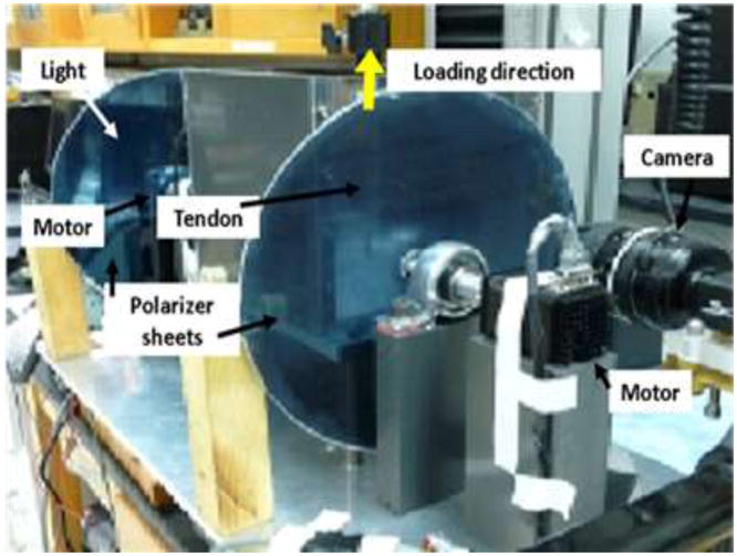
Angled side-view of the tendon tensile testing setup showing polarized light and imaging system: light source, rotating cross-polarized sheets, stepper motors, and camera.
4. 3 Data analysis
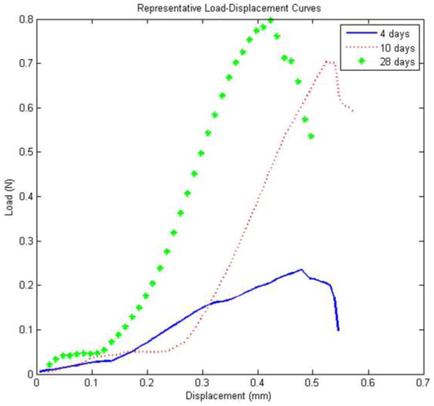
The following parameters were measured: transition stress (stress at the intersection of the toe- and linear-regions of the load-displacement curve), transition strain (strain measured at the transition point), local optical toe- and linear-moduli, cross-sectional area, and the spread of the local collagen fiber distribution at several points throughout the mechanical testing process. Local strain was measured optically and stress was calculated as force divided by initial area. A structural fiber recruitment model was used to determine the transition point for each age group (intersection of the toe- and linear-regions of the load-displacement curve). 21 The transition-region was modeled at 50% fiber recruitment and a point in the linear-region was modeled at 75% fiber recruitment utilizing the load-displacement curves (Fig. 2). Fiber alignment was calculated from the image sets as previously described. 13, 14 Briefly, images of the tendon surface were divided into rectangular areas. Care was taken to maintain a consistent resolution across all age groups for alignment analysis. Pixel intensities were summed by area per image and plotted against angle of polarizer rotation. A sine wave was fitted to the intensity-angle to determine the angle corresponding to the minimum pixel intensity, which represents the average direction of the area’s collagen fiber alignment.
Figure 2.
A representative load-displacement curve from each age group (4, 10 and 28 days) is shown demonstrating the decrease in transition strain between 28 and 10 day tendons.
Circular variance, transition stress, transition strain, toe-region modulus, and the linear-region stiffness and modulus were determined. Circular variance (VAR), a measure of the distribution of collagen fiber alignment, was calculated for fiber distributions before and after preconditioning (5, 10, 20 cycles), at the transition region (intersection of toe- and linear-regions: determined using a structural fiber recruitment model at 50% fiber recruitment),21 and the linear-region strain (at 75% fiber recruitment). Fiber re-alignment during preconditioning was evaluated by comparing VAR values from before preconditioning (BP) and after preconditioning (AP) for each preconditioning protocol. Similar methods were used to determine fiber re-alignment during the toe- and linear-regions of the stress-strain curve.
4. 4 Statistical analysis
For all parameters, two-way ANOVAs were used to evaluate interactions between 1) protocol and location and 2) age and location. A one-way ANOVA was used when the interaction was not significant followed by post-hoc tests. In addition, multiple comparisons were evaluated with Bonferroni corrections. Mechanical parameters were evaluated using parametric statistics and the data is presented as mean ± standard deviation (SD). Changes in parameters were compared for tendon location (midsubstance vs. insertion) and tendon age. Alternatively, Shapiro-Wilk tests indicated non-normally distributed data for VAR values and as a result, non-parametric statistical tests were used for evaluating fiber re-alignment. VAR data was analyzed as paired comparisons and is presented as median ± interquartile range. Changes in fiber alignment (Friedman test) were compared for tendon location (midsubstance vs. insertion) and for the mechanical test region (preconditioning, toe- and linear-region). Finally, the Spearman rank correlation coefficients were calculated to evaluate correlations between mechanical and alignment parameters.
5. Results
5. 1 Protocol
No differences in collagen fiber re-alignment or mechanics were found between protocols with varying number of preconditioning cycles. Therefore to increase power, data was pooled across the protocols for the remaining analyses.
5. 2 Re-Alignment
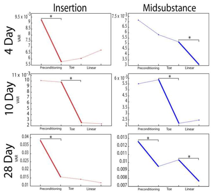
At 4 days old, the midsubstance location re-aligned in the linear-region while the insertion site re-alignment occurred during preconditioning (Fig. 3). At 10 days old, both the midsubstance and insertion site locations re-aligned in the toe-region of the stress-strain curve. At 28 days old, collagen fiber re-alignment occurred during preconditioning for both locations as well as during the linear-region for the midsubstance. Locally, the insertion site of the tendon was more disorganized than the midsubstance only before preconditioning in 4 day old tendons (Fig. 3). The insertion site was more disorganized than the tendon midsubstance at all mechanical testing points, except at the 10 day transition point, in 10 and 28 day old tendons (Table 1).
Figure 3.
Circular variance (VAR) values for representative samples demonstrate that re-alignment (decrease in VAR) occurred during preconditioning for 4 days (top) insertion site, at the linear-region for 4 day midsubstance, at the toe-region for both 10 day (middle) locations, and during preconditioning for both 28 day (bottom) locations. * = p<0. 025
Table I.
P-values comparing local collagen fiber distributions throughout the mechanical test.
| Region of Test | Age
|
||
|---|---|---|---|
| 4 days | 10 days | 28 days | |
| Before | 0. 0084* | 0. 0002* | 0. 0009* |
| After | 0. 15 | 0. 0008* | 0. 0003* |
| Trans | 0. 32 | 0. 031 | 0. 0008* |
| Linear | 0. 15 | 0. 0006* | 0. 0005* |
Indicates significant difference at p<0. 025, compares distributions at the insertion site and midsubstance locations throughout the mechanical test.
5. 3 Mechanics
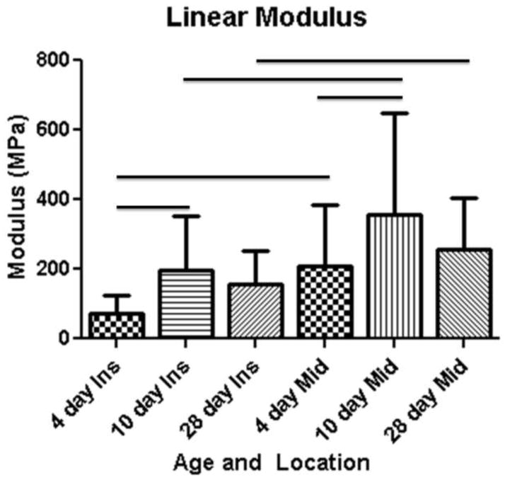
Local differences in toe modulus were found at 4 days and 28 days old with the midsubstance location having a higher modulus than the insertion site (Fig. 4). Additionally, there was an increase in toe modulus at the midsubstance location from 10 days to 28 days (Table 2). Surprisingly, the linear modulus was greater at the tendon midsubstance than at the insertion site for all age groups (4, 10 and 28 days). Additionally, there was an increase in linear modulus at the insertion site and the midsubstance locations from 4 days to 10 days old (Fig. 4). The transition stress for the insertion site location decreased from 10 days old to 28 days old (Table 2). The transition strain at the insertion site was higher than at the midsubstance location at 4 days old (Fig. 5). Additionally, the transition strain at the insertion site location decreased with age for all time points and decreased from 10 to 28 days at the midsubstance. Stiffness increased with age across all time points. Cross sectional-area was larger at the insertion site location than the midsubstance at both 10 and 28 days old. Increases in cross-sectional area with age were found at both the midsubstance and insertion site locations (Table 2).
Figure 4.
A lower linear modulus is present at the insertion site compared to the midsubstance location at all time points. An increase in linear modulus is seen between 4 and 10 days at both locations.
TABLE II.
Mechanical parameters throughout postnatal development
| Age (Days) | Location | Initial Area (mm2) | Toe Modulus (MPa) | Linear Modulus (MPa) | Transition Strain (mm/mm) | Transition Stress (MPa) |
|---|---|---|---|---|---|---|
| 4 | Insertion | 0. 016±0. 01 | 43. 7±28. 6a | 74. 1±50. 8a | 0. 064±0. 04a | 3. 38±1. 81 |
| 10 | Insertion | 0. 030±0. 01ab | 74. 0±77. 6 | 198. 5±153. 9ab | 0. 036±0. 26b | 3. 48±2. 82 |
| 28
|
Insertion | 0. 075±0. 04ac | 60. 8±46. 3a | 157±94. 6a | 0. 024±0. 02c | 2. 18±1. 30c |
| 4 | Midsub | 0. 012±0. 01 | 91. 6±74. 9 | 209. 7±174. 2 | 0. 03±0. 02 | 4. 84±3. 09 |
| 10 | Midsub | 0. 027±0. 01b | 75. 8±68. 7 | 357. 5±292. 9b | 0. 025±0. 02 | 3. 46±2. 66 |
| 28 | Midsub | 0. 049±0. 02c | 170. 0±159. 2c | 258. 3±147. 3 | 0. 013±0. 01c | 3. 35±2. 50 |
Data represented as mean ± st dev.
Significant comparisons between midsubstance (abbreviated “midsub”) and insertion site location;
Significantly different between 4 and 10 days.
Significantly different between 10 and 28 days. Significance set at p<0. 025.
Figure 5.
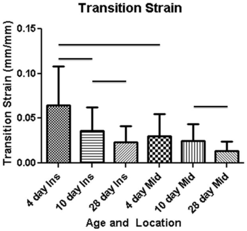
Transition strain decreases with age at the insertion site and from 10 to 28 days at the midsubstance. A local decrease in transition strain from the insertion site to the midsubstance location was noted at 4 days.
5. 4 Correlation
A negative and significant correlation was found between collagen fiber alignment and mechanics for both the before (rs=−0. 24, p=0. 001) and after preconditioning (rs=−0. 20, p=0. 007) VAR values and linear-region modulus.
6. Discusssion
This study quantified mechanical properties throughout neonatal development in the mouse SST. As expected, mechanical properties increased throughout development. Specifically, increases in the toe- and linear-moduli as well as cross-sectional area were seen throughout development. The results in the developmental SST support similar findings from previous work in the midsubstance of the mouse Achilles tendon. 1 Both the SST and Achilles tendon demonstrated an increase in linear modulus from 4 to 10 days but no significant differences between 10 and 28 days (Fig. 4). Additionally, a decrease in transition strain was noted from 10 to 28 days at the midsubstance location in the SST and Achilles tendon. 1 Transition strain also decreased with all time points at the insertion site in the present study (Fig. 5). The large transition strain found at early developmental ages suggests that the structural changes that occur during the toe-region of the mechanical test may take longer in developmental tendon. The toe-region of the mechanical test is thought to be explained by uncrimping of collagen fibers. It has been speculated that early developmental tendons possess a higher frequency of collagen fiber crimps potentially explaining the elongated toe-region. 8 Additionally, it is possible that the immature collagen fibril network (lack of long, continuous collagen fibrils and established collagen cross-links) limit effective communication between neighboring fibrils, resulting in a slower response to mechanical load through both collagen fiber re-alignment and uncrimping.
Interestingly, this study shows that significant collagen fiber re-alignment occurs at different points in the mechanical loading protocol, indicating that the tendon’s ability to structurally respond to load through re-orientation of collagen fibers in the direction of load is dependent on developmental age and more importantly, level of tissue organization or maturity. As hypothesized, fiber re-alignment occurred during preconditioning at both locations in 28 day tendon (Fig. 3). However, no changes in fiber alignment were demonstrated during the toe-region at 28 days for either location. In the linear-region, however, re-alignment was found to occur at the midsubstance location but not at the insertion site. These results show that the 28 day tendon responded structurally at the first sign of load, despite the magnitude of load for the preconditioning protocols, and demonstrated similar behavior to the mature rat SST. 16
At 10 days, collagen fiber re-alignment occurred during the toe-region of the mechanical test at both locations (Fig. 3). Interestingly, the re-alignment behavior at 4 days was location-dependent. The midsubstance of the tendon re-aligned in the linear-region and the insertion site re-aligned during preconditioning (Fig. 3). These results suggest that early developmental tendons require either higher loads or a longer exposure to load before responding structurally. The delay in structural response could potentially be explained by the immature networks of collagen fibrils that have been previously demonstrated at 4 and 10 days in mouse tendon. 31 Four day mouse tendons have been shown to consist of disconnected collagen fibril intermediates, while at 10 days collagen fibrils have linearly fused but still consist primarily of small diameter fibrils. 31 The results from the present study at 4 days suggest that the ability of small collagen fibril intermediates to communicate and transmit load may be insufficient. It is possible that fibers are unable to communicate to each other due to the lack of a developed collagen fiber network and collagen-matrix interactions that may help transmit and support load between the insertion site and tendon midsubstance. Changes in collagen cross-links, crimp morphology and proteoglycan content in tendon are thought to be related to the mechanical properties of tendon in transmitting tensional forces. 11 In particular, the lack of mature collagen fiber cross-links may prevent the transmission of tensile forces between adjacent collagen fibrils, resulting in a longer toe-region and lack of communication between the insertion site and tendon midsubstance during early postnatal development. Additionally, while linear fusions are present in collagen fibrils at 10 days postnatal, the fibril network is still underdeveloped. However, long, continuous collagen fibrils are one proposed mechanism by which tendons transmit load. 22 While the exact mechanism of load transfer is still under debate, the present study supports this, suggesting that elongated collagen fibrils may allow the tendon to uniformly respond to load as seen in the 10 day uniform re-alignment during the toe-region. Linear growth of collagen fibrils has been suggested to be integral in the development of mechanical stability and has been correlated to increases in ultimate tensile strength and elastic modulus. 27 The increases in collagen fibril length could be a consequence of fibril fusion and crosslink formation enabling for quicker communication and transmission of loads between collagen fibrils, resulting in a shorter transition region and increased modulus values. Additionally, a higher strain was necessary to transition from the toe- to linear-region of the stress-strain curve at 10 days compared to 28 days (Figs. 2&5). This indicates that although fibrils are linearly growing, they still require longer exposure to loads, or higher loads, to reach the linear-region of the mechanical test.
Local differences in mechanical properties, collagen fiber distribution and collagen fiber re-alignment behavior were examined throughout neonatal development. At 4 days, differences in collagen fiber distribution and collagen fiber re-alignment behavior suggest that the insertion site and tendon midsubstance develop at different rates. The lack of organized and mature tissue at the insertion site is a potential explanation for the local differences seen at 4 days old. Previous work has shown that at 3 days old the tendon insertion site is composed primarily of type 2 collagen as opposed to the 4-zone insertion seen in mature tendon. 12 Additionally, as previously discussed, the collagen fibril network at 4 days has been demonstrated to be small, disconnected fibril intermediates that may not be able to communicate or transmit load from one tendon location to another. It is possible that the mechanical properties measured in 4 day tendon (particularly at the insertion site location) are primarily representative of the tendon extracellular matrix (and not the disconnected collagen fibrils) (Fig. 2).
In addition to demonstrating different local re-alignment behaviors, 4 day old tendons were more disorganized at the insertion site compared to the tendon midsubstance only before preconditioning. Remarkably, following re-alignment during preconditioning at the insertion site, differences in collagen fiber distribution were no longer detectable between the midsubstance and insertion (Table 1). Collagen fibers at midsubstance location may be more developed as indicated by their higher linear modulus and more organized collagen distribution compared to the insertion site. The 4 day tendon midsubstance behaves as expected with a delayed re-alignment response compared to mature SST and 10 day tendon. These results suggest that the midsubstance collagen fiber network is more developed than the insertion site and can gradually recruit fibers in the direction of load (as represented by the long transition-region seen at 4 days), while the insertion site is composed primarily of extracellular matrix and a disconnected group of fibril intermediates. One possible explanation for this is that the tendon midsubstance may experience tension from the surrounding muscle throughout development. 26 Previous work has noted that the development of functionally distinct fibrocartilages occurs at different rates in the rat Achilles and quadriceps tendons. 24, 25 It is possible that the local differences seen in mechanics and alignment behavior can be explained by the midsubstance and insertion site developing at different rates. The insertion site might need to be more developed before successfully transmitting loads or providing communication between the midsubstance and insertion site locations.
Local differences in linear modulus and collagen fiber distribution were found at 10 days. Weaker mechanical properties and more disorganized collagen fiber distributions were found at the insertion site compared to the midsubstance location. However, while differences were present in mechanics and collagen fiber distributions, the two locations demonstrated similar collagen fiber realignment behavior (Fig. 3) and no differences in toe-region modulus. The insertion site may be more developed at 10 days compared to 4 days, allowing for communication and load transmission. Previous studies have shown that the transition zone and layers of fibrocartilage near the insertion site were evident, regardless of mechanical stimuli, at 7 days postnatal. 29 Additionally, type X collagen and mineralized fibrocartilage have been found at the insertion site location in 14 day old tendon. 29 Ten day tendons may be beginning to experience complex loads commonly occurring in mature animals at the tendon-to-bone insertion site, as demonstrated by the more disorganized collagen fiber distribution and lower modulus compared to the midsubstance location.
Additionally, local differences in mechanical and organizational properties were found at 28 days. Decreased modulus and more disorganized collagen fiber distributions were present at the insertion site compared to the midsubstance location. Previous work demonstrated that by 21 days, the characteristic 4 zone insertion site is fully developed. 12 Additionally, it has been demonstrated that mechanical stimuli are necessary to form the transitional fibrocartilaginous insertion site. 29 These previous studies, together with the strong local differences shown in the present study for mechanical properties and fiber distribution, suggest that the insertion site experiences complex multi-axial loads at 28 days old. Surprisingly, a difference in re-alignment behavior was found at 28 days between the insertion site and midsubstance locations (Fig. 3). Previous work examining mature rat SST and subscapularis tendon found identical re-alignment behavior between the insertion site and midsubstance. 15, 16 The 28 day tendon midsubstance was found to re-align in the linear-region of the mechanical test in addition to during preconditioning in this study (Fig. 3). It is uncertain if the differences in re-alignment behavior are unique to mouse SST or a result of the immaturity of 28 day tendons. Postnatal mouse tendons are not considered mature until 3–4 months old, therefore one potential explanation for the discrepancy between collagen fiber re-alignment behavior seen at 28 days of age versus the response of mature rat tendon is that the collagen fiber-fiber, fiber-matrix interactions and cross-link networks are not yet fully mature. Additional cross-links and fiber-matrix interactions will provide a more complex, interconnected network of fibrils, increasing mechanical strength and the potential for load transmission across the tendon. While a fully mature network is expected to allow the fibers to communicate and transmit load to other fibers at a fast rate and to respond to load quicker, the additional bonds formed between the collagen fibers and the extracellular matrix will also provide additional resistance for the fibers to rotate toward the direction of loading. It is possible that a larger number of fibers rotate initially during preconditioning in the 28 day tendons, while collagen fibers from mature tendons may continue to recruit and re-align in the direction of load throughout the remainder of the mechanical test. Another potential explanation is that the tendon is beginning to fail at the insertion site and that collagen fibers are damaged or broken therefore preventing the tendon from demonstrating additional re-alignment. It is expected that mature mouse tendons’ collagen fibers will fail at higher loads than the 28 day fibers. It is possible that the 28 day tendons did not demonstrate significant re-alignment in the toe- and linear-regions as while some populations of fibers re-aligned in the direction of loading, those initially recruited fibers were catastrophically failing.
This study also determined that a low (rs=−0. 24) but significant (p=0. 001) correlation exists between mechanical and organizational properties. A negative correlation was identified between linear modulus and the before preconditioning and after preconditioning circular variance values, suggesting that increased alignment (decr VAR) is correlated with increased mechanical properties. This finding further supports previous work in the human SST that found a negative correlation between linear modulus and VAR values. 13
This study is not without assumptions which must be understood. First, toe- and linear-region VAR values for each age group were determined using the average strain quantified from the individual sample fiber recruitment model results in order to make comparisons at one consistent strain value per developmental age rather than individually determining an appropriate value to represent the toe- and linear-region for each individual sample and at each location. We are confident that the average values are within the parameters to represent the toe- and linear-regions of each age group and that the chosen method improves consistency within this experiment in addition to allowing for comparisons with future experiments examining additional mechanisms of structural changes. Secondly, the crossed polarizer method used in this study can only measure fibers ±45° from the tendon long axis instead of the full 180° range that would contain all possible fiber orientations. An angle value correction was applied based on the assumption that fibers must reorient toward the direction of loading as has been performed previously. 13 Finally, collagen fibril diameter has not been examined in the mouse supraspinatus tendon at all ages throughout development. The ages chosen for this study were based on results from the mouse Achilles tendon and results may vary by tendon. However, a broad range was utilized that likely spans the relevant ages.
In conclusion, this study demonstrated that tendon collagen fiber re-alignment may be dependent upon developmental age and that collagen fibril development may affect the tendon’s ability to structurally respond to load on a higher hierarchical level. Additionally, the study identified a negative correlation between linear modulus and collagen fiber distribution before and after preconditioning across all developmental time points. Further, this study provides valuable insights regarding the development of the supraspinatus tendon insertion site in the mouse. Future studies will investigate local changes in collagen fiber crimp throughout development to determine if crimp behavior can further explain the observed changes in transition strain and re-alignment. Additionally, further work is necessary to examine collagen fibril development in the mouse SST and to examine mechanical properties on smaller hierarchical scales.
Acknowledgments
This study was supported by NIH/NIAMS. We also thank David P. Beason, Jennica J. Tucker and Elizabeth Feeney for assistance.
References
- 1.Ansorge HL, Adams S, Birk DE, Soslowsky LJ. Mechanical, compositional, and structural properties of the post-natal mouse achilles tendon. Ann Biomed Eng. 39:1904–1913. doi: 10.1007/s10439-011-0299-0. [DOI] [PMC free article] [PubMed] [Google Scholar]
- 2.Ansorge HL, Meng X, Zhang G, Veit G, Sun M, Klement JF, Beason DP, Soslowsky LJ, Koch M, Birk DE. Type xiv collagen regulates fibrillogenesis: Premature collagen fibril growth and tissue dysfunction in null mice. J Biol Chem 2. 2009;84:8427–8438. doi: 10.1074/jbc.M805582200. [DOI] [PMC free article] [PubMed] [Google Scholar]
- 3.Benjamin M, Kumai T, Milz S, Boszczyk BM, Boszczyk AA, Ralphs JR. The skeletal attachment of tendons--tendon “Entheses”. Comp Biochem Physiol A Mol Integr Physiol. 2002;133:931–945. doi: 10.1016/s1095-6433(02)00138-1. [DOI] [PubMed] [Google Scholar]
- 4.Benjamin M, Newell RL, Evans EJ, Ralphs JR, Pemberton DJ. The structure of the insertions of the tendons of biceps brachii, triceps and brachialis in elderly dissecting room cadavers. J Anat. 1992;180(Pt 2):327–332. [PMC free article] [PubMed] [Google Scholar]
- 5.Birk DE, Nurminskaya MV, Zycband EI. Collagen fibrillogenesis in situ: Fibril segments undergo post-depositional modifications resulting in linear and lateral growth during matrix development. Dev Dyn. 1995;202:229–243. doi: 10.1002/aja.1002020303. [DOI] [PubMed] [Google Scholar]
- 6.Birk DE, Zycband EI, Woodruff S, Winkelmann DA, Trelstad RL. Collagen fibrillogenesis in situ: Fibril segments become long fibrils as the developing tendon matures. Dev Dyn. 1997;208:291–298. doi: 10.1002/(SICI)1097-0177(199703)208:3<291::AID-AJA1>3.0.CO;2-D. [DOI] [PubMed] [Google Scholar]
- 7.Derwin KA, Soslowsky LJ, Green WD, Elder SH. A new optical system for the determination of deformations and strains: Calibration characteristics and experimental results. J Biomech. 1994;27:1277–1285. doi: 10.1016/0021-9290(94)90281-x. [DOI] [PubMed] [Google Scholar]
- 8.Diamant J, Keller A, Baer E, Litt M, Arridge RG. Collagen; ultrastructure and its relation to mechanical properties as a function of ageing. Proc R Soc Lond B Biol Sci. 1972;180:293–315. doi: 10.1098/rspb.1972.0019. [DOI] [PubMed] [Google Scholar]
- 9.Favata M. Ph D thesis. University of Pennsylvania; 2006. Scarless healing in the fetus: Implications and strategies for postnatal tendon repair. [Google Scholar]
- 10.Festing MF. Design and statistical methods in studies using animal models of development. ILAR J. 2006;47:5–14. doi: 10.1093/ilar.47.1.5. [DOI] [PubMed] [Google Scholar]
- 11.Franchi M, Trire A, Quaranta M, Orsini E, Ottani V. Collagen structure of tendon relates to function. ScientificWorldJournal. 2007;7:404–420. doi: 10.1100/tsw.2007.92. [DOI] [PMC free article] [PubMed] [Google Scholar]
- 12.Galatz L, Rothermich S, VanderPloeg K, Petersen B, Sandell L, Thomopoulos S. Development of the supraspinatus tendon-to-bone insertion: Localized expression of extracellular matrix and growth factor genes. J Orthop Res. 2007;25:1621–1628. doi: 10.1002/jor.20441. [DOI] [PubMed] [Google Scholar]
- 13.Lake SP, Miller KS, Elliott DM, Soslowsky LJ. Tensile properties and fiber alignment of human supraspinatus tendon in the transverse direction demonstrate inhomogeneity, nonlinearity, and regional isotropy. J Biomech. 43:727–732. doi: 10.1016/j.jbiomech.2009.10.017. [DOI] [PMC free article] [PubMed] [Google Scholar]
- 14.Lake SP, Miller KS, Elliott DM, Soslowsky LJ. Effect of fiber distribution and realignment on the nonlinear and inhomogeneous mechanical properties of human supraspinatus tendon under longitudinal tensile loading. J Orthop Res. 2009;27:1596–1602. doi: 10.1002/jor.20938. [DOI] [PMC free article] [PubMed] [Google Scholar]
- 15.Miller KSSJT, Trasolini NA, Soslowsky LJ. The upper band of the subscapularis tendon in the rat has inferior mechanical properties. Transactions of the Orthopaedic Research Society. 2011 [Google Scholar]
- 16.Miller KS, Edelstein L, Soslowsky LJ. Effect of preconditioning on collagen fiber recruitment: Inhomogeneous properties of the rat supraspinatus tendon. Proceeding of the ASME 2010 Summer Bioengineering Conference; 2010. [Google Scholar]
- 17.Moore MJ, De Beaux A. A quantitative ultrastructural study of rat tendon from birth to maturity. J Anat. 1987;153:163–169. [PMC free article] [PubMed] [Google Scholar]
- 18.Nakagawa Y, Majima T, Nagashima K. Effect of ageing on ultrastructure of slow and fast skeletal muscle tendon in rabbit achilles tendons. Acta Physiol Scand. 1994;152:307–313. doi: 10.1111/j.1748-1716.1994.tb09810.x. [DOI] [PubMed] [Google Scholar]
- 19.Oryan A, Shoushtari AH. Histology and ultrastructure of the developing superficial digital flexor tendon in rabbits. Anat Histol Embryol. 2008;37:134–140. doi: 10.1111/j.1439-0264.2007.00811.x. [DOI] [PubMed] [Google Scholar]
- 20.Parry DA, Craig AS, Barnes GR. Tendon and ligament from the horse: An ultrastructural study of collagen fibrils and elastic fibres as a function of age. Proc R Soc Lond B Biol Sci. 1978;203:293–303. doi: 10.1098/rspb.1978.0106. [DOI] [PubMed] [Google Scholar]
- 21.Peltz CD, Sarver JJ, Dourte LM, Wurgler-Hauri CC, Williams GR, Soslowsky LJ. Exercise following a short immobilization period is detrimental to tendon properties and joint mechanics in a rat rotator cuff injury model. J Orthop Res. 28:841–845. doi: 10.1002/jor.21059. [DOI] [PMC free article] [PubMed] [Google Scholar]
- 22.Provenzano PP, Vanderby R., Jr Collagen fibril morphology and organization: Implications for force transmission in ligament and tendon. Matrix Biol. 2006;25:71–84. doi: 10.1016/j.matbio.2005.09.005. [DOI] [PubMed] [Google Scholar]
- 23.Quinn KP, Winkelstein BA. Preconditioning is correlated with altered collagen fiber alignment in ligament. J Biomech Eng. 2011;133:064506. doi: 10.1115/1.4004205. [DOI] [PubMed] [Google Scholar]
- 24.Ralphs JR, Tyers RN, Benjamin M. Development of functionally distinct fibrocartilages at two sites in the quadriceps tendon of the rat: The suprapatella and the attachment to the patella. Anat Embryol (Berl) 1992;185:181–187. doi: 10.1007/BF00185920. [DOI] [PubMed] [Google Scholar]
- 25.Rufai A, Benjamin M, Ralphs JR. Development and ageing of phenotypically distinct fibrocartilages associated with the rat achilles tendon. Anat Embryol (Berl) 1992;186:611–618. doi: 10.1007/BF00186984. [DOI] [PubMed] [Google Scholar]
- 26.Schweitzer R, Zelzer E, Volk T. Connecting muscles to tendons: Tendons and musculoskeletal development in flies and vertebrates. Development. 137:2807–2817. doi: 10.1242/dev.047498. [DOI] [PMC free article] [PubMed] [Google Scholar]
- 27.Silver FH, Freeman JW, Seehra GP. Collagen self-assembly and the development of tendon mechanical properties. J Biomech. 2003;36:1529–1553. doi: 10.1016/s0021-9290(03)00135-0. [DOI] [PubMed] [Google Scholar]
- 28.Sverdlik A, Lanir Y. Time-dependent mechanical behavior of sheep digital tendons, including the effects of preconditioning. J Biomech Eng. 2002;124:78–84. doi: 10.1115/1.1427699. [DOI] [PubMed] [Google Scholar]
- 29.Thomopoulos S, Kim HM, Rothermich SY, Biederstadt C, Das R, Galatz LM. Decreased muscle loading delays maturation of the tendon enthesis during postnatal development. J Orthop Res. 2007;25:1154–1163. doi: 10.1002/jor.20418. [DOI] [PubMed] [Google Scholar]
- 30.Thomopoulos S, Williams GR, Gimbel JA, Favata M, Soslowsky LJ. Variation of biomechanical, structural, and compositional properties along the tendon to bone insertion site. J Orthop Res. 2003;21:413–419. doi: 10.1016/S0736-0266(03)00057-3. [DOI] [PubMed] [Google Scholar]
- 31.Zhang G, Young BB, Ezura Y, Favata M, Soslowsky LJ, Chakravarti S, Birk DE. Development of tendon structure and function: Regulation of collagen fibrillogenesis. J Musculoskelet Neuronal Interact. 2005;5:5–21. [PubMed] [Google Scholar]


