Abstract
The discovery of G-quadruplexes and other DNA secondary elements has increased the structural diversity of DNA well beyond the ubiquitous double helix. However, it remains to be determined whether tertiary interactions can take place in a DNA complex that contains more than one secondary structure. Using a new data analysis strategy that exploits the hysteresis region between the mechanical unfolding and refolding traces obtained by a laser-tweezers instrument, we now provide the first convincing kinetic and thermodynamic evidence that a higher order interaction takes place between a hairpin and a G-quadruplex in a single-stranded DNA fragment that is found in the promoter region of human telomerase. During the hierarchical unfolding or refolding of the DNA complex, a 15-nucleotide hairpin serves as a common species among three intermediates. Moreover, either a mutant that prevents this hairpin formation or the addition of a DNA fragment complementary to the hairpin destroys the cooperative kinetic events by removing the tertiary interaction mediated by the hairpin. The coexistence of the sequential and the cooperative refolding events provides direct evidence for a unifying kinetic partition mechanism previously observed only in large proteins and complex RNA structures. Not only does this result rationalize the current controversial observations for the long-range interaction in complex single-stranded DNA structures, but this unexpected complexity in a promoter element provides additional justification for the biological function of these structures in cells.
Introduction
Protein and RNA are well known to assume tertiary structures with biological functions. While single-domain proteins show cooperative, two-state folding and unfolding processes,1 intermediates are more common in the folding and unfolding of complex proteins2, 3 and RNA structures.4, 5 In RNA, local interactions between secondary elements are responsible for the hierarchical nature of the structure.4-6 Until recently, in complete contrast to protein and RNA, it was believed that DNA serves solely as the repository for genetic information and does not have direct biological functions. Recent insight into non-B-DNA structures, such as G-quadruplexes,7 is challenging this stereotypical profile of DNA. There is now compelling evidence that G-quadruplexes exist naturally in cells,8-10 and they can serve as transcription regulators for various genes, including c-MYC8 and c-KIT.11
G-quadruplexes, which consist of four runs of two or more contiguous guanines connected through G-tetrads,12 are found to be abundant in promoter elements throughout the human genome.13 Although still controversial,14 in vitro evidence points to the existence of high-order interactions between neighboring G-quadruplexes15, 16 in single-stranded DNA (ss) sequences, such as those of telomeres, that can host multiple G-quadruplexes. This suggests that, like RNA or protein, tertiary DNA structures may also form. In spite of their significant structural differences, protein and RNA share similar energy minima in the folding energy landscape representative of a large molecule.17 This leads to alternative folding pathways for both species, a process known as the kinetic partition mechanism.17 It would be interesting to see whether this unifying mechanism can apply to complex DNA structures as well.
Using a mechanical unfolding and refolding approach in a home-built laser-tweezers instrument,18, 19 we have observed a complex structure in an ssDNA fragment from the core promoter of human telomerase reverse transcriptase (hTERT). This structure is consistent with that determined by DMS footprinting and circular dichroism (CD),20 which reveals a G-quadruplex with a long hairpin-containing loop. We have shown here that unfolding/refolding events of this structure follow either cooperative or sequential pathways. This observation directly supports the kinetic partition mechanism previously seen only in the folding of complex protein2, 3 and RNA structures.4, 5 Among the three intermediates we have observed for the sequential unfolding/refolding pathways, the 15-nucleotide (nt) hairpin is the critical species that serves as a nucleus in the refolding. Removal of this hairpin by mutation or hybridizing with a complementary strand dramatically reduces the cooperative unfolding and entirely abrogates the cooperative refolding events. Comparison of the change in free energy of unfolding (ΔGunfold) between the sequential and cooperative pathways reveals a tertiary interaction (~6 kcal/mol) between the G-quadruplex and the hairpin. The partition of the DNA complex into a conformation with or without this tertiary interaction may result in cooperative or sequential unfolding/refolding pathways. This helps to explain the current controversial observations on the existence of DNA tertiary interactions.14-16
Results and Discussion
The hTERT 5-12 structure follows either cooperative or sequential unfolding/refolding pathways
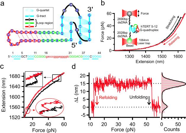
To investigate the unfolding and refolding processes of a complex DNA structure with potential tertiary interactions, we focused on a DNA fragment (Figure 1a), 5′-GGG GGC TGG GCC GGG gac ccg gga ggg gtc GGG ACG GGG CGG GG, from the core promoter (−90 to −46, or hTERT 5-12) of hTERT. We sandwiched this ssDNA fragment between 2690- and 2028-bp double-stranded DNA (dsDNA) handles, which were immobilized to the two optically trapped beads (Figure 1b, inset). A force-ramp assay was carried out at the force-loading rate of 5.5 pN/s in 10 mM Tris buffer with 100 mM K+ (pH 7.4) to collect force-extension (F-X) curves (see Experimental Section). To analyze unfolding or refolding transitions that are represented by a sudden change in the tension or extension in the F-X curves (Figure 1b), we switched the F-X coordinates (Figure 1c), which were then transformed to a plot of change-in-contour-length (ΔL) vs force, or ΔL-F plot (Figure 1d, left), using a worm-like-chain model (see Experimental Section). The histogram derived from each trace allows clear identification of a transition process (ΔL = 15 nm population) from background (ΔL = 0 nm population) (Figure 1d, right). The ΔL determined in this manner matches remarkably well with that averaged from single F-X curve measurements (see Experimental Section and Figure S1). After analyzing 2448 plots, two types of transitions were observed. One was a large, single-step transition with an average ΔL of 16 ± 2 nm (Figure 1d and Figure 2), which represents cooperative unfolding or refolding; the other was composed of a series of smaller steps, which indicates sequential kinetic pathways (Figure 2a). As clearly demonstrated here, this novel approach allows straightforward and intuitive determination of small transitions that are often obscured in conventional F-X curves (Figure S1). The ΔL of the cooperative transitions was consistent with unfolding of the 42-nt G-quadruplex-hairpin hybrid previously determined (Figure 1a, see Experimental Section for calculation).20
Figure 1.
Cooperative unfolding and refolding in the hTERT 5-12 fragment. (a) The structure formed in the hTERT 5-12 fragment. (b) Force-extension curves of a DNA construct that contains the hTERT fragment. Black and red data points (100 Hz bandwidth) represent stretching and relaxing processes, respectively. The inset shows the experimental setup, in which a single DNA construct is tethered between two beads trapped by laser tweezers. (c) Extension-force curves show cooperative unfolding (black) and refolding (red) events. (d) Difference in the extension, Δx, between the unfolding and refolding traces in (c) is converted to the change-in-contour-length (ΔL) between 10 and 60 pN (the ΔL-F plot). A histogram of ΔL from the ΔL-F plot is shown to the right. The black curve represents a two-peak Gaussian fitting.
Figure 2.
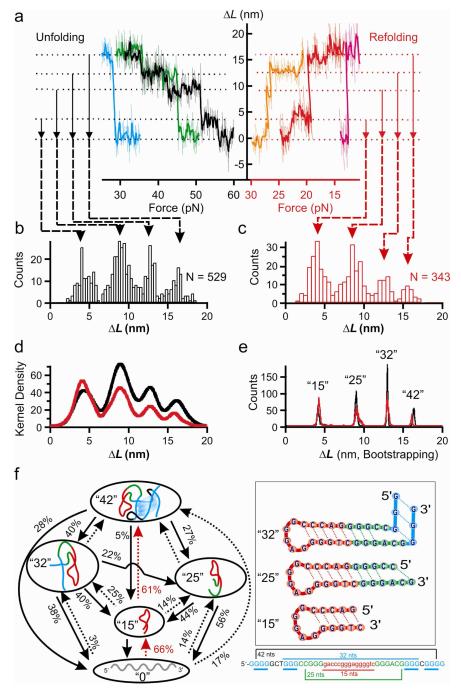
Three intermediates in the sequential unfolding/refolding transitions. The ΔL-F plots for typical unfolding and refolding transitions (a). Data with 100 Hz (thin traces) and 10 Hz (thick traces) bandwidths are plotted together. Population histograms for fully folded (“42”); 32-nt (“32”), 25-nt (“25”), and 15-nt (“15”) intermediates; and fully unfolded (“0”) species are shown in (b) and (c). (d) Kernel density estimation of the unfolding (black) and refolding (red) processes. (e) Histograms (black for the unfolding and red for the refolding) of the population based on bootstrapping analyses. (f) Schematic of unfolding (black solid arrows) or refolding (dotted arrows, major pathways are highlighted with red arrows) transitions for structures (shown to the right) in the hTERT 5-12 fragment. Percentage value depicts the probability for a particular transition.
Population analysis revealed a significant portion (28% in unfolding and 17% in refolding, Figure 2f) of the events undergoing cooperative transitions. These events suggest the existence of a higher order interaction in the complex DNA structure,5, 21 likely between the G-quadruplex and the hairpin. With the current unfolding geometry (Figure 1b), the G-quadruplex should be ruptured first, which would simultaneously destroy the tertiary interaction between the G-quadruplex and the hairpin, leading to the immediate unfolding of the hairpin whose rupture force is lower than that for the G-quadruplex.19, 20, 22
Two possible scenarios may explain the 17% cooperative refolding. First, the G-quadruplex folded before the hairpin. Limited by the refolding trajectory (Figure 1b), we cannot follow subsequent formation kinetics of the hairpin. However, since the folding of the G-quadruplex is slower than that for the hairpin,19, 22 together with the direct measurement of the two refolding rate constants (shown below), such a refolding order is unlikely. In the second scenario, the hairpin and G-quadruplex refolded simultaneously. This may occur as the hairpin facilitates the preassembly of the four G-rich tracts into a G-quadruplex. When these experiments were repeated in a 10 mM Tris buffer (pH 7.4) with 100 mM Li+, the 16-nm (or the 42-nt) population disappeared (Figure S2), which confirmed that the presence of a G-quadruplex is required for tertiary structure formation in the DNA complex, as Li+ is known to inhibit quadruplex formation.
The 15-nt hairpin is the key intermediate in the unfolding and refolding pathways
The majority of unfolding (72%) or refolding (83%) events were sequential. These observations strongly suggest that the complex DNA structure is hierarchical, which has been observed previously only in complex proteins and RNAs.4, 5 To reveal the number of species in sequential processes, we measured ΔL for the transition between each folded structure and the unfolded ssDNA. The ΔL histogram reveals four populations for both unfolding (Figure 2b, N = 529) and refolding (Figure 2c, N = 343) processes. The four populations were confirmed by the probability density estimation for all transitions based on a Gaussian kernel calculation23 (Figure 2d, see Experimental Section). To accurately determine the ΔL for each species, we then used a bootstrapping analysis to identify the most likely peaks from the kernel density estimation (see Experimental Section). Figure 2e shows that the size of each structure exactly matches both the unfolding and refolding trajectories, suggesting that the same species are involved in both processes. After converting ΔL into the number of nucleotides (nt) (see Experimental Section), a fully folded conformation with 42-nt and three intermediates that contain 32-, 25-, and 15-nt was clearly identified (Figure 2e). The 5′-end to 3′-end unfolding geometry (Figure 1b) led us to propose intermediate structures in Figure 2f. While they all resemble hairpins, the 32-nt species has the longest stem, in which the last three Hoogsteen guanine pairs are residues from the G-quadruplex. The 25-nt species contains a stem with two bulged nucleotides (see Figure S3a for a stabilized conformation from simulation), whereas the 15-nt species has the canonical hairpin structure with a pentaloop and a stem comprising five Watson-Crick base pairs.
Figure 2f summarizes all the observed transitions among the three intermediates (“32”, “25”, and “15”), the fully folded structure (“42”), and the unfolded ssDNA (“0”) with the probability for each process. For clarity, Figure 3a summarizes the major unfolding/refolding events with a probability >25%.
Figure 3.
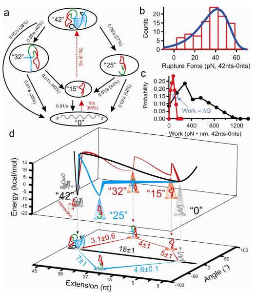
Kinetics and thermodynamics of the major transitions. (a) Unfolding (black solid arrows) or refolding (red dotted arrows) transitions (>25% probability) labeled with rate constants. (b) Histogram of the unfolding force for the “42”→“0” transition. Blue curve is the Dudko fitting24. (c) Histogram of the work associated with the unfolding (black) or refolding (red) of the “42”→“0” transition. The cross point is the change in free energy (ΔG) for the transition.27, 28 (d) Free energy vs extension (nt) and angle (°) (see Experimental Section) for “42”→“0” (black), “42”→“25”→“0” (blue), and “42”→“32”→“15”→“0” (red) trajectories. The color circle above “42” depicts the folded structure without tertiary interactions. Bottom projection shows ΔG (mean ± s.d., kcal/mol) between different species.
The fully folded G-quadruplex-hairpin hybrid undergoes three major unfolding trajectories: the cooperative unfolding (28%), through the 32-nt species (40%), or through the 25-nt intermediate (27%). Interestingly, the same 15-nt hairpin appeared in the latter two routes. Calculation revealed that starting with the fully folded structure, ~28% of all unfolding trajectories encountered the 15-nt hairpin intermediate. During refolding, the importance of the 15-nt hairpin was more obvious (Figure 3a): 66% of the population folded through this species. In contrast, only 14% of the population folded back to the 25-nt intermediate, while 17% underwent cooperative refolding. Once the 15-nt hairpin was formed, the majority of the hairpin (61%) directly folded to the G-quadruplex-hairpin hybrid. The coexistence of sequential and cooperative refolding pathways directly supports the kinetic partition mechanism previously associated only with complex proteins or RNA structures.17
Kinetics and thermodynamic analyses reveal a tertiary interaction between the hairpin and the G-quadruplex
The critical role of the 15-nt hairpin was again manifested in the kinetic analyses, in which only major transitions (>25% probability) were processed for accurate determination of rate constants (Figure 3a). For a representative cooperative unfolding event (“42→”0”), the Dudko model24 (Figure 3b) and the Evans model25 (Figure S4) provided identical kunfold of 0.02 s−1. In addition, the Dudko model revealed an activation energy, ΔG‡ = 29 kcal/mol, for this event (Figure 3b). These values were then used to calculate the unfolding activation energy for other processes (see Experimental Section and Table 1).
Table 1.
Thermodynamics, kinetics, and unfolding/refolding angles of the major transitions in the hTERT 5-12 fragment without the 15-nt hairpin complement at 23 °C.
| Transition | x‡ (nm) | ΔG (kcal/mol) |
ΔG‡ (kcal/mol) |
Angle (°) |
|---|---|---|---|---|
| 42~0 | 0.33 ± 0.01 | 18 ± 1 | 29 ± 2 | 0.0 |
| 42~32 | 0.33 ± 0.02 | 3.1 ± 0.6 | 29 ± 3 | 86.7 |
| 42~25 | 0.23 ± 0.02 | 7 ± 1 | 28 ± 3 | −80.0 |
| 32~15 | 0.38 ± 0.02 | 4 ± 1 | 10 ± 1 | −82.0 |
| 25~0 | 0.37 ± 0.03 | 4.6 ± 0.1 | 9 ± 2 | 78.9 |
| 15~0 | 0.41 ± 0.02 | 5 ± 1 | 10 ± 1 | −76.3 |
Similar refolding analyses indicate that the 15-nt hairpin has the fastest refolding kinetics (k = 9 s−1, Figure 3a), suggesting a seeding role of this hairpin to the fully folded G-quadruplex-hairpin hybrid. Although a long loop discourages the formation of a G-quadruplex,26 the formation of the 15-nt hairpin in the loop apparently remediates this effect by gathering distal G-rich tracts for the G-quadruplex assembly. The fact that the hairpin folds much faster than the G-quadruplex (9 vs 2 s−1, Figure 3a) is consistent with the literature19, 22 and supports our argument that a tertiary interaction between the G-quadruplex and the hairpin is responsible for cooperative transitions (see above).
The tertiary interaction was further analyzed from a perspective of change in free energy (ΔG) using the Crooks theorem27, 28 (see Figure 3c for the “42”→“0” transition; for the remainder, see Figure S5). ΔG42→0 for the cooperative process (18 kcal/mol, the black process in Figure 3d) was significantly higher than those for sequential pathways (12.1 and 11.6 kcal/mol for “42”→“32”→“15”→“0” (red) and “42”→“25”→“0” (blue) trajectories, respectively, Figure 3d). This result suggests that folded structures can partition into a conformation with or without a tertiary interaction, which is on the order of 6 kcal/mol. We surmise that the long-range interaction can result from the docking of the hairpin to the G-quadruplex, which may be aided by the base-pairing between the hairpin loop and one of the G-quadruplex loops. Figure 3d summarizes the 3D free energy landscape vs distance (nt) and angle (°) for major trajectories (see Table 1 and Figure S6 for details).
Tertiary interaction is perturbed by a DNA fragment complementary to the 15-nt hairpin
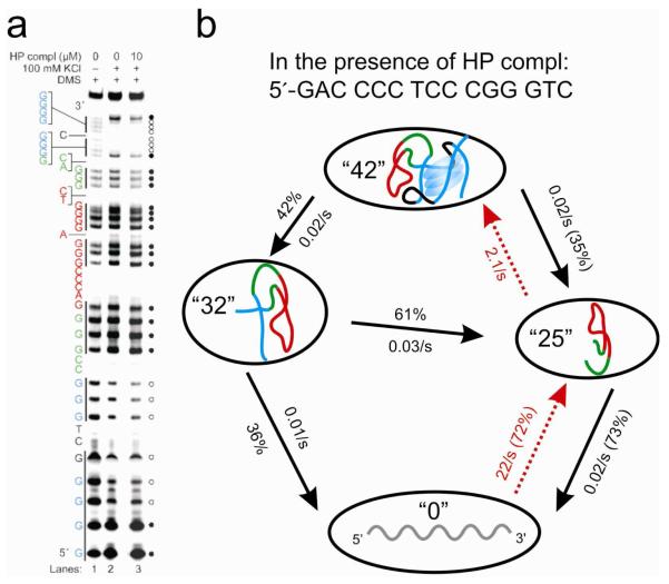
The seeding role of the 15-nt hairpin in the refolding processes was further confirmed in the experiments in which 10 μM ssDNA complementary to the 15-nt hairpin was incubated with 0.2 nM hTERT 5-12 fragment (see Experimental Section). Footprinting has shown no significant change in the DNA structure in the presence of the ssDNA complement (Figure 4a). However, the probability for pathways involving the 15-nt hairpin was much reduced (Figure 4b and Figure S7). In contrast, the 32-nt and 25-nt species became predominant during unfolding. During the refolding processes that became less likely to occur (Figure S7), the 25-nt species evolved to be the predominant species with the fastest rate (Figure 4b, and Figure S8). These results were expected as the 15-nt hairpin was disturbed after hybridizing with the ssDNA. The fact that the 25-nt intermediate became predominate during refolding further supports the “seeding” role of the 15-nt hairpin, without which the 25-nt species would function as an alternative to bring together distal G-rich tracts for the quadruplex assembly. Computer simulation confirmed that the 25-nt species is stable in the presence of the ssDNA complement (Figure S3b).
Figure 4.
Effect of the 15-nt hairpin complement (HP compl) on the hTERT 5-12 fragment. (a) DMS footprinting of the hTERT fragment without (lane 2) and with (lane 3) HP compl shows that the complement has no effect on the footprinting pattern. Circles to the right of the gel indicate protection (open circles) and cleavage (filled circles) by DMS. Vertical black lines to the left indicate G-tracts. (b) Major transitions (>25% probability) of the hTERT fragment in the presence of the HP compl (unfolding events are shown with black solid arrows; refolding events are shown with red dotted arrows).
The addition of the ssDNA fragment obliterated the cooperative refolding and dramatically reduced the cooperative unfolding events (Figure 4b and Figure S7). This strongly supports that the tertiary interaction between the G-quadruplex and the hairpin is responsible for the cooperative transitions. With the loss of the 15-nt hairpin structure, the premise for the long-range interaction no longer exists, which leads to the drastic reduction of the cooperative unfolding or refolding processes. The observation that tertiary interaction is responsible for the cooperative unfolding/refolding transitions is in full agreement with previous findings.29-35
Further support for the critical role of the 15-nt hairpin came from the unfolding and refolding of a mutant, 5′-GGG GGC TGG GCC GGG gac ccg gga gaa act GGG ACG GGG CGG GG (Figure S9). The bold, underlined sequence in this fragment is the mutation to prevent the formation of the 15-nt hairpin, which was confirmed by the laser-tweezers experiments (Figure S9a and S9b). In addition, only 4% of fully folded structure was observed among all populations, and the cooperative unfolding constituted 4% of all events (cooperative refolding was not observed). These results corroborated the previous finding that the 15-nt hairpin plays a pivotal role for the tertiary interaction in the DNA complex.
Conclusions
In summary, the mechanical unfolding and refolding of a complex DNA structure in the single-stranded hTERT promoter fragment suggest a tertiary interaction between the G-quadruplex and the hairpin in the structure. Among the three intermediates identified in the sequential or cooperative unfolding/refolding pathways, the 15-nt hairpin demonstrates its indispensable role in these processes. That this structure shows both cooperative and hierarchical unfolding/refolding pathways not only supports the unifying kinetic parallel mechanism previously seen only in RNA and complex proteins, it also resolves current contradictory observations on the presence of higher order DNA interactions, as folded structures can partition into a conformation with or without tertiary interactions. While this study does not take into account other proximal interactions that might occur with an adjacent G-quadruplex in the hTERT promoter,20 our results provide fundamental structural and kinetic insights into complex DNA structures with tertiary interactions in a single-stranded DNA context in vitro. Since telomerase is a critical enzyme involved in cancer and senescence,36 we anticipate the higher order interaction and the intermediates revealed here to be instrumental in the development of drugs for cancer or other age-related diseases by targeting the hTERT promoter regions.
Experimental Section
DNA engineering
The DNA construct used in the single-molecule assay consists of a single-stranded G-quadruplex-forming sequence in the promoter region of human telomerase (5′-GGG GGC TGG GCC GGG gac ccg gga ggg gtc GGG ACG GGG CGG GG, hTERT 5-12) sandwiched between two dsDNA handles. To reduce the interference from dsDNA handles on the G-quadruplex-forming sequence, five deoxythymidine residues were introduced at each end of the hTERT 5-12 fragment. The DNA construct was prepared according to procedures described previously.37 Briefly, an ssDNA/dsDNA hybrid with two restriction enzyme sites, EagI and XbaI, was ligated to two dsDNA handles. One of the dsDNA handles (2690 bp) was prepared by EagI and SacI (New England Biolabs) digestions of a pEGFP vector (Clontech, Mountain View, CA) followed by agarose gel purification. The digoxigenin was then introduced at the SacI end by terminal deoxynucleotidyl transferase (New England Biolabs). The other dsDNA handle was a 2028 bp PCR product amplified from the pBR322 plasmid (New England Biolabs). This handle has a biotin at one end and an XbaI site at the other. Final ligation between the hTERT 5-12 containing the dsDNA–ssDNA hybrid and the two dsDNA handles was achieved by T4 DNA ligase (New England Biolabs).
Single-molecule force-ramp assay
To immobilize the hTERT 5-12 containing the DNA construct on the surface of anti-digoxigenin antibody–coated polystyrene beads, 0.1 ng (3.5 × 10⁻¹⁷ mol) DNA was mixed with 1 μL beads (2.10 μm in diameter, 0.5% w/v) in 10 μL of a 10 mM Tris buffer with 100 mM KCl (pH 7.4). The mixture was diluted to 1 mL with the same buffer after gently shaking at room temperature for 30 minutes. The DNA-immobilized beads were injected into a custom-made chamber and made ready for laser tweezers experiments.
We used home-built dual-trap 1064-nm laser tweezers to carry out the force-ramp assay18, 19 at 23 °C. One laser focus (mobile trap) grabbed the bead labeled with the DNA, while another focus (static trap) trapped a streptavidin-coated bead (1.87 μm diameter, Spherotech). The mobile trap, controlled by a movable mirror, brought the two beads close to each other, which allowed the tethering of the DNA construct between the two beads through affinity interactions. After this, the mobile trap moved two beads apart to a force of 60 pN, followed by relaxing to 0 pN at a load rate of 5.5 pN/s and a data collection rate of 1000 Hz.
For assays in the presence of a 15-nt hairpin complement, the hTERT 5-12 DNA construct was incubated with anti-digoxigenin beads in the same way as mentioned above, except that 10 μM 15-nt hairpin complement was added into the incubation buffer. This concentration was maintained in the flow buffer during force-ramp assays in laser tweezers.
Plot of change in contour length vs force
Each extension-force plot was separated into a stretching (black) and a relaxing (red) region (Figure 1c). The extension during the stretching was subtracted from that during the relaxing at a particular force. The resulting change in extension (Δx) was then converted to the change in contour length (ΔL) through the Worm-Like-Chain (WLC) model:19, 38, 39
where x is the end-to-end distance (or extension), L is the contour length, F is the force, S is the stretching modulus (1226 pN), P is the persistent length (51.95 nm), kB is the Boltzmann constant, and T is absolute temperature. In our case, the fully unfolded structure in the hTERT 5-12 is an ssDNA, which can be fit by a Freely-Jointed-Chain (FJC) model.40, 41 However, our construct contains two dsDNA handles (4700 bp) with combined contour length two orders of magnitude longer than that of the 54-nt fragment (including the hTERT 5-12 sequence with flanking deoxythymidines); therefore, the dsDNA element should predominate in the equation that describes the entire DNA construct. A numerical calculation on a fitting equation based on the sequential WLC and FJC models indeed showed that the FJC component in our DNA construct only contributed less than 1% from 0 pN to 60 pN, which was the force range used in our experiments. Thus, the WLC model for dsDNA can be used to obtain an apparent contour length for the entire DNA construct. The difference in the apparent contour length between the DNA construct that contains a folded structure (the stretching curve before the rupture event) and that which contains an unfolded structure (the relaxing curve before the refolding event) represents the ΔL of the folded structure. A similar rationale has been used to calculate the ΔL of proteins in the single-molecule AFM experiments.38
To validate this new method, we performed the ΔL measurement using conventional procedures,38 in which a single ΔL value was obtained from one set of F-X curves. As shown in Figure S1a (inset), the conventional method and the hysteresis region–based new analysis yielded identical ΔL values.
Kernel density estimation and bootstrapping analysis
The probability density for each transition between a folded species and the unfolded ssDNA, p, is estimated according to the following Gaussian kernel expression,23
where ΔL and σ are the change in contour length for the transition and its associated standard error, respectively, and x is defined by the range ΔL ± 3σ. The σ was the average standard error for the two regions flanking the transition event (see Figure S10 for the histograms). The probability density for each step was then added to construct the kernel density estimation (Figure 2d). Bootstrapping analysis was performed by a random re-sampling procedure. The three highest peaks from each kernel density estimation were identified by an Igor6® program. After 3000 times of re-sampling, a histogram was constructed for these selected peaks (Figure 2e). This approach yielded the unfolding ΔL centered at 16.2 ± 0.2 nm, 13.0 ± 0.1 nm, 9.0 ± 0.2 nm, and 4.1 ± 0.1 nm. For refolding, the ΔL values are centered at 16.0 ± 0.2 nm, 13.0 ± 0.1 nm, 9.0 ± 0.2 nm, and 4.1 ± 0.2 nm, respectively (Figure 2e).
Calculation of the nucleotides in a folded structure
On the basis of the transition between fully folded and fully unfolded conformations (ΔL = 16.1 ± 0.2 nm for the average of unfolding and refolding transitions, Figure 2e), the single nucleotide contour length, Lns, was calculated to be 0.40 nm using the following,5, 19, 37, 42
where ΔL is the change in contour length, n is the number of nucleotides, and the end-to-end distance (x) is estimated to be 1 nm (PDB: 2KZD).43 This Lsn is within the range of values obtained from our previous results and references (0.36–0.45 nm).19, 44, 45 The same equation was used to calculate the number of nucleotides involved in an intermediate structure. For the unfolding transition with ΔL = 13.0 ± 0.1 nm (the corresponding refolding ΔL = 13.0 ± 0.1 nm), this equation reveals that 32 nucleotides are involved in the structure (n= 32) when Lsn = 0.40 nm and x = 1.2 nm (the distance between Hoogsteen G–G pairs43) are used in the calculation. The use of x = 1.2 nm is validated as the “32” intermediate (Figure 2f, right) has its two terminal guanine residues joined by the Hoogsteen pair. For the unfolding transitions with ΔL = 9.0 ± 0.2 nm and 4.1 ± 0.1 nm (the corresponding refolding ΔL are 9.0 ± 0.2 nm and 4.1 ± 0.2 nm, respectively), calculations reveal 25 and 15 nucleotides in the structures, respectively, based on Lsn = 0.40 nm and x = 1.8 nm (the diameter of a dsDNA with Watson-Crick base pairs46). The use of x = 1.8 nm is again validated since both 25-nt and 15-nt intermediates have their ends joined by Watson-Crick base pairs (Figure 2f).
Calculation of unfolding (kf→u) or refolding (ku→f) rate constants
The calculation of the unfolding (kf→u) or refolding (ku→f) rate constant was based on the Evans model47 (Figures S4 and S8):
where r is the load rate (5.5 pN/s) and N(F,r) and U(F,r) represent the fraction of folded and unfolded populations for unfolding and refolding processes, respectively, at the specific force F and the load rate, x‡ is the distance from the folded state to the transition state, kB is the Boltzmann constant, and T is absolute temperature.
3D free energy landscape construction
The change in free energy, ΔG , for a particular transition was calculated by the Crooks theorem (Figure S5):27, 48
where kB is the Boltzmann constant, T is absolute temperature, and PU(W) and PR(−W) represent the probability of population with a work W for unfolding and refolding transitions, respectively. The work was calculated with the following equation:
where F and Δx represent the rupture force and the rupture distance, respectively, for a folded species.
The rupture force histogram of the transition from fully folded state (or “42”) to fully unfolded state (or “0”) was fitted to the Dudko model:24
where , koff is the unfolding rate constant at zero force, x‡ is the activation distance from the folded state to the transition state, ΔG‡ is the activation energy, and v is the factor depicting the shape of the energy barrier (v = 1/2 for a sharp, cusp-like shape barrier while v = 2/3 for a softer, cubic shape). The average from the v = 1/2 and v = 2/3 fittings was used to construct the energy landscape in Figure 3d, Table 1, and Figure S6.
The activation energy, ΔG‡ , for other major transitions was calculated by the Arrhenius equation:
where k is the rate constant obtained either by the Dudko model or Evans model discussed above, kB is the Boltzmann constant, and T is absolute temperature. The prefactor, A = 5.03 × 10¹⁹ s−1, was obtained from the ΔG‡ of the “42”→“0” unfolding process by the Dudko model discussed above. For unfolding of the intermediates that resemble hairpins, the prefactor49 of A =105 s−1 was used to obtain the activation energy (Figure 3d, Table 1, and Figure S6) .
Unfolding angles were calculated according to the proposed structure in the hTERT 5-12 fragment20 (Figure 1a). NMR structures (PDB 2KZD and PDB 1NGU)43, 46 were used to represent the G-quadruplex and the hairpin elements in the hTERT, respectively. First, coordinates of the two phosphate atoms flanking the two ends of a particular species (either 42, 32, 25, or 15 nt) were projected onto a y-z plane. For example, coordinates for the fully folded species (42-nt) were obtained from P31 and P617 atoms in PDB 2KZD. After projection onto the y-z plane, coordinates of (5.016, −9.293) and (−3.040, −14.274) were obtained. The line between these two points was then designated as the 0 degree angle and served as a reference. After the two ends of other species were projected onto the same plane, the unfolding angle of the species was measured by the angle between its projected line and the reference line. To orientate the hairpin (PDB 1NGU)46 and the G-quadruplex (PDB 2KZD)43 in the structure described in Figure 1a, we adjusted the angle of the 25-nt species (measured from the hairpin structure) to the same value of the 25-nt intermediate measured from the G-quadruplex structure. This adjustment was applied to determine the unfolding angle of the 15-nt species measured from the hairpin (PDB 1NGU).46
The angular, kinetic, and energetic values are summarized in Table 1.
DMS footprinting
PAGE-purified oligonucleotides were 5′-end 32P labeled, diluted to 1 μM, and annealed in the presence or absence of varying concentrations of 15-nts by heating at 70 °C and slow cooling over 2 hr. A 10% native polyacrylamide gel was used to separate the annealed and linear species, which were excised and extracted using 10 mM Tris-HCl. In the presence of 100 mM KCl, the G-quadruplex was induced by heating to 65 °C for 10 min and slow cooling over 2 hr. Two micrograms of calf thymus DNA was added to each sample, and the samples were treated with 0.5% DMS as a final concentration for 10 min at room temperature. Each reaction was quenched by adding 3.5 μL of β-mercaptoethanol, and 10 μL of a 10% glycerol solution was added prior to running on a 10% native polyacrylamide gel. The unimolecular oligonucleotide species were separated by EMSA, excised, extracted in dH2O, and precipitated at −20 °C in the presence of 74% ethanol, 100 mM NaOAc, and 6.5 μg/mL yeast tRNA as final concentrations. The oligonucleotides were then subjected to treatment with 20% piperidine (v/v in 10 mM Tris-HCl) at 90 °C for 18 min, and the resulting cleavage products were separated on a 16% sequencing gel. The protection of each residue against DMS treatment for samples in the buffer with 100 mM KCl was evaluated by comparing the gel density of a band with that of the corresponding band for samples in the buffer without KCl.20
Molecular modeling
Structures of the 25-nt species (Figure 2f) with and without the 15-nt complimentary sequence (Figure 4b) were constructed using the Insight II / Biopolymer modeling program.50 Charges were assigned using the consistent valence force field (CVFF). Sodium counter ions were added to the structures, and the entire system was soaked in a 10 Å layer of TIP3P water molecules. The structures were then minimized separately using 30000 steps of Discover 3.0 minimization within Insight II. This was followed by molecular dynamics simulations with 50 picoseconds equilibration and 950 ps simulations at 300 K. Frames were collected every 1000 femtoseconds during the simulation. Trajectories were analyzed on the basis of potential energy. Average structure was created using 20 lowest potential energy frames, and this average structure was refined using 100000 steps of Discover 3.0 minimization. These minimized average structures were then used for modeling studies. The complex between hTERT 25-nt and its complimentary 15-nt sequence was created using interactive docking and refined using a similar procedure as described above.
Supplementary Material
Acknowledgements
HM thanks support from NIH (DK081191-01) and NSF (CHE-1026532). LHH acknowledges support from NIH (GM085585) and the National Foundation for Cancer Research (VONHOFF0601). We thank Dr. Tracy Brooks for helpful discussions in processing the data. We are grateful to Dr. David Bishop for proofreading and editing the text for submission.
Footnotes
Supporting Information Available: Figures S1 to S10. This material is available free of charge via the Internet at http://pubs.acs.org.
References
- 1.Thirumalai D, O’Brien EP, Morrison G, Hyeon C. Theoretical Perspectives on Protein Folding. Annu. Rev. Biophys. 2010;39:159–83. doi: 10.1146/annurev-biophys-051309-103835. [DOI] [PubMed] [Google Scholar]
- 2.Cecconi C, Shank EA, Bustamante C, Marqusee S. Direct Observation of the Three-State Folding of a Single Protein Molecule. Science. 2005;309:2057–2060. doi: 10.1126/science.1116702. [DOI] [PubMed] [Google Scholar]
- 3.Peng Q, Li H. Atomic force microscopy reveals parallel mechanical unfolding pathways of T4 lysozyme: Evidence for a kinetic partitioning mechanism. Proc. Nat. Acad. Sci. USA. 2008;105:1885–1890. doi: 10.1073/pnas.0706775105. [DOI] [PMC free article] [PubMed] [Google Scholar]
- 4.Onoa B, Dumont S, Liphardt J, Smith SB, Tinoco I, Jr., Bustamante C. Identifying kinetic barriers to mechanical unfolding of the T-thermophila ribozyme. Science. 2003;299(5614):1892–1895. doi: 10.1126/science.1081338. [DOI] [PMC free article] [PubMed] [Google Scholar]
- 5.Greenleaf WJ, Frieda KL, Foster DAN, Woodside MT, Block SM. Direct Observation of Hierarchical Folding in Single Riboswitch Aptamers. Science. 2008;319:630–633. doi: 10.1126/science.1151298. [DOI] [PMC free article] [PubMed] [Google Scholar]
- 6.Li PT, Vieregg J, Tinoco I., Jr. How RNA unfolds and refolds. Annu Rev Biochem. 2008;77:77–100. doi: 10.1146/annurev.biochem.77.061206.174353. [DOI] [PubMed] [Google Scholar]
- 7.Brooks TA, Kendrick S, Hurley LH. Making sense of G-quadruplex and i-motif functions in oncogene promoters. FEBS Journal. 2010;277:3459–3469. doi: 10.1111/j.1742-4658.2010.07759.x. [DOI] [PMC free article] [PubMed] [Google Scholar]
- 8.Brooks TA, Hurley LH. The role of supercoiling in transcriptional control of MYC and its importance in molecular therapeutics. Nat Rev Cancer. 2009;9:849–861. doi: 10.1038/nrc2733. [DOI] [PubMed] [Google Scholar]
- 9.Paeschke K, Capra JA, Zakian VA. DNA replication through G-quadruplex motifs is promoted by the Saccharomyces cerevisiae Pif1 DNA helicase. Cell. 2011;145(5):678–691. doi: 10.1016/j.cell.2011.04.015. [DOI] [PMC free article] [PubMed] [Google Scholar]
- 10.Decorsière A, Cayrel A, S. V, Millevoi S. Essential role for the interaction between hnRNP H/F and a G quadruplex in maintaining p53 pre-mRNA 3′-end processing and function during DNA damage. Genes & Development. 2011;25:220–225. doi: 10.1101/gad.607011. [DOI] [PMC free article] [PubMed] [Google Scholar]
- 11.Balasubramanian S, Hurley LH, Neidle S. Targeting G-quadruplexes in gene promoters: a novel anticancer strategy? Nat. Rev. Drug Discovery. 2011;10:261–275. doi: 10.1038/nrd3428. [DOI] [PMC free article] [PubMed] [Google Scholar]
- 12.Qin Y, Hurley LH. Structures, folding patterns, and functions of intramolecular DNA G-quadruplexes found in eukaryotic promoter regions. Biochimie. 2008;90:1149–1171. doi: 10.1016/j.biochi.2008.02.020. [DOI] [PMC free article] [PubMed] [Google Scholar]
- 13.Huppert JL, Balasubramanian S. Prevalence of quadruplexes in the human genome. Nucleic Acids Res. 2005;33(9):2908–2916. doi: 10.1093/nar/gki609. [DOI] [PMC free article] [PubMed] [Google Scholar]
- 14.Yu H-Q, Miyoshi D, Sugimoto N. Characterization of Structure and Stability of Long Telomeric DNA G-Quadruplexes. J. Am. Chem. Soc. 2006;128:15461–15468. doi: 10.1021/ja064536h. [DOI] [PubMed] [Google Scholar]
- 15.Petraccone L, Trent JO, Chaires JB. The Tail of the Telomere. J. Am. Chem. Soc. 2008;130:16530–16532. doi: 10.1021/ja8075567. [DOI] [PMC free article] [PubMed] [Google Scholar]
- 16.Schonhoft JD, Bajracharya R, Dhakal S, Yu Z, Mao H, Basu S. Direct experimental evidence for quadruplex-quadruplex interaction within the human ILPR. Nucleic Acids Res. 2009;37:3310–3320. doi: 10.1093/nar/gkp181. [DOI] [PMC free article] [PubMed] [Google Scholar]
- 17.Thirumalai D, Woodson SA. Kinetics of Folding of Proteins and RNA. Acc. Chem. Res. 1996;29:433–439. [Google Scholar]
- 18.Mao H, Luchette P. An Integrated Laser Tweezers Instrument for Microanalysis of Individual Protein Aggregates. Sensors and Actuators B. 2008;129:764–771. [Google Scholar]
- 19.Yu Z, Schonhoft JD, Dhakal S, Bajracharya R, Hegde R, Basu S, Mao H. ILPR G-Quadruplexes Formed in Seconds Demonstrate High Mechanical Stabilities. J. Am. Chem. Soc. 2009;131:1876–1882. doi: 10.1021/ja806782s. [DOI] [PubMed] [Google Scholar]
- 20.Palumbo SL, Ebbinghaus SW, Hurley LH. Formation of a Unique End-to-End Stacked Pair of G-Quadruplexes in the hTERT Core Promoter with Implications for Inhibition of Telomerase by G-Quadruplex-Interactive Ligands. J Am Chem Soc. 2009;131:10878–10891. doi: 10.1021/ja902281d. [DOI] [PMC free article] [PubMed] [Google Scholar]
- 21.Shakhnovich E. Protein Folding Thermodynamics and Dynamics: Where Physics, Chemistry, and Biology Meet. Chem. Rev. 2006;106:1559–1588. doi: 10.1021/cr040425u. [DOI] [PMC free article] [PubMed] [Google Scholar]
- 22.Woodside MT, Anthony PC, Behnke-Parks WM, Larizadeh K, Herschlag D, Block SM. Direct Measurement of the Full, Sequence-Dependent Folding Landscape of a Nucleic Acid. Science. 2006;314:1001–1004. doi: 10.1126/science.1133601. [DOI] [PMC free article] [PubMed] [Google Scholar]
- 23.Cheng W, Arunajadai SG, Moffitt JR, Tinoco I, Jr., Bustamante C. Single-base pair unwinding and asynchronous RNA release by the hepatitis C virus NS3 helicase. Science. 2011;333(6050):1746–9. doi: 10.1126/science.1206023. [DOI] [PMC free article] [PubMed] [Google Scholar]
- 24.Dudko O, Hummer G, Szabo A. Intrinsic Rates and Activation Free Energies from Single-Molecule Pulling Experiments. Phys. Rev. Lett. 2006;96:108101. doi: 10.1103/PhysRevLett.96.108101. [DOI] [PubMed] [Google Scholar]
- 25.Evans E. Probing the relation between force-lifetime and chemistry in single molecular bonds. Annu. Rev. Biophys. Biomol. Struct. 2001;30:105–28. doi: 10.1146/annurev.biophys.30.1.105. [DOI] [PubMed] [Google Scholar]
- 26.Risitano A, Fox KR. Influence of Loop Size on the Stability of Intramolecular DNA Quadruplexes. Nucl. Acids Res. 2004;32:2598–2606. doi: 10.1093/nar/gkh598. [DOI] [PMC free article] [PubMed] [Google Scholar]
- 27.Crooks GE. Entropy production fluctuation theorem and the nonequilibrium work relation for free-energy differences. Phys. Rev. E. 1999;60:2721–2726. doi: 10.1103/physreve.60.2721. [DOI] [PubMed] [Google Scholar]
- 28.Collin D, Ritort F, Jarzynski C, Smith SB, Tinoco IJ, Bustamante C. Verification of the Crooks fluctuation theorem and recovery of RNA folding free energies. Nature. 2005;437:231–234. doi: 10.1038/nature04061. [DOI] [PMC free article] [PubMed] [Google Scholar]
- 29.Shakhnovich E. Protein folding thermodynamics and dynamics: where physics, chemistry, and biology meet. Chem Rev. 2006;106(5):1559–88. doi: 10.1021/cr040425u. [DOI] [PMC free article] [PubMed] [Google Scholar]
- 30.Shakhnovich EI. Proteins with selected sequences fold into unique native conformation. Phys Rev Lett. 1994;72(24):3907–3910. doi: 10.1103/PhysRevLett.72.3907. [DOI] [PubMed] [Google Scholar]
- 31.Maddox J. Does folding determine protein configuration? Nature. 1994;370(6484):13. doi: 10.1038/370013a0. [DOI] [PubMed] [Google Scholar]
- 32.Go N, Taketomi H. Respective roles of short- and long-range interactions in protein folding. Proc Natl Acad Sci U S A. 1978;75(2):559–63. doi: 10.1073/pnas.75.2.559. [DOI] [PMC free article] [PubMed] [Google Scholar]
- 33.Abkevich VI, Gutin AM, Shakhnovich EI. Impact of local and non-local interactions on thermodynamics and kinetics of protein folding. J Mol Biol. 1995;252(4):460–71. doi: 10.1006/jmbi.1995.0511. [DOI] [PubMed] [Google Scholar]
- 34.Govindarajan S, Goldstein RA. Optimal local propensities for model proteins. Proteins. 1995;22(4):413–8. doi: 10.1002/prot.340220411. [DOI] [PubMed] [Google Scholar]
- 35.Zuo G, Wang J, Wang W. Folding with downhill behavior and low cooperativity of proteins. Proteins. 2006;63(1):165–73. doi: 10.1002/prot.20857. [DOI] [PubMed] [Google Scholar]
- 36.Urquidi V, Tarin D, Goodison S. Role of telomerase in cell senescence and oncogenesis. Annu Rev Med. 2000;51:65–79. doi: 10.1146/annurev.med.51.1.65. [DOI] [PubMed] [Google Scholar]
- 37.Dhakal S, Schonhoft JD, Koirala D, Yu Z, Basu S, Mao H. Coexistence of an ILPR i-Motif and a Partially Folded Structure with Comparable Mechanical Stability Revealed at the Single-Molecule Level. J. Am. Chem. Soc. 2010;132:8991–8997. doi: 10.1021/ja100944j. [DOI] [PMC free article] [PubMed] [Google Scholar]
- 38.Carrion-Vazquez M, Oberhauser AF, Fisher TE, Marszalek P, Li H, Fernandez JM. Mechanical design of proteins studied by single-molecule force spectroscopy and protein engineering. Prog. Biophy. Mol. Biol. 2000;74:63–91. doi: 10.1016/s0079-6107(00)00017-1. [DOI] [PubMed] [Google Scholar]
- 39.Baumann CG, Smith SB, Bloomfield VA, Bustamante C. Ionic effects on the elasticity of single DNA molecules. Proc. Natl. Acad. Sci. USA. 1997;94:6185–6190. doi: 10.1073/pnas.94.12.6185. [DOI] [PMC free article] [PubMed] [Google Scholar]
- 40.Smith SB, Cui YJ, Bustamante C. Overstretching B-DNA: The elastic response of individual double-stranded and single-stranded DNA molecules. Science. 1996;271(5250):795–799. doi: 10.1126/science.271.5250.795. [DOI] [PubMed] [Google Scholar]
- 41.Dessinges MN, Maier B, Zhang Y, Peliti M, Bensimon D, Croquette V. Stretching Single Stranded DNA, a Model Polyelectrolyte. Phys Rev Lett. 2002;89:248102. doi: 10.1103/PhysRevLett.89.248102. [DOI] [PubMed] [Google Scholar]
- 42.Dietz H, Rief M. Exploring the energy landscape of GFP by single-molecule mechanical experiments. Proc. Nat. Acad. Sci. USA. 2004;101(46):16192–16197. doi: 10.1073/pnas.0404549101. [DOI] [PMC free article] [PubMed] [Google Scholar]
- 43.Lim KW, Lacroix L, Yue DJ, Lim JK, Lim JM, Phan AT. Coexistence of two distinct G-quadruplex conformations in the hTERT promoter. J Am Chem Soc. 2010;132(35):12331–42. doi: 10.1021/ja101252n. [DOI] [PubMed] [Google Scholar]
- 44.Mills JB, Vacano E, Hagerman PJ. Flexibility of Single-Stranded DNA: Use of Gapped Duplex Helices to Determine the Persistence Lengths of Poly(dT) and Poly(dA) J. Mol. Biol. 1999;285:245–257. doi: 10.1006/jmbi.1998.2287. [DOI] [PubMed] [Google Scholar]
- 45.Laurence TA, Kong X, Jager M, Weiss S. Probing structural heterogeneities and fluctuations of nucleic acids and denatured proteins. Proc. Nat. Acad. Sci. USA. 2005;102:17348–17353. doi: 10.1073/pnas.0508584102. [DOI] [PMC free article] [PubMed] [Google Scholar]
- 46.Shiflett PR, Taylor-McCabe KJ, Michalczyk R, Silks LA, Gupta G. Structural studies on the hairpins at the 3′ untranslated region of an anthrax toxin gene. Biochemistry. 2003;42(20):6078–89. doi: 10.1021/bi034128f. [DOI] [PubMed] [Google Scholar]
- 47.Li PT, Collin D, Smith SB, Bustamante C, Tinoco I., Jr. Probing the mechanical folding kinetics of TAR RNA by hopping, force-jump, and force-ramp methods. Biophys J. 2006;90(1):250–60. doi: 10.1529/biophysj.105.068049. [DOI] [PMC free article] [PubMed] [Google Scholar]
- 48.Collin D, Ritort F, Jarzynski C, Smith SB, Tinoco I, Jr., Bustamante C. Verification of the Crooks fluctuation theorem and recovery of RNA folding free energies. Nature. 2005;437(7056):231–4. doi: 10.1038/nature04061. [DOI] [PMC free article] [PubMed] [Google Scholar]
- 49.Woodside MT, Behnke-Parks WM, Larizadeh K, Travers K, Herschlag D, Block SM. Nanomechanical Measurements of the Sequence-Dependent Folding Landscapes of Single Nucleic Acid Hairpins. Proc. Natl. Acad. Sci. USA. 2006;103:6190–6195. doi: 10.1073/pnas.0511048103. [DOI] [PMC free article] [PubMed] [Google Scholar]
- 50.Insight II 2005L. Molecular Modeling Software, Accelrys Inc.; 9685 Scranton Rd., San Diego, CA 92121. [Google Scholar]
Associated Data
This section collects any data citations, data availability statements, or supplementary materials included in this article.


