Abstract
Skeletal muscles are formed from two cell lineages, myogenic and fibroblastic. Mesoderm-derived myogenic progenitors form muscle cells whereas fibroblastic cells give rise to the supportive connective tissue of skeletal muscles, such as the tendons and perimysium. It remains unknown how myogenic and fibroblastic cell-cell interactions affect cell fate determination and the organization of skeletal muscle. In the present study, we investigated the functional significance of cell-cell interactions in regulating skeletal muscle development. Our study shows that cranial neural crest (CNC) cells give rise to the fibroblastic cells of the tongue skeletal muscle in mice. Loss of Tgfbr2 in CNC cells (Wnt1-Cre;Tgfbr2flox/flox) results in microglossia with reduced Scleraxis and Fgf10 expression as well as decreased myogenic cell proliferation, reduced cell number and disorganized tongue muscles. Furthermore, TGF-β2 beads induced the expression of Scleraxis in tongue explant cultures. The addition of FGF10 rescued the muscle cell number in Wnt1-Cre;Tgfbr2flox/flox mice. Thus, TGF-β induced FGF10 signaling has a critical function in regulating tissue-tissue interaction during tongue skeletal muscle development.
Keywords: Cranial neural crest cell, occipital somite, cell proliferation, differentiation, tongue development, TGF-β, FGF10, Scleraxis, mouse
Introduction
During skeletal muscle development, two mesenchymal cell lineages are required. Myogenic cells give rise to mature muscle cells, and fibroblastic cells give rise to the surrounding connective tissue such as perimysium and tendon. In the craniofacial region, skeletal muscle cells are derived from unsegmented paraxial mesoderm, and the surrounding connective tissue comes from cranial neural crest (CNC)-derived cells (Noden, 1988). In the trunk region, on the other hand, skeletal muscle cells are derived from somite-derived cells, segmented paraxial mesoderm cells, and the surrounding connective cells come from lateral mesoderm cells (Chevallier et al., 1977). The cell origins of the tongue are essentially a hybrid. Myogenic cells in the tongue are derived from occipital somites and connective tissue is derived from CNC cells (Noden, 1983; Noden and Francis-West, 2006). To date, these conclusions have been based on chick/quail recombination experiments (Noden1983), and the extent to which mammalian development conserves these mechanisms has remained unknown.
The Cre-LoxP system provides a tool for mapping cell fate and deleting specific genes with spatial and temporal specificity. We previously reported CNC cell fate analysis using Wnt1-Cre;R26R mice (Chai et al., 2000). During tongue skeletal muscle development, myogenic cells and the surrounding CNC cells can be distinguished using the Cre-LoxP system. In the present study, we investigate the relative contribution of CNC and myogenic cell lineages in the developing tongue primordium using Wnt1-Cre to target CNC cells and Myf5-Cre to target myogenic cells (Chai et al., 2000; Tallquist et al., 2000).
Chick/quail recombination experiments have previously demonstrated that CNC cells surround the myogenic cell lineage at an early stage, but do not penetrate into the myogenic core (Bogusch, 1986; Noden, 1986; Noden and Francis-West, 2006). Early in development, CNC cells secrete BMP and Wnt inhibitors, which induce myogenic differentiation in the branchial arch (Tzahor et al., 2003). Borue and Noden proposed a passive displacement model based on the interface between CNC and myogenic cells in later developmental stages (Borue and Noden, 2004). Finally, CNC cells give rise to tissue surrounding skeletal muscles such as perimysium, epimysium, endomysium, and tendon (Couly et al., 1992; Evans and Noden, 2006), however, the molecular mechanism involved in regulating their development is still unknown.
Transforming Growth Factor-β (TGF-β) is composed of three isoforms in mammals, TGF-β1, -β2, and -β3. TGF-β ligands bind to a TGF-β type II receptor (TGFβRII) and then Type II and Type I receptors form a hetero-tetramer. Subsequently, Smad2/3 are phosphorylated by the receptor complex and bind to Smad4, the common Smad. This Smad complex then translocates into the nucleus to regulate downstream target genes (Massague, 1998; Wu and Hill, 2009). TGF-β signaling is involved in multiple biological functions, such as cell proliferation, differentiation, extracellular matrix synthesis, and cell migration during embryonic development, wound healing, and carcinogenesis (Hosokawa et al., 2005; Massague, 1998; Massague and Gomis, 2006). Previous studies indicate that TGF-β1 and TGFβRII are co-expressed in undifferentiated mesenchymal cells (Lawler et al., 1994). Furthermore, TGF-β1 is expressed in the surrounding tissue at late stages of skeletal muscle development (McLennan, 1993). The function of TGF-β signaling during tongue muscle formation in vivo is still unknown. In the present study, we investigate the function of TGF-β signaling in CNC cells during tongue muscle development.
Loss of Tgfbr2 (which encodes for TGFβRII) in CNC cells results in microglossia and disorganized tongue muscles. Specifically, there is compromised FGF10 signaling in CNC-derived cells and retardation of myogenic cell proliferation activity. Our data suggests that TGF-β induced FGF signaling regulates tissue-tissue interactions to control tongue muscle development.
Materials and Methods
Generation of Wnt1-Cre;Tgfbr2flox/flox mutant mice
Wnt1-Cre transgenic mice have been described previously (Chai et al., 2000). We crossed Wnt1-Cre;Tgfbr2flox/+ with Tgfbr2flox/flox mice to generate Wnt1-Cre;Tgfbr2flox/flox mice, which were genotyped using PCR primers as previously described (Ito et al., 2003).
Two-component genetic system for marking myogenic and CNC-derived cells
The R26R reporter allele has been described previously (Soriano, 1999). We mated Wnt1-Cre or Myf5-Cre mice with R26R mice to generate Wnt1-Cre;R26R or Myf5-Cre;R26R embryos in which CNC- or myogenic-derived cells could be detected, respectively. Detection of β-galactosidase activity in sections was carried out as previously described (Chai et al., 2000). Wnt1-Cre;Tgfbr2flox/+ mice were crossed with Tgfbr2flox/flox;R26Rflox/flox mice to produce embryos with the genotype of Wnt1-Cre;Tgfbr2flox/flox;R26Rflox/+. β-galactosidase analysis was carried out as previously described (Chai et al., 2000).
Histological analysis
Tissues were fixed in 4% paraformaldehyde in phosphate buffered saline, paraffin embedded, sectioned at 7 μm and stained with hematoxylin/eosin. For immunostaining, tissue sections were incubated with anti-TGF-β IIR (1:100, Santa Cruz biotechnology Inc, Santa Cruz, CA) and anti-Myogenin [MF 20, 1:100, DSHB (Developmental Studies Hybridoma Bank), Iowa City, IA] following standard procedures. For LacZ and BrdU/MHC double labeling, bromodeoxyuridine (BrdU; Sigma, St Louis, MO) was injected intraperitoneally into mice at E12.5 and E13.5 at a dose of 100 μg BrdU per gram of body weight. Mice were sacrificed 1 hour after injection and pups were harvested. Tissues were fixed in 0.05 % glutaraldehyde and processed into cryosections. We first performed LacZ staining and then anti-BrdU or MHC (Myosin Heavy Chain) (1:100, DSHB, Iowa City, IA) staining, following manufacturer's directions (BrdU antibody: Zymed, South San Francisco, CA) or standard procedure (MHC immunostaining).
FDG (Fluorescence di-β-D-Galactopyranoside) staining for β-galactosidase and Fluorescence Activated Cell Sorting (FACS)
At E12.5, we digested the tongue primordium with TrypLE Express (Invitrogen, Grand Island, NY) and collected the cells. FDG staining and FACS analysis were performed as reported previously (Zhao et al., 2006).
In situ hybridization
Sectioned in situ hybridization was performed according to standard procedure (Wilkinson, 1998). Several negative controls (sense probe and no probe) were run in parallel with the experimental reaction. Fgf10 RNA probe was generated as reported previously (Bellusci et al., 1997).
Cell culture system
Timed-pregnant mice were sacrificed on postcoital day 12.5 and staged according to somite age. Tongue primordium was removed from the first branchial arch and cut into tissue blocks. These tissue blocks were seeded on 35 mm culture plates (BD biology, San Jose, CA) and cultured with 0.5 ml of growth medium (DMEM with 40 % FBS) (Invitrogen, Carlsbad, CA) at 37 °C overnight. The next day, an additional 1.5 ml of growth medium was added (Oh et al., 2004). After 2 days, the tissue blocks were removed and the remaining primary cells were cultured under the growth medium for 2 more days. After that, the medium was switched to differentiation medium (DMEM with 5 % horse serum) and 10 ng/ml FGF10 (R&D systems Inc, Minneapolis, MN) was added (Harada et al., 2002), followed by cell culture for ten days. Eight fields (20×) from each genotype were used for quantification of muscle cell number (Doherty et al., 2005).
Organ culture of wild type and Wnt1-Cre;Tgfbr2flox/flox mutant tongue explants and bead implantation experiments
Timed-pregnant mice were sacrificed on postcoital day 12.5. Tongue explants were cultured in serum-less, chemically-defined BGJB medium according to standard methods (Chai et al., 1994). For delivery of TGF-β2, we used affi-gel blue beads (BioRad, Hercules, CA), diameter 50–80 μm. The beads were washed in phosphate-buffered saline (PBS) and then incubated for 1 hour at room temperature in 10 μg/ml TGF-β2 (R&D, Minneapolis, MN). Control beads were incubated in 0.1% BSA. TGF-β or BSA-containing beads were placed in the tongue explant.
Real-time quantitative RT-PCR
The mRNA level of Fgf10 was analyzed by real-time quantitative RT-PCR (Bio-Rad iCycler system). Tongue primordium was dissected at E12.5 and total RNA was extracted. The mRNAs were reverse-transcribed into cDNAs by using SuperScript™ First-Strand (Invitrogen). The real-time PCR was performed using a SYBR super mix kit (Bio-Rad) as reported previously (Oka et al., 2007). Fgf10 and Tgfbr2 primers were generated as reported previously (Oka et al., 2007; Sakaue et al., 2002).
In vivo transplantation of cultured cells
We isolated myogenic and cranial neural crest cells by FACS as described above. CNC and myoblast cells were combined as depicted schematically in Figure S3A-C. Cultured cells (1.0 x10⁶) were mixed with 3D scaffold collagen composition (BD, Bedfold, MA), and the scaffold with cell mixture was then transplanted subcutaneously into the dorsal surface of 4-week-old female immunocompromised beige mice (NIH-bg-nu/ν-xid, Harlan Sprague Dawley, Indianapolis, IN, http://www.harlan.com).
Results
The relationship of mesoderm- and CNC-derived cells and expression of TGF-β type II receptor in the tongue primordium
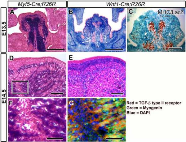
To investigate cell lineages in the tongue primordium, we examined myogenic or CNC-derived cells in Myf5-Cre;R26R and Wnt1-Cre;R26R mice, respectively. In Myf5-Cre;R26R mice, LacZ positive cells were detected in the center of the tongue primordium (Figure 1A). In contrast, the LacZ expression pattern in Wnt1-Cre;R26R mice was the reverse of that of Myf5-Cre;R26R mice (Figure 1B). This pattern suggests that CNC cells circumscribe somite-derived myogenic progenitor cells at E13.5 (Figure 1A and 1B). Furthermore, the expression pattern of Myosin Heavy Chain (MHC), a marker for myogenic differentiation, coincided with LacZ-positive cells in Myf5-Cre;R26R mice and LacZ-negative cells in Wnt1-Cre;R26R mice (Figure 1A–C). We also found that tongue skeletal muscle cells in Myf5-Cre;R26R mice appeared to form bundle structure, whereas CNC-derived connective tissue cells in Wnt1-Cre;R26R mice populated the region between the presumptive Myf5 positive bundles at E14.5 (Figure 1D and 1E). Furthermore, we assessed the expression pattern of TGF-β type II receptor during tongue development. We detected TGF-β IIR expression in CNC-derived cells (Figure 1G, red) and myogenic progenitors (Figure 1G, green).
Figure 1. The interface between cranial neural crest and myogenic cells in the tongue primordium.
LacZ staining and immunostaining of frontal sections from Myf5-Cre;R26R (A, D, and F) and Wnt1-Cre;R26R (B, C, E, and G) mice. (A) LacZ positive cells (Myf5-Cre;R26R) form the core of the tongue primordium at E13.5. (B) LacZ positive cells (Wnt1-Cre;R26R) circumscribe the LacZ negative core at E13.5. (C) Myosin heavy chain (MHC) immunostaining and LacZ staining of E13.5 Wnt1-Cre;R26R mice. MHC positive cells are detectable in the center of the tongue primordium. LacZ positive cells (Wnt1-Cre;R26R) circumscribe the MHC positive cells. (D, F) LacZ positive cells (Myf5-Cre;R26R) form myofibril-like structures at E14.5. F is enlarged from the box in D. (E) LacZ positive cells are detectable between muscle cell-like structures at E14.5. (G) Immunofluorescence of TGF-β type II receptor (red) and myogenin (green). Myf5 negative cells, which are CNC-derived cells, are immunopositive for TGF-β type II receptor (red). Myogenin positive cells (green) coincide with Myf5 positive cells (see F), and some are immunopositive for TGF-β type II receptor (yellow). Scar bars: 300 μm in A, B, 100 μm in C–E, 50 μm in F,G.
Loss of TGF-β signaling in CNC cells produce microglossia and disorganization of muscle cell alignment
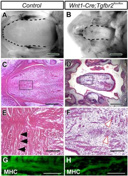
The loss of TGF-β signaling in CNC cells resulted in a severe defect in tongue development. Wnt1-Cre;Tgfbr2flox/flox mice exhibited microglossia when compared with Wnt1-Cre;Tgfbr2flox/+ (control) mice (Figure 2A and 2B), which were indistinguishable from wild type mice (data not shown). We detected well-organized muscle cells in control mice by histological analysis (Figure 2C and 2E). In contrast, muscle cells were disorganized and present in low density in Wnt1-Cre;Tgfbr2flox/flox mice (Figure 2D and 2F). We detected that MHC expression in muscle cells of Wnt1-Cre;Tgfbr2flox/flox mice was similar per cell to that of control mice, although muscle cells were decreased in number and curved (Figure 2G and 2H). We previously reported that the oral space of Wnt1-Cre;Tgfbr2flox/flox mice was smaller than that of control mice because of their small mandible and cleft palate (Ito et al., 2003; Oka et al., 2007). To test the hypothesis that these physiological differences affected the tongue development process, we performed tongue organ culture to remove any physical constraints on tongue growth. After isolation of the tongue primordium at E12.0, there was no structure inhibiting tongue growth via physiological force. Following seven days culture, we found that the size of the tongue primordium from mutant mice (n=6) was smaller than that from control mice (n=6) (Figure S1A and S1B). The maximal length and width of tongues from Wnt1-Cre;Tgfbr2flox/flox mice were reduced by 21.1±2.5 % and 31.7±1.7%, respectively. Thus, we conclude that the tongue phenotype of Wnt1-Cre;Tgfbr2flox/flox mice was a primary defect and not a secondary defect due to constraints such as a small mandible. Because TGF-β signaling is lost only in CNC cells, our data suggest that TGF-β signaling regulates tissue-tissue interaction during skeletal muscle development.
Figure 2. Wnt1-Cre;Tgfbr2flox/flox mice exhibit microglossia and muscle cell disorganization.
Macroscopic appearance (A and B), hematoxylin and eosin staining (C–F), and myosin heavy chain (MHC) immunohistochemistry (G and H) of tongues from Wnt1-Cre; Tgfbr2flox/+ (control) (A, C, E, and G) and Wnt1-Cre; Tgfbr2flox/flox (B, D, F, and H) newborn mice. (A and B) Wnt1-Cre;Tgfbr2flox/flox mice exhibit microglossia (dotted lines indicate the outline of tongue). (C–F) Hematoxylin and eosin staining of control mice shows well-organized myofibrils (C, E; black arrowheads), but muscle cells are disorganized and scattered in Wnt1-Cre;Tgfbr2flox/flox mice (D, F white arrowheads indicate the diminished muscle cells). E and F are enlarged from the boxes in C and D, respectively. (G and H) Muscle cells are positive for MHC expression (green) in both control and Wnt1-Cre; Tgfbr2flox/flox mice. Scale bars: 1 mm in A, B, 500 μm in C, D, 100 μm in E, F, 50 μm in G, H.
Loss of TGF-β signaling in CNC cells results in reduced myogenic cell proliferation
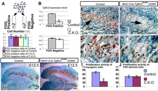
During tongue skeletal muscle development, myogenic cells migrate from the occipital somite to the tongue primordium through the hypoglossal cord (Huang et al., 1999). We found that myogenic precursor cells started to invade the tongue primordium around E12.0 (data not shown). To compare the cell population in Wnt1-Cre;Tgfbr2flox/flox;R26R and Wnt1-Cre;Tgfbr2flox/+;R26R (control) mice, we performed Fluorescence Activated Cell Sorting (FACS) using FDG staining for β-galactosidase. Through the sorting process, we separated cranial neural crest derived cells (FDG positive) and non-cranial neural crest cells (FDG negative) (Figure 3A). We found that the cell numbers of both FDG–positive and –negative cells in Wnt1-Cre;Tgfbr2flox/flox mice were indistinguishable from those of control mice. After FACS, we assessed Tgfbr2 gene expression in CNC cells and non-CNC cells in Wnt1-Cre;Tgfbr2flox/flox;R26R samples. Real-time PCR data demonstrated that Tgfbr2 gene expression was deleted only in CNC cells of Wnt1-Cre;Tgfbr2flox/flox mice (Figure 3B). Furthermore, to assess myogenic cell migration in Wnt1-Cre;Tgfbr2flox/flox mice, we analyzed MHC expression in the tongue primordium at E12.5. We detected MHC positive cells throughout the anterior to posterior regions of the tongue primordium in Wnt1-Cre;Tgfbr2flox/flox and control mice (Figure 3C and 3D). Thus, myogenic cell migration into the tongue primordium in Wnt1-Cre;Tgfbr2flox/flox mice was indistinguishable from that of control mice. In order to investigate the cellular mechanism responsible for microglossia, we examined cell proliferation activity in Wnt1-Cre;Tgfbr2flox/flox mice. To determine whether CNC or myogenic cell lineages exhibited reduced cell proliferation, we performed anti-BrdU and LacZ double staining. In Wnt1-Cre;Tgfbr2flox/+;R26R (control) mice, both CNC cells (Figure 3E, open arrow) and myogenic cells (Figure 3E and 3G, black arrows) were positive for cell proliferation activity. In contrast, proliferation activity was reduced in myogenic cells of Wnt1-Cre;Tgfbr2flox/flox;R26R mice (Figure 3F and 3H, black arrows), although CNC cells maintained proliferation activity similar to that of control mice (Figure 3F, open arrow). We also analyzed the number of BrdU positive and negative CNC and myogenic cells of the tongue primordium (Figure 3I and 3J). This quantitative analysis confirmed that cell proliferation activity was significantly decreased in the myogenic cell lineage but not in the CNC cell lineage of Wnt1-Cre;Tgfbr2flox/flox mice.
Figure 3. Loss of Tgfbr2 adversely affects myogenic cell proliferation.
(A) Schematic diagram of Fluorescence Activated Cell Sorting (FACS) by Fluorescence di-β-D-Galactopyranoside (FDG) staining. The graph indicates the number of FDG-positive or -negative cells from Wnt1-Cre;Tgfbr2flox/+ (Control) or Wnt1-Cre;Tgfbr2flox/flox mice. (B) Tgfbr2 gene expression level in either FDG -positive or – negative cells. Student t-tests were used for statistical analysis. *: P<0.05. (N=2). (C and D) MHC immunostaining and LacZ staining of E12.5 sagittal sections from Wnt1-Cre;Tgfbr2flox/+;R26R (Control) and Wnt1-Cre;Tgfbr2flox/flox;R26R mice. MHC positive cells are detectable at the tip of the tongue primordium in Control and Wnt1-Cre; Tgfbr2flox/flox;R26R mice. (E–H) BrdU staining with LacZ staining of E13.5 sections from Wnt1-Cre; Tgfbr2flox/+;R26R (E, G, Control) and Wnt1-Cre; Tgfbr2flox/flox;R26R (F, H, conditional knock out mice. G and H are enlarged from E and F, respectively. (E and G) BrdU positive cells in control mice are distributed between CNC cells (blue staining, white arrow) and myogenic cells (non-blue cells, black arrow). (F and H) BrdU positive myogenic cells are scattered in Wnt1-Cre; Tgfbr2flox/flox;R26R mice (black arrows). (I and J) BrdU labeling index in each myogenic cell (G) and CNC cells (H). Five randomly selected, non-overlapping samples were used from each experimental group. Student t-tests were used for statistical analysis. *: P<0.05. (N=5). Scale bars: 200 μm in C, D, E, and F.
Skeletal muscle organization is compromised in Wnt1-Cre;Tgfbr2flox/flox mice
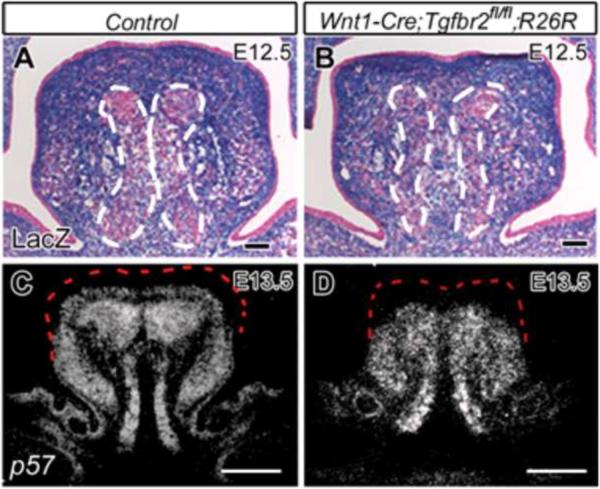
We assessed whether skeletal muscle organization was altered in Wnt1-Cre;Tgfbr2flox/flox mice. At E12.5, myogenic cells derived from occipital somites formed a myogenic core in the center of the tongue primordium of Wnt1-Cre;Tgfbr2flox/+;R26R (control) mice (Figure 4A, delineated by the dotted line). The myogenic core was also detectable in the tongue primordium of Wnt1-Cre;Tgfbr2flox/flox;R26R mice (Figure 4B), implying that the early stage of myogenic formation in Wnt1-Cre;Tgfbr2flox/+ mice was unaffected. Division of the myogenic core had occurred in control mice at E13.5, based on the expression pattern of p57, a marker for myogenic cell differentiation (Zhang et al., 1999) (Figure 4C), whereas division of myogenic core failed to occur in Wnt1-Cre;Tgfbr2flox/flox mice (Figure 4D). Thus, we conclude that although myogenic differentiation at early stages was normal as indicated by MHC staining (See Fig. 3D), the division process was compromised in Wnt1-Cre;Tgfbr2flox/flox mice.
Figure 4. Compromised patterning of myogenic cells in Wnt1-Cre;Tgfbr2flox/flox mice.
(A and B) LacZ staining of Wnt1-Cre;Tgfbr2flox/+;R26R mice (A) and Wnt1-Cre; Tgfbr2flox/flox;R26R mice (B) at E12.5. White dotted line indicates the margin of myogenic core. (C and D) In situ hybridization of p57 from Wnt1-Cre;Tgfbr2flox/+ mice (C) and Wnt1-Cre;Tgfbr2flox/flox mice (D). Red dotted line indicates the margin of tongue primordium. Scale bars: 200 μm in A–D.
Cell autonomous requirement for TGF-β signaling during the differentiation of CNC-derived cells in the tongue primordium
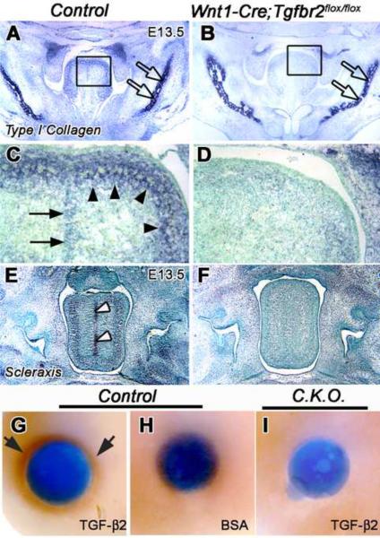
We hypothesized that TGF-β signaling controls the fate of CNC-derived cells and, therefore, analyzed cell differentiation status in Wnt1-Cre;Tgfbr2flox/flox mice. Type I collagen, the main component of connective tissue, was expressed in the dense lamina propria and septum of the tongue in control mice (Figure 5A, 5C), whereas this staining appeared diminished in Wnt1-Cre;Tgfbr2flox/flox mice (Figure 5B, 5D). In contrast, expression of type I collagen in the mandible was indistinguishable between control and Wnt1-Cre;Tgfbr2flox/flox mice (Figure 5A, 5B, open arrows). Scleraxis (Scx), a bHLH transcription factor, is expressed in the mature tendons of limbs and trunk as well as their progenitors, and plays an important role in regulating tendon differentiation (Brent et al., 2003). Scx was expressed in the perimysium, epimysium, and septum that surround tongue muscle fibers of control mice (Figure 5E, white arrowheads). In Wnt1-Cre;Tgfbr2flox/flox mice, Scx staining was diminished in the tendon of the tongue muscle (Figure 5F). We hypothesized that TGF-β signaling induces Scx expression during tongue development. Our study showed that TGF-β2 soaked beads induced Scx expression in the tongue primordium of control mice (Figure 5G), but BSA soaked beads had no effect (Figure 5H). In contrast, TGF-β2 soaked beads failed to induce Scx expression in Wnt1-Cre;Tgfbr2flox/flox mice (Figure 5I). These data suggest that TGF-β signaling is required to play a cell-autonomous role in inducing Scx expression and in regulating CNC differentiation during tongue development.
Figure 5. Scleraxis (Scx) and type I collagen expression in CNC-derived cells in the tongue.
In situ hybridization of type I collagen and Scleraxis (Scx) in control (A,C,E) and Wnt1-Cre;Tgfbr2flox/flox (B,D,F) mice at E13.5. (A–D) Type I collagen is expressed in the dense lamina propria (arrowhead) and septum (arrow) but its expression is greatly reduced in the Tgfbr2 mutant sample. Open arrow points to type I collagen expression in the developing mandible. (E and F) Scx is expressed in the tongue septum of control mice (white arrowheads), but not in the tongue of Tgfbr2 mutant mice. (G–I) Explants from tongue primordia of E12.5 control and Wnt1-Cre;Tgfbr2flox/flox;R26R mice treated with beads for 12 hours. The control sample treated with TGF-β2 beads (G) is positive for Scx expression (black arrows), but BSA beads do not induce Scx expression (H). The conditional knockout sample treated with TGF-β2 beads is negative for Scx expression (I).
Loss of TGF-β signaling in CNC cells results in a reduction of muscle cell number in vitro
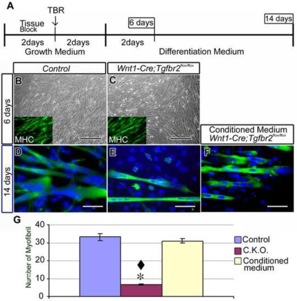
To investigate the relationship between cranial neural crest and myogenic cells, we analyzed a primary cell culture of tongue primordium from Wnt1-Cre;Tgfbr2flox/flox mice. The cells harvested from Wnt1-Cre;Tgfbr2flox/flox mice were indistinguishable from those of control mice when cultured in growth medium, which inhibited myogenic differentiation (data not shown), and after changing from growth medium to differentiation medium (total 6 days culture) (Figure 6B, 6C). Moreover, Wnt1-Cre;Tgfbr2flox/flox and control cells both expressed myosin heavy chain (MHC), a marker of myogenic differentiation, at this stage (Figure 6B, 6C, inserts). Thus, primary myogenic differentiation appears normal in Wnt1-Cre;Tgfbr2flox/flox mice. After 14 days culture, the number of muscle cells in Wnt1-Cre;Tgfbr2flox/flox mice was decreased when compared with that from control mice (Figure 6D, 6E, 6G). These observations suggest that CNC cells might have a critical function in muscle cell formation. To test this hypothesis, we applied conditioned medium from the control culture to the Wnt1-Cre;Tgfbr2flox/flox primary culture. This addition resulted in an increased number of muscle cells in the Wnt1-Cre;Tgfbr2flox/flox primary cell culture (Figure 6F, 6G). This stimulation indicates that there may be secreted factors from cranial neural crest cells that are required for myogenic development.
Figure 6. Compromised muscle cell formation in primary cell culture of Wnt1-Cre; Tgfbr2flox/flox mice.
(A) Schematic diagram of the timetable of primary cell culture. TBR: Tissue Block Remove. (B and C) After 6 days culture, the appearance of cells from Wnt1-Cre; Tgfbr2flox/+ (control) and Wnt1-Cre;Tgfbr2flox/flox mice is indistinguishable under the phase microscope. Inserts show immunostaining of myosin heavy chain (MHC). (D–F) MHC immunostaining (MHC appears green, DAPI appears blue) after 14 days of Wnt1-Cre; Tgfbr2flox/+ (control) and Wnt1-Cre;Tgfbr2flox/flox primary cell culture and Wnt1-Cre; Tgfbr2flox/flox cells treated with conditioned medium from the control culture. After14 days of culture, the number of muscle cells in the Wnt1-Cre; Tgfbr2flox/flox culture (E) is reduced compared to control (D). The addition of conditioned medium to the Wnt1-Cre; Tgfbr2flox/flox culture (F) increased the number of muscle cells. (G) Quantitation of muscle cell number from D–F. Scale bars: 300 μm in A–E, 50 μm in G–I.
FGF10 functions downstream of TGF-β signaling to regulate tongue development
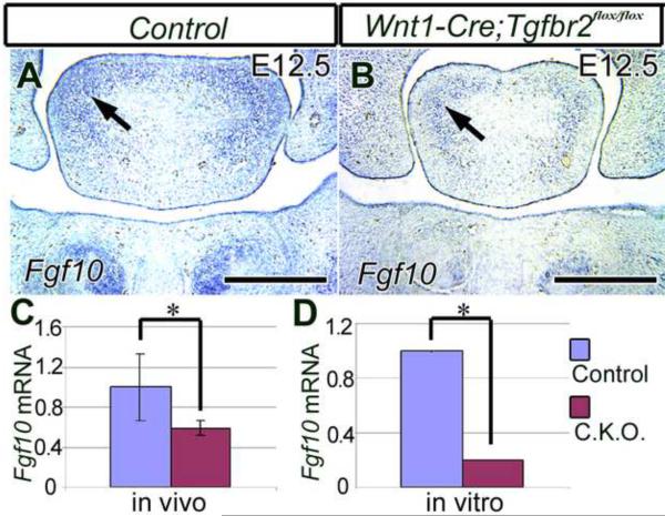
To elucidate the molecular mechanism of TGF-β signaling during tongue development, we performed microarray analyses to compare gene expression profiles between wild type and Tgfbr2 mutant tongue at E12.5. We decided to focus on E12.5 because we detected cell proliferation defects in the developing tongue of Wnt1-Cre;Tgfbr2flox/flox mutant mice beginning at E13.5 (see Figure S2 and 3D). In our initial set of three microarray experiments, we detected a significant reduction in Fgf10 expression in the tongue primordium of Wnt1-Cre;Tgfbr2flox/flox mice (data not shown). Both FGFs and their receptors are expressed during tongue development (Nie, 2005). Furthermore, FGF signaling has been shown to be critical for skeletal myogenesis during Xenopus and chick embryonic development (Itoh et al., 1996; Standley et al., 2001). We detected Fgf10 expression in CNC-derived cells in tongue primordium of control mice at E12.5 (Figure 7A). In the Wnt1-Cre;Tgfbr2flox/flox mutant, there was a reduction in Fgf10 expression in the CNC-derived cells surrounding the myogenic core (Figure 7B). In order to test whether there was a significant reduction in the expression level of Fgf10, we performed real-time RT-PCR analysis. We found that there was a significant reduction in Fgf10 expression in the tongue primordium and in primary culture of tongue primordium in Wnt1-Cre;Tgfbr2flox/flox mice (Figure 7C and 7D). Thus, our data suggest that there is a cell autonomous requirement for TGF-β signaling to induce Fgf10 expression in CNC-derived cells during tongue development.
Figure 7. Fgf10 expression analysis in the developing tongue.
(A and B) In situ hybridization of Fgf10 mRNA in E12.5 Wnt1-Cre;Tgfbr2flox/+ (control) and Wnt1-Cre; Tgfbr2flox/flox mice. Fgf10 mRNA is expressed in cranial neural crest cells around a myogenic core in the tongue primordium (arrows) of control and Wnt1-Cre; Tgfbr2flox/flox mice. (C and D) Quantitative analysis of Fgf10 expression in Wnt1-Cre;Tgfbr2flox/+ and Wnt1-Cre;Tgfbr2flox/flox mice using real-time PCR. The expression level of Fgf10 in Wnt1-Cre;Tgfbr2flox/flox mice at E12.5 is reduced in vivo (C). Fgf10 expression is also reduced in primary cell culture samples in vitro (D). Student t-tests were used for statistical analysis. *: P<0.05. (N=5). Scale bars: 300 μm in A, B.
Exogenous FGF10 rescues myogenic cell proliferation in Tgfbr2 mutant mice
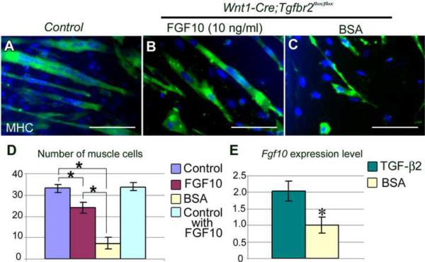
Mutation in Tbx1 results in down-regulation of FGF10 expression, which affects the patterning of CNC cells in the mandibular arch and results in defects in branchiomeric myogenesis in mice (Kelly et al., 2004). To test the hypothesis that FGF10 acts downstream of TGF-β to control myogenic cell proliferation, we performed rescue experiments using the primary tongue cell culture model. Following four days of culture in growth medium, we switched the cultured tongue cells into differentiation medium for another ten days. Cells from the control sample expanded their population and differentiated into myogenic cells, as validated by positive MHC staining (Figure 8A). In Wnt1-Cre;Tgfbr2flox/flox mutant samples, there was a significant reduction (p<0.05) in the number of muscle cells. Significantly, the addition of exogenous FGF10 in the differentiation medium was able to increase the number of muscle cells (Figure 8B). In contrast, addition of BSA failed to restore the number of muscle cells in the Wnt1-Cre;Tgfbr2flox/flox mutant tongue cell culture (Figure 8C). Statistical analyses revealed that exogenous FGF10 was able to restore the number of muscle cells in the Wnt1-Cre;Tgfbr2flox/flox cell culture substantially, but not completely to wild type levels, although addition of FGF10 had no effect on control samples (Figure 8D). These results suggest that additional members of the FGF family, or other factors, may also mediate TGF-β signaling to control the proliferation and survival of myogenic cells during tongue development. In a parallel approach, we placed beads carrying FGF10 or BSA in cultured tongue explants. The number of muscle cells in Wnt1-Cre;Tgfbr2flox/flox mutant tongue explants treated with FGF10 beads increased close to that of control mice explants, whereas BSA beads failed to restore the number of muscle cells in mutant tongue explants (data not shown). Finally, to test whether TGF-β2 could induce Fgf10 expression, we applied TGF-β2 ligand or BSA into primary tongue cell cultures of control mice, immediately following the switch from growth medium to differentiation medium. Fgf10 expression was induced two hours after the addition of TGF-β, whereas BSA addition had no effect (Figure 8E). Taken together, our data suggest that TGF-β-induced FGF signaling plays an important role in regulating tissue-tissue interaction and myogenic cell proliferation during tongue development.
Figure 8. Exogenous FGF10 rescues the reduction of muscle cells in Wnt1-Cre;Tgfbr2flox/flox mice.
(A–C) Immunostaining of MHC (green) in Wnt1-Cre; Tgfbr2flox/+ primary cell culture (Control, A) and Wnt1-Cre;Tgfbr2flox/flox primary cell cultures with FGF10 (B) or BSA (C) added into the medium. (D) Quantitative analysis of the number of muscle cells detectable in sample fields (20×). Student t-tests were used for statistical analysis. *: P<0.05. (N=5). (E) Fgf10 gene expression level analyzed by real-time PCR. Exogenous TGF-β2 ligand induces Fgf10 expression. Student t-tests were used for statistical analysis. *: P<0.05. (N=2). Scale bar: 100 μm in A–C.
Neural crest cells are required for organizing tongue muscle cells
To test the reciprocal relationship between muscle cells and tendon cells (derived from CNC cells), we used a tissue regeneration approach. We separated myogenic and CNC cells from E12.5 control and Wnt1-Cre;Tgfbr2flox/flox tongue primordia using cell sorting and combined cells as depicted in Figure S3A–C. The first sample contained CNC cells and myoblasts from control mice, the second was CNC cells from control mice and myoblasts from conditional knockout mice, and the third was CNC cells from conditional knockout mice and myoblasts from control mice. These cell mixtures were seeded in a collagen scaffold and the scaffold was inserted beneath the back skin of host mice for 2 weeks. We detected muscle cells along the scaffold in the first sample (Figure S3D and S3G). The second sample also resulted in muscle cell formation in which the myoblast cells had been derived from conditional knockout mice (Figure S3E and S3H). In contrast, we observed disorganized MHC (myosin heavy chain) positive cells in the third sample, even though the myoblasts were derived from control mice. Our results suggest that CNC cells play a crucial role in organizing myogenic cells. Furthermore we examined the transplanted CNC cells to eliminate the possibility that the phenotype we observed was due to a difference in survival. We detected CNC cells from control mice around muscle cells in the first and second groups (Figure S3J and S3K) and the CNC cells from conditional knockout mice in the scaffold (third group, Figure S3L), scattered but not organized around the myogenic cells. These observations suggest that CNC cells possess a function important for muscle cell formation and organization.
Discussion
In this present study, we provide the first evidence that TGF-β signaling has a critical function in regulating skeletal muscle development through tissue-tissue interactions. We find that loss of TGF-β signaling in CNC cells results in microglossia with defects in CNC-derived connective tissue and tongue muscle development. Specifically, TGF-β is required for Scleraxis and Fgf10 expression in CNC-derived cells and cell proliferation and organization in their neighboring myogenic progenitors. Thus, we demonstrate that the CNC-derived cells surrounding the myogenic progenitors have a critical function in regulating the development of tongue muscle.
Tissue-tissue interactions in regulating tongue muscle development
Craniofacial organs offer unique opportunities for the investigation of reciprocal tissue-tissue interactions in regulating embryonic development. For instance, tooth development involves reciprocal interactions between the oral epithelium and the CNC-derived ectomesenchyme tissues (Kettunen et al., 2000). Muscles in the craniofacial region derive from two mesenchymal cell lineages; one consists of mesoderm-derived myogenic progenitors and their surrounding tissues derive from cranial neural crest cells. The intimate relationship between these two cell lineages suggests reciprocal interactions between CNC and myogenic cells in the process of skeletal muscle development. Furthermore, because of our ability to generate CNC or myogenic cell specific inactivation of a gene of interest, we have a unique opportunity to investigate the functional significance of tissue-tissue interactions in regulating craniofacial skeletal muscle development.
The tongue is a muscular organ and performs important physiological functions in our daily lives. Using our two component genetic model for indelibly marking the progenies of CNC cells, we show in this study that tendons and connective tissues are derived from CNC cells in the tongue and are critical for the organization and function of tongue. Previous studies have shown that, in invertebrates such as Drosophila, the ectoderm is the origin for tendons (Schnorrer and Dickson, 2004). Chick/quail experiments indicates that the tendon and surrounding tissue of craniofacial muscles are composed of CNC cells in the craniofacial region (Noden, 1988). In vertebrates, tendons are derived from the lateral mesoderm, except in the craniofacial region where CNC-derived cells give rise to tendon and connective tissue in the skeletal muscles.
In the mouse model, Scleraxis (Scx) is considered a marker for tendon cells (Brent et al., 2003). Tendon formation is diminished in Scx knockout mice (Murchison et al., 2007). Scx is a basic helix-loop-helix transcription factor involved in controlling collagen expression. Pro-α1(I) collagen (COLa1) is one of the chains composed of Type I collagen (van der Rest and Garrone, 1991). There are TSE1 (Tendon-specific elements) and TSE2 regions in the promoter region of COLa1 (Terraz et al., 2002). SCX binds to TSE1 and TSE2 and controls the expression of COLa1 (Lejard et al., 2007). Studies of TSE1 and TSE2 LacZ transgenic mice indicate that these regions express in the fibroblast cells giving rise to tendon cells (Terraz et al., 2002). The LacZ staining in these mice is detectable not only in the tendons of the trunk region but also in the tendons of craniofacial muscles such as the masseter. Our present study reveals that Scx and Type I collagen expression is compromised following the loss of TGF-β signaling and that TGF-β ligand can induce Scx expression. Taken together, we conclude that TGF-β-mediated Scleraxis and Type I collagen expression is critical in regulating the fate of CNC cells during craniofacial skeletal muscle development.
Skeletal muscle development involves cell migration, proliferation, differentiation, and organization stages (Birchmeier and Brohmann, 2000; Buckingham et al., 2003; Mackenzie et al., 1998; Noden and Francis-West, 2006). During the migration stage, myoblast precursor cells can migrate into their presumptive location even if the CNC cells are ablated (Huang et al., 1999; von Scheven et al., 2006). This result implies that CNC cells are not required for the migration of myogenic precursors towards their presumptive destinations, including the branchial arch and tongue primordium. At the differentiation stage, CNC cells in the branchial arch of chicks secrete BMP and Wnt inhibitors, which induce myogenic differentiation (Tzahor et al., 2003). However, it is still unclear how CNC cells control the organization of myogenic cells. During skeletal muscle development, muscle cells and surrounding tissue, such as tendon cells, both appear to be involved in the formation of the attachment to the skeletal muscle. For instance, the loss of stripe, which is involved in early steps of tendon differentiation, results in the disruption of the entire somatic muscle pattern (Frommer et al., 1996). A defect in tendon formation causes compromised muscle formation in Drosophila and zebrafish (Frommer et al., 1996; Kudo et al., 2004). The loss of Periostin, an adhesion molecule, in the myoseptum causes a differentiation defect in myoblasts (Kudo et al., 2004). These results imply that the reciprocal interaction between myogenic and surrounding cells, including tendon cells, is critical as a regulatory mechanism of skeletal muscle development.
TGF-β mediated FGF signaling controls tissue-tissue interactions during tongue development
Previous studies have shown that FGF ligands and their receptors are required for skeletal muscle development (de Alvaro et al., 2005; Flanagan-Steet et al., 2000). For instance, the viral expression of truncated Fgf type I receptor causes a skeletal muscle defect in the chick limb (Flanagan-Steet et al., 2000). FGF signaling controls myogenic cell differentiation status through MAP kinase (de Alvaro et al., 2005; Gonzalez et al., 2004; Naya and Olson, 1999). We demonstrate here that secreted factor(s) from CNC cells are critical for myogenic development, because conditioned cell culture medium can rescue the reduction of muscle cell number in the Wnt1-Cre;Tgfbr2flox/flox sample. Fgf10 is a candidate for this secreted factor. Fgf10 expression is reduced in Wnt1-Cre;Tgfbr2flox/flox mice, and exogenous FGF10 reverses the reduction of tongue muscle cell number in vitro. The Fgf10 conventional knockout mouse is another model that has revealed reciprocal interactions between two cell lineages. Fgf10 null mice exhibited a reduction of cell proliferation activity in the dental epithelium, even though Fgf10 expression is detected only in the dental mesenchyme. Exogenous FGF10 reverses the reduction of cell proliferation in Fgf10 null mice (Harada et al., 2002). FGF10 has also been shown to be critical for mediating tissue-tissue interactions during lung and palate development (Bellusci et al., 1997; Bhushan et al., 2001; Rice et al., 2004). Taken together, our study and others show that a paracrine function of FGF10 is critical for embryonic organ development.
Our conditional knockout mouse model helped us to explore the reciprocal relationship among tissue layers during organogenesis. Specifically, this study demonstrates that the loss of Tgfbr2 in non-myogenic cells results in the disorganization of muscle cells. In contrast, conditional inactivation of Tgfbr2 in myogenic specific cell lineages exhibit organized muscle cells (our unpublished data), although myogenic differentiation is compromised as reported in a previous cell culture study (Filvaroff et al., 1994). We have also investigated how the cells surrounding the myoblast contribute to muscle cell organization during their formation using a tissue regeneration experimental approach. Our data indicate that TGF-β signaling is crucial in CNC-derived cells, but not in myoblasts, for organizing muscle cells during tongue muscle development.
We have presented the first study investigating the function of CNC cells during skeletal muscle development using a genetic, rather than surgical or tissue transplantation approach. Although cell line experiments have revealed that many genes are critical for skeletal muscle development in a cell autonomous manner (de Alvaro et al., 2005; Deng et al., 1994), they have yet to provide information about the functional significance of tissue-tissue interactions during muscle development. Previously, Drosophila or zebrafish studies have been used to reveal the mechanism of skeletal muscle organization (Frommer et al., 1996; Kudo et al., 2004). Our in vivo system allows us to investigate the functional significance of genes in the spatial complexity of the mouse muscular organ and has provided evidence for the importance of TGF-β signaling in regulating tissue-tissue interactions during tongue muscle development. In summary, TGF-β signaling regulates fibroblast differentiation by controlling Scleraxis expression in a cell autonomous manner. TGF-β signaling controls myogenic cell proliferation via secreted factors from CNC cells and tissue-tissue interactions during tongue morphogenesis. Disruption of this interaction may lead to dysmorphogenesis.
Supplementary Material
(A and B) Macroscopic appearance of tongue organ culture from E12.0 Wnt1-Cre;Tgfbr2flox/+ (Control) and Wnt1-Cre; Tgfbr2flox/flox mice after 7 days culture.
(A and B) BrdU incorporation of tongue primordium in Wnt1-Cre;Tgfbr2flox/+ (Control) and Wnt1-Cre;Tgfbr2flox/flox mice at E12.5. (C) BrdU incorporation index. BrdU positive cell ratio of Wnt1-Cre;Tgfbr2flox/flox mice is the same as that of control mice. Scale bars: 200 μm in A and B.
(A–C) Schema of cell recombination experiment. (D–E) Hematoxylin and eosin staining. (G–I) Immunostaining for Myosin heavy chain (MHC). (J–L) Lac Z staining. (A, D, G, J) Both cranial neural crest (CNC) cells and myoblasts are derived from control mice. Organized muscle cells are positive for MHC (G, arrows) and LacZ positive cells surround muscle cells (J, arrowheads). (B, E, H, K) Myoblasts of conditional knockout mice combined with CNC cells of control mice. MHC positive muscle cells were detectable (H, arrows) surrounded by lacZ positive cells (K, arrowheads). (C, F, I, L) Myoblasts of control mice combined with CNC cells of Tgfbr2 mutant mice. MHC positive cells were not organized (I, arrow). LacZ positive cell are scattered (L,arrowheads).
Acknowledgments
We thank Julie Mayo for critical reading of the manuscript, J. L. R. Rubenstein for reagents, and H. Moses for the Tgfbr2flox/flox mice. This study was supported by grants from the National Institute of Dental and Craniofacial Research, NIH (DE014078, DE012711, and DE017007) to Yang Chai.
Footnotes
Publisher's Disclaimer: This is a PDF file of an unedited manuscript that has been accepted for publication. As a service to our customers we are providing this early version of the manuscript. The manuscript will undergo copyediting, typesetting, and review of the resulting proof before it is published in its final citable form. Please note that during the production process errors may be discovered which could affect the content, and all legal disclaimers that apply to the journal pertain.
References
- Bellusci S, Grindley J, Emoto H, Itoh N, Hogan BL. Fibroblast growth factor 10 (FGF10) and branching morphogenesis in the embryonic mouse lung. Development. 1997;124:4867–4878. doi: 10.1242/dev.124.23.4867. [DOI] [PubMed] [Google Scholar]
- Bhushan A, Itoh N, Kato S, Thiery JP, Czernichow P, Bellusci S, Scharfmann R. Fgf10 is essential for maintaining the proliferative capacity of epithelial progenitor cells during early pancreatic organogenesis. Development. 2001;128:5109–5117. doi: 10.1242/dev.128.24.5109. [DOI] [PubMed] [Google Scholar]
- Birchmeier C, Brohmann H. Genes that control the development of migrating muscle precursor cells. Curr Opin Cell Biol. 2000;12:725–730. doi: 10.1016/s0955-0674(00)00159-9. [DOI] [PubMed] [Google Scholar]
- Bogusch G. On the spatial relationship between fibroblasts and myogenic cells during early development of skeletal muscles. Acta Anat (Basel) 1986;125:225–228. doi: 10.1159/000146167. [DOI] [PubMed] [Google Scholar]
- Borue X, Noden DM. Normal and aberrant craniofacial myogenesis by grafted trunk somitic and segmental plate mesoderm. Development. 2004;131:3967–3980. doi: 10.1242/dev.01276. [DOI] [PubMed] [Google Scholar]
- Brent AE, Schweitzer R, Tabin CJ. A somitic compartment of tendon progenitors. Cell. 2003;113:235–248. doi: 10.1016/s0092-8674(03)00268-x. [DOI] [PubMed] [Google Scholar]
- Buckingham M, Bajard L, Chang T, Daubas P, Hadchouel J, Meilhac S, Montarras D, Rocancourt D, Relaix F. The formation of skeletal muscle: from somite to limb. J Anat. 2003;202:59–68. doi: 10.1046/j.1469-7580.2003.00139.x. [DOI] [PMC free article] [PubMed] [Google Scholar]
- Chai Y, Jiang X, Ito Y, Bringas P, Jr., Han J, Rowitch DH, Soriano P, McMahon AP, Sucov HM. Fate of the mammalian cranial neural crest during tooth and mandibular morphogenesis. Development. 2000;127:1671–1679. doi: 10.1242/dev.127.8.1671. [DOI] [PubMed] [Google Scholar]
- Chai Y, Mah A, Crohin C, Groff S, Bringas P, Jr., Le T, Santos V, Slavkin HC. Specific transforming growth factor-beta subtypes regulate embryonic mouse Meckel's cartilage and tooth development. Dev Biol. 1994;162:85–103. doi: 10.1006/dbio.1994.1069. [DOI] [PubMed] [Google Scholar]
- Chevallier A, Kieny M, Mauger A. Limb-somite relationship: origin of the limb musculature. J Embryol Exp Morphol. 1977;41:245–58. [PubMed] [Google Scholar]
- Couly GF, Coltey PM, Le Douarin NM. The developmental fate of the cephalic mesoderm in quail-chick chimeras. Development. 1992;114:1–15. doi: 10.1242/dev.114.1.1. [DOI] [PubMed] [Google Scholar]
- de Alvaro C, Martinez N, Rojas JM, Lorenzo M. Sprouty-2 overexpression in C2C12 cells confers myogenic differentiation properties in the presence of FGF2. Mol Biol Cell. 2005;16:4454–4461. doi: 10.1091/mbc.E05-05-0419. [DOI] [PMC free article] [PubMed] [Google Scholar]
- Deng CX, Wynshaw-Boris A, Shen MM, Daugherty C, Ornitz DM, Leder P. Murine FGFR-1 is required for early postimplantation growth and axial organization. Genes Dev. 1994;8:3045–3057. doi: 10.1101/gad.8.24.3045. [DOI] [PubMed] [Google Scholar]
- Doherty KR, Cave A, Davis DB, Delmonte AJ, Posey A, Earley JU, Hadhazy M, McNally EM. Normal myoblast fusion requires myoferlin. Development. 2005;132:5565–5575. doi: 10.1242/dev.02155. [DOI] [PMC free article] [PubMed] [Google Scholar]
- Evans DJ, Noden DM. Spatial relations between avian craniofacial neural crest and paraxial mesoderm cells. Dev Dyn. 2006;235:1310–1325. doi: 10.1002/dvdy.20663. [DOI] [PubMed] [Google Scholar]
- Filvaroff EH, Ebner R, Derynck R. Inhibition of myogenic differentiation in myoblasts expressing a truncated type II TGF-beta receptor. Development. 1994;120:1085–1095. doi: 10.1242/dev.120.5.1085. [DOI] [PubMed] [Google Scholar]
- Flanagan-Steet H, Hannon K, McAvoy MJ, Hullinger R, Olwin BB. Loss of FGF receptor 1 signaling reduces skeletal muscle mass and disrupts myofiber organization in the developing limb. Dev Biol. 2000;218:21–37. doi: 10.1006/dbio.1999.9535. [DOI] [PubMed] [Google Scholar]
- Frommer G, Vorbruggen G, Pasca G, Jackle H, Volk T. Epidermal egr-like zinc finger protein of Drosophila participates in myotube guidance. Embo J. 1996;15:1642–1649. [PMC free article] [PubMed] [Google Scholar]
- Gonzalez I, Tripathi G, Carter EJ, Cobb LJ, Salih DA, Lovett FA, Holding C, Pell JM. Akt2, a novel functional link between p38 mitogen-activated protein kinase and phosphatidylinositol 3-kinase pathways in myogenesis. Mol Cell Biol. 2004;24:3607–3622. doi: 10.1128/MCB.24.9.3607-3622.2004. [DOI] [PMC free article] [PubMed] [Google Scholar]
- Harada H, Toyono T, Toyoshima K, Yamasaki M, Itoh N, Kato S, Sekine K, Ohuchi H. FGF10 maintains stem cell compartment in developing mouse incisors. Development. 2002;129:1533–1541. doi: 10.1242/dev.129.6.1533. [DOI] [PubMed] [Google Scholar]
- Hosokawa R, Urata MM, Ito Y, Bringas P, Jr., Chai Y. Functional significance of Smad2 in regulating basal keratinocyte migration during wound healing. J Invest Dermatol. 2005;125:1302–1309. doi: 10.1111/j.0022-202X.2005.23963.x. [DOI] [PubMed] [Google Scholar]
- Huang R, Zhi Q, Izpisua-Belmonte JC, Christ B, Patel K. Origin and development of the avian tongue muscles. Anat Embryol (Berl) 1999;200:137–152. doi: 10.1007/s004290050268. [DOI] [PubMed] [Google Scholar]
- Ito Y, Yeo JY, Chytil A, Han J, Bringas P, Jr., Nakajima A, Shuler CF, Moses HL, Chai Y. Conditional inactivation of Tgfbr2 in cranial neural crest causes cleft palate and calvaria defects. Development. 2003;130:5269–5280. doi: 10.1242/dev.00708. [DOI] [PubMed] [Google Scholar]
- Itoh N, Mima T, Mikawa T. Loss of fibroblast growth factor receptors is necessary for terminal differentiation of embryonic limb muscle. Development. 1996;122:291–300. doi: 10.1242/dev.122.1.291. [DOI] [PubMed] [Google Scholar]
- Kelly RG, Jerome-Majewska LA, Papaioannou VE. The del22q11.2 candidate gene Tbx1 regulates branchiomeric myogenesis. Hum Mol Genet. 2004;13:2829–2840. doi: 10.1093/hmg/ddh304. [DOI] [PubMed] [Google Scholar]
- Kettunen P, Laurikkala J, Itaranta P, Vainio S, Itoh N, Thesleff I. Associations of FGF-3 and FGF-10 with signaling networks regulating tooth morphogenesis. Dev Dyn. 2000;219:322–332. doi: 10.1002/1097-0177(2000)9999:9999<::AID-DVDY1062>3.0.CO;2-J. [DOI] [PubMed] [Google Scholar]
- Kudo H, Amizuka N, Araki K, Inohaya K, Kudo A. Zebrafish periostin is required for the adhesion of muscle fiber bundles to the myoseptum and for the differentiation of muscle fibers. Dev Biol. 2004;267:473–487. doi: 10.1016/j.ydbio.2003.12.007. [DOI] [PubMed] [Google Scholar]
- Lawler S, Candia AF, Ebner R, Shum L, Lopez AR, Moses HL, Wright CV, Derynck R. The murine type II TGF-beta receptor has a coincident embryonic expression and binding preference for TGF-beta 1. Development. 1994;120:165–175. doi: 10.1242/dev.120.1.165. [DOI] [PubMed] [Google Scholar]
- Lejard V, Brideau G, Blais F, Salingcarnboriboon R, Wagner G, Roehrl MH, Noda M, Duprez D, Houillier P, Rossert J. Scleraxis and NFATc regulate the expression of the pro-alpha1(I) collagen gene in tendon fibroblasts. J Biol Chem. 2007;282:17665–17675. doi: 10.1074/jbc.M610113200. [DOI] [PubMed] [Google Scholar]
- Mackenzie S, Walsh FS, Graham A. Migration of hypoglossal myoblast precursors. Dev Dyn. 1998;213:349–358. doi: 10.1002/(SICI)1097-0177(199812)213:4<349::AID-AJA1>3.0.CO;2-6. [DOI] [PubMed] [Google Scholar]
- Massague J. TGF-beta signal transduction. Annu Rev Biochem. 1998;67:753–91. doi: 10.1146/annurev.biochem.67.1.753. [DOI] [PubMed] [Google Scholar]
- Massague J, Gomis RR. The logic of TGFbeta signaling. FEBS Lett. 2006;580:2811–2820. doi: 10.1016/j.febslet.2006.04.033. [DOI] [PubMed] [Google Scholar]
- McLennan IS. Localisation of transforming growth factor beta 1 in developing muscles: implications for connective tissue and fiber type pattern formation. Dev Dyn. 1993;197:281–290. doi: 10.1002/aja.1001970406. [DOI] [PubMed] [Google Scholar]
- Murchison ND, Price BA, Conner DA, Keene DR, Olson EN, Tabin CJ, Schweitzer R. Regulation of tendon differentiation by scleraxis distinguishes force-transmitting tendons from muscle-anchoring tendons. Development. 2007;134:2697–2708. doi: 10.1242/dev.001933. [DOI] [PubMed] [Google Scholar]
- Naya FJ, Olson E. MEF2: a transcriptional target for signaling pathways controlling skeletal muscle growth and differentiation. Curr Opin Cell Biol. 1999;11:683–688. doi: 10.1016/s0955-0674(99)00036-8. [DOI] [PubMed] [Google Scholar]
- Nie X. Apoptosis, proliferation and gene expression patterns in mouse developing tongue. Anat Embryol (Berl) 2005;210:125–132. doi: 10.1007/s00429-005-0009-5. [DOI] [PubMed] [Google Scholar]
- Noden DM. The role of the neural crest in patterning of avian cranial skeletal, connective, and muscle tissues. Dev Biol. 1983;96:144–165. doi: 10.1016/0012-1606(83)90318-4. [DOI] [PubMed] [Google Scholar]
- Noden DM. Patterning of avian craniofacial muscles. Dev Biol. 1986;116:347–356. doi: 10.1016/0012-1606(86)90138-7. [DOI] [PubMed] [Google Scholar]
- Noden DM. Interactions and fates of avian craniofacial mesenchyme. Development. 1988;103(Suppl):121–140. doi: 10.1242/dev.103.Supplement.121. [DOI] [PubMed] [Google Scholar]
- Noden DM, Francis-West P. The differentiation and morphogenesis of craniofacial muscles. Dev Dyn. 2006;235:1194–1218. doi: 10.1002/dvdy.20697. [DOI] [PubMed] [Google Scholar]
- Oh J, Takahashi R, Adachi E, Kondo S, Kuratomi S, Noma A, Alexander DB, Motoda H, Okada A, Seiki M, et al. Mutations in two matrix metalloproteinase genes, MMP-2 and MT1-MMP, are synthetic lethal in mice. Oncogene. 2004;23:5041–5048. doi: 10.1038/sj.onc.1207688. [DOI] [PubMed] [Google Scholar]
- Oka K, Oka S, Sasaki T, Ito Y, Bringas P, Jr., Nonaka K, Chai Y. The role of TGF-beta signaling in regulating chondrogenesis and osteogenesis during mandibular development. Dev Biol. 2007;303:391–404. doi: 10.1016/j.ydbio.2006.11.025. [DOI] [PMC free article] [PubMed] [Google Scholar]
- Rice R, Spencer-Dene B, Connor EC, Gritli-Linde A, McMahon AP, Dickson C, Thesleff I, Rice DP. Disruption of Fgf10/Fgfr2b-coordinated epithelial-mesenchymal interactions causes cleft palate. J Clin Invest. 2004;113:1692–1700. doi: 10.1172/JCI20384. [DOI] [PMC free article] [PubMed] [Google Scholar]
- Sakaue H, Konishi M, Ogawa W, Asaki T, Mori T, Yamasaki M, Takata M, Ueno H, Kato S, Kasuga M, et al. Requirement of fibroblast growth factor 10 in development of white adipose tissue. Genes Dev. 2002;16:908–912. doi: 10.1101/gad.983202. [DOI] [PMC free article] [PubMed] [Google Scholar]
- Schnorrer F, Dickson BJ. Muscle building; mechanisms of myotube guidance and attachment site selection. Dev Cell. 2004;7:9–20. doi: 10.1016/j.devcel.2004.06.010. [DOI] [PubMed] [Google Scholar]
- Soriano P. Generalized lacZ expression with the ROSA26 Cre reporter strain. Nat Genet. 1999;21:70–71. doi: 10.1038/5007. [DOI] [PubMed] [Google Scholar]
- Standley HJ, Zorn AM, Gurdon JB. eFGF and its mode of action in the community effect during Xenopus myogenesis. Development. 2001;128:1347–1357. doi: 10.1242/dev.128.8.1347. [DOI] [PubMed] [Google Scholar]
- Tallquist MD, Weismann KE, Hellstrom M, Soriano P. Early myotome specification regulates PDGFA expression and axial skeleton development. Development. 2000;127:5059–5070. doi: 10.1242/dev.127.23.5059. [DOI] [PubMed] [Google Scholar]
- Terraz C, Brideau G, Ronco P, Rossert J. A combination of cis-acting elements is required to activate the pro-alpha 1(I) collagen promoter in tendon fibroblasts of transgenic mice. J Biol Chem. 2002;277:19019–19026. doi: 10.1074/jbc.M200125200. [DOI] [PubMed] [Google Scholar]
- Tzahor E, Kempf H, Mootoosamy RC, Poon AC, Abzhanov A, Tabin CJ, Dietrich S, Lassar AB. Antagonists of Wnt and BMP signaling promote the formation of vertebrate head muscle. Genes Dev. 2003;17:3087–3099. doi: 10.1101/gad.1154103. [DOI] [PMC free article] [PubMed] [Google Scholar]
- van der Rest M, Garrone R. Collagen family of proteins. Faseb J. 1991;5:2814–2823. [PubMed] [Google Scholar]
- von Scheven G, Alvares LE, Mootoosamy RC, Dietrich S. Neural tube derived signals and Fgf8 act antagonistically to specify eye versus mandibular arch muscles. Development. 2006;133:2731–2745. doi: 10.1242/dev.02426. [DOI] [PubMed] [Google Scholar]
- Wilkinson DG. In Situ Hybridaization: A Practical Approach. Oxford University Press; Oxford, UK: 1998. [Google Scholar]
- Wu MY, Hill CS. TGF-β superfamily signaling in embryonic development and homeostasis. Dev. Cell. 2009;16:329–343. doi: 10.1016/j.devcel.2009.02.012. [DOI] [PubMed] [Google Scholar]
- Zhang P, Wong C, Liu D, Finegold M, Harper JW, Elledge SJ. p21(CIP1) and p57(KIP2) control muscle differentiation at the myogenin step. Genes Dev. 1999;13:213–224. doi: 10.1101/gad.13.2.213. [DOI] [PMC free article] [PubMed] [Google Scholar]
- Zhao P, Caretti G, Mitchell S, McKeehan WL, Boskey AL, Pachman LM, Sartorelli V, Hoffman EP. Fgfr4 is required for effective muscle regeneration in vivo. Delineation of a MyoD-Tead2-Fgfr4 transcriptional pathway. J Biol Chem. 2006;281:429–438. doi: 10.1074/jbc.M507440200. [DOI] [PMC free article] [PubMed] [Google Scholar]
Associated Data
This section collects any data citations, data availability statements, or supplementary materials included in this article.
Supplementary Materials
(A and B) Macroscopic appearance of tongue organ culture from E12.0 Wnt1-Cre;Tgfbr2flox/+ (Control) and Wnt1-Cre; Tgfbr2flox/flox mice after 7 days culture.
(A and B) BrdU incorporation of tongue primordium in Wnt1-Cre;Tgfbr2flox/+ (Control) and Wnt1-Cre;Tgfbr2flox/flox mice at E12.5. (C) BrdU incorporation index. BrdU positive cell ratio of Wnt1-Cre;Tgfbr2flox/flox mice is the same as that of control mice. Scale bars: 200 μm in A and B.
(A–C) Schema of cell recombination experiment. (D–E) Hematoxylin and eosin staining. (G–I) Immunostaining for Myosin heavy chain (MHC). (J–L) Lac Z staining. (A, D, G, J) Both cranial neural crest (CNC) cells and myoblasts are derived from control mice. Organized muscle cells are positive for MHC (G, arrows) and LacZ positive cells surround muscle cells (J, arrowheads). (B, E, H, K) Myoblasts of conditional knockout mice combined with CNC cells of control mice. MHC positive muscle cells were detectable (H, arrows) surrounded by lacZ positive cells (K, arrowheads). (C, F, I, L) Myoblasts of control mice combined with CNC cells of Tgfbr2 mutant mice. MHC positive cells were not organized (I, arrow). LacZ positive cell are scattered (L,arrowheads).


