Abstract
Background:
Tissue healing is a complex process that involves local and systemic responses. The use of low level laser therapy for wound healing has been shown to be effective in modulating both local and systemic response.
Aim:
The aim of this study was to accelerate and facilitate wound healing and reduce scar formation and wound contraction of an open wound by a low level laser.
Materials & Methods:
Twenty adult male rabbits, lepus cuniculus demostica, were brought from a Basrah local market and raised under proper management conditions in Basrah Veterinary Medicine College. The age of these rabbits ranged between 8-10 months and their body weight was 1.5-2 Kg. The rabbits were divided into two groups, group I (Control) and group II (Treated). General anesthesia was provided by a mixture of Xylazine and Ketamine at a ratio of 1:0.5m intramuscularly. Selected sites were shaved, cleaned and disinfected. A wound of 4-cm length and 3-cm depth was made on the gluteal region; six hours later, the wound was treated with gallium aluminum and an arsenide diode laser with a power output of 10m at a wavelength of 890nm in pulsed nods, with a frequency of 20 KLTZ. The wound exposure to the laser was once a day at 890 nm wavelength for 5 minutes over a 7-day period. Histopathological study was obtained regarding the wound depth and edge of the skin on the 3rd, 7th and 14th days.
Results:
The histopathological finding of group I at three days postoperative showed hemorrhage with inflammatory cell infiltration, mainly neutrophils as well as congested blood vessels in the gap. At seven days, the gap contained necrotized neutrophils together with hemolysis and granulation tissue under the dermis tissue. Hemolysis was seen between the muscle fibers. At 14 days, there was irregular fibrous connective tissue proliferation with congested blood vessels seen in the gap with mononuclear cell infiltration. In group II at three days postoperative, severe inflammatory cell infiltration was observed, mainly neutrophils with proliferation of fibroblasts from a few fibrous connective tissues. On the 7th day, the main lesion was characterized by severe granulation tissue that consisted of proliferation of fibrous connective tissue and congested blood vessels in the gap of the incision with mononuclear cell infiltration.
Conclusions:
The study found that low level laser therapy (II) was effective in open wounds, which showed better regeneration and faster restoration of structural and functional integrity as compared to the control group.
Keywords: Laser, wound healing, histopathological, low level laser therapy, open wounds, integrity.
Introduction
wound is a physical bodily injury characterized by disruption of the normal continuity of body structures. Wounding may cause injury to superficial cutaneous structures and to the structures underlying the skin. Systemic treatment and local application of different chemical, material or physical methods will improve wound healing[1].
Tissue healing is a complex process that involves local and systemic responses. The use of Low Level Laser Therapy (LLLT) for wound healing has been shown to be effective in modulating both local and systemic response[2]. Over 75 trillion cells in a human body all need electrons to communicate with each other. Laser light delivers these required electrons directly to the cells and enhances both their ability to communicate with each other and their ability to produce ATP (energy).Additionally, LLLT causes the release of healing enzymes, which were required for optimal functioning, as well as the production of endorphins, which are natural pain killers[3,4].
The aim of this study was to accelerate and facilitate wound healing and reduce scar formation and wound contraction of an open wound by a Low Level Laser, both morphologically and histopathologically.
Materials and Methods
Twenty healthy male rabbits (lepus cuniculus demostica) were brought from a Basrah local market and kept under proper management conditions at Basrah Veterinary College. Their ages ranged between 8-10 months with a body weight of 1.5-2 kg. The animals were divided into two groups, group I (control) and group II (treated group), with 10 rabbits in each group.
A mixture of Xylazine and ketamine at ratio of 1:0.5 was injected IM (intramuscularly) to induce general anesthesia (Xylazine, Sanofi; Sante Nutrition, Laballasrere-3301, Libonne Codex, France; Ketamine, Rotex-medicr GMBH, Germany). The gluteal areas were shaved, cleaned and disinfected with surgical sprit (for an open wound by an incision of 4 cm in length and of 3 cm in width).
In each rabbit, to remove all skin thickness six hours prior to the operation, the wound in group I (untreated) was irrigated with normal saline and left without treatment, while the second group (treated group) was exposed for 5 minutes to a gallium aluminum-arsenide diode laser (Omega Universal Technologies, London, UK.) with a power output of 10m at a wavelength of 890 nm in pulsed nods and a frequency of 20 KLTZ. For seven days, however, the laser irradiation process was performed by special restraint of the rabbit by keeping the diode head of the laser device perpendicular to the wound and at a 5-mm distance from the wound surface. The irradiations were done spot by spot to cover the entire wound area. With the laser beam (1 cm in diameter with power of 4w(watt) and 50Hz frequency), histological evaluation was collected from the wound area including a part of the skin edge on the 3rd, 4th and 14th days postoperative. The specimen was preserved in 10% formalin solution for slide preparation, which was later stained with hematoxlin eosin stain.
Results
In group I (untreated), the histopathological readings of the prepared slides of the 3rd day postoperative showed hemorrhage with inflammatory cells infiltration, which are mainly neutrophils, in addition to congested blood vessels in the gap (Figure 1). On the 7th day, the gap contained neutralized neutrophils with hemolysis and granulation on tissue formation under the dermis tissue (Figure 2). Hemolysis was also seen between the muscles fibers. However, the 14th day slide findings showed irregular fibrous connective tissue in proliferation with congested blood vessels in the gap with infiltration of mononuclear cells. There was also moderate dense cellular fibrous connective tissue with a few mononuclear cells replacing the gap and covered by thick, larger cellular epidermis (Figures 3 & 4).
Fig. 1.
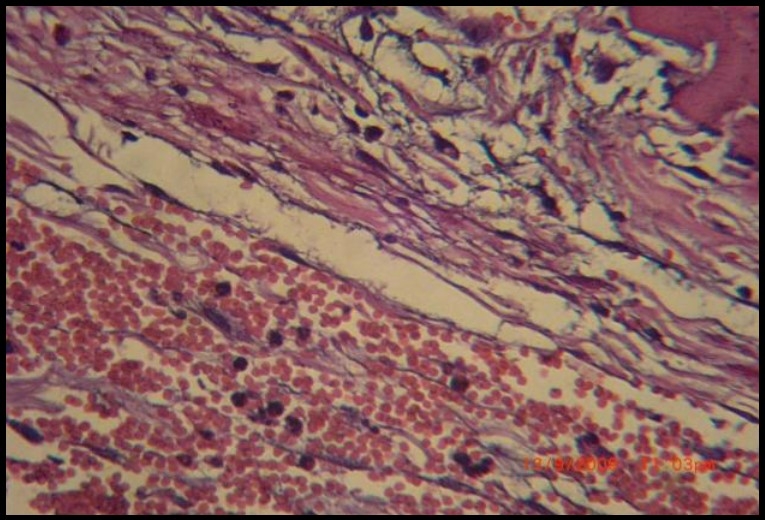
Control group at 3 days post-operation shown hemorrhage with inflammatory cell infiltration (H&E 40X).
Fig. 2.
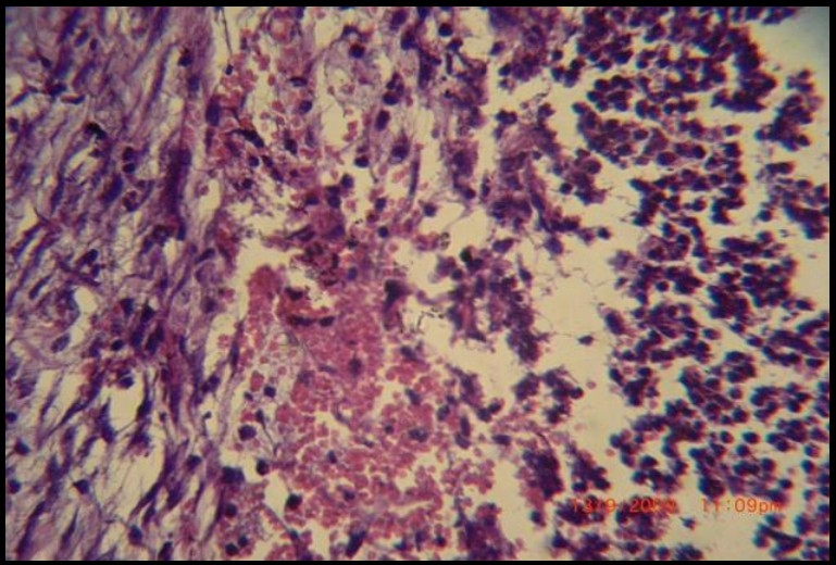
Control group at 7 day post operation shows the gap contains necrotized neutrophils with hemolysis RBC (H&E 40X).
Fig. 3.
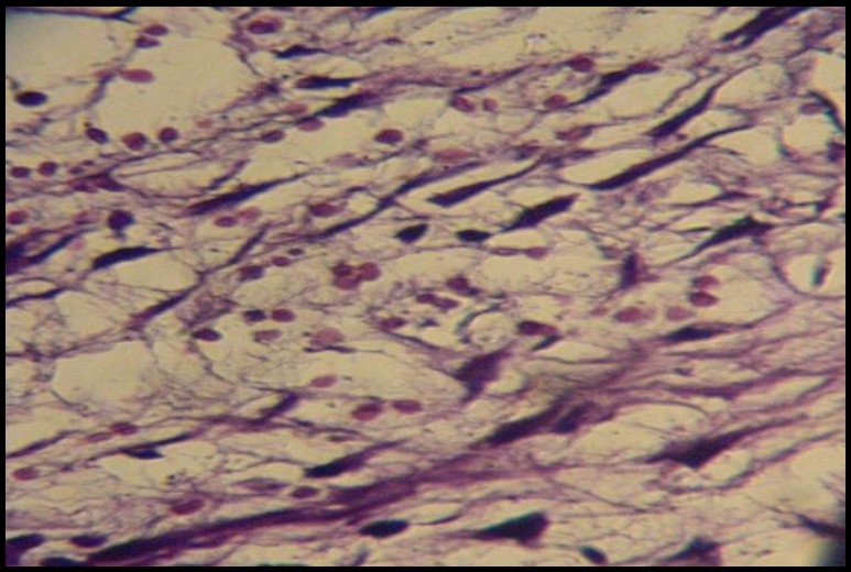
Control group at 14 days post operation shows irregular fibrous connective tissue proliferation with congested blood vessel as well as mononuclear cell infiltration(H&E 40X).
Fig. 4.
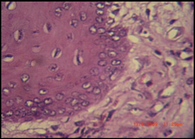
Control group at 14 days shows dense moderates cellular fibrous connective tissue with few mononuclear replaced the gap(D) and cover by very thickness cellular epidermal layer (H&E 40X).
In group II (treated), the 3rd day postoperative slide indicated severe infiltration of inflammatory cells, mainly neutrophils with proliferation of fibroblasts forming a few fibrous connective tissues (Figure 5). The lesion on the 7th day was characterized by granulation tissue, which formed from proliferated fibrous connective tissue and congested blood vessels and mononuclear cell infiltration filling the incision gap (Figure 6). The section prepared on the 14th day revealed debris material surrounded by a thick layer of connective tissue and dense collage, but less cellular fibrous connective tissue was present in the dermis covered by a thick epidermal layer (Figures 7 & 8).
Figure 5.
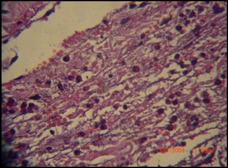
Treated group at 3 days post operation shows proliferation of fibroblast with few fibrous connective tissue as well as sever inflammatory cell infiltration (H&E 40X).
Fig. 6.
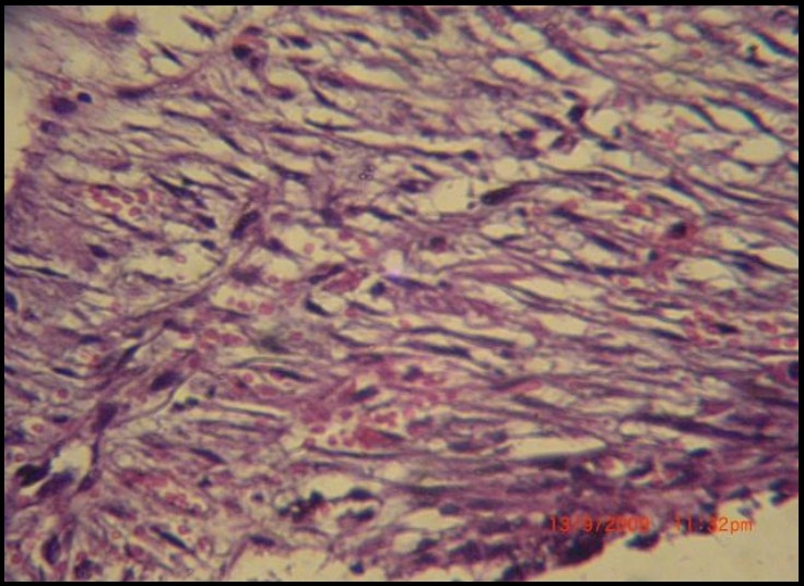
Treated group at 7days post operation shows severs granulation tissue with mononuclear cell infiltrated (H&E 40X).
Fig. 7.
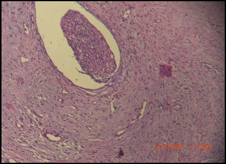
Treated group at 14 days post operation revealed space contains debris material surrounding by sever thickness of fibrous connective tissue (H&E 40X).
Fig. 8.
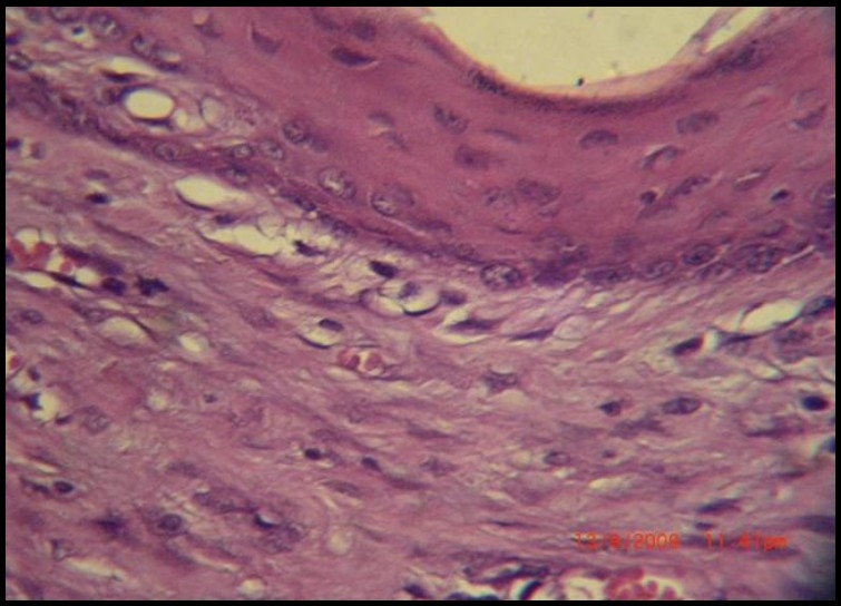
Treated group at14 days shows dense collagens, less cellular fibrous connective tissue present in the dermis covered by thick epidermal layer (H&E 40X).
In this group, the wound was completely closed and a skin layer was formed with less scar formation and shrinking of the size of the wound of up to 2 Cm. When compared to group I, the wound was not yet completely closed and scar formation was noted. There was no noticeable shrinkage in the size of the wound.
Discussion
The results showed clear promotion of healing in the treated group in comparison with the control group. The histopathological examination observed that the good response of this group may be related to stimulation of inflammatory cell or activation of the chemotactic factor by irradiation with this dose. This observation agreed with a previous study[5], in which by using LLLT, the high phagocytic activity of macrophages was observed as early as 6 hours. LLLT can facilitate wound healing, which may be due to acute inflammation is resolved more rapidly and the proliferation phase of healing begins earlier[6], therefore, the LLLT decreased the inflammatory reaction of wound healing.
During the healing stage, some studies revealed the presence of neutrophils at the site of injury at 48-96 hours and reached a peak in 72 hours[7,8]. These cells have an important role in healing during phagocytes; they then lyse and enter the cement material[7,8]. The results of these studies are in agreement with the current study.
In general, fibroblasts are known to be essential in the healing of tissue injuries including surgical wounds, the epithelialization and granulation tissue formation was created in the repair stage; fibroblasts begin to synthesize collagen and ground substances[3,9]. The laser-treated group in comparison with the other group showed higher numbers of fibroblast proliferation in the early stage. These observations confirm the results achieved by a previous study[4] that indicated a possibility of laser-induced fibroblast proliferation during healing mechanism. The effect of laser stimulation of fibroblasts on wound regeneration is by the maintenance of a high mitotic activity of the fibroblast in the later healing period[10], in which it was demonstrated that LLLT preferentially stimulates resting cells rather than proliferating ones.
The macroscopic and microscopic results in group II could be due to the increased collagen production by fibroblasts. This in turn affects LLLT in enhancing the job of mononuclear cells through production of leukotrien-B4, which is derived from arachnoid acid and production of interleukin-8, which promote fibroblasts[5]. These results are similar to those in this study.
The exact mechanism by which LLLT facilitates wound healing is largely unknown. However, several theories may help explain the enhanced wound contraction observed in this study. In vitro studies have shown an increase in fibroblast proliferation after irradiation, suggesting that LLLT therapy may facilitate fibroplasia during the repair phase of tissue healing. Our data support this suggestion. We did not observe differences between the LLLT and control groups until day 6, when wounds would have been well into the repair phase of soft tissue healing. However, it should also be noted that other investigators found no in vitro changes in fibroblast proliferation after LLLT. The disparity could be due to changes in specific laser irradiation settings, such as wavelength, duration, power, and intensity.
Facilitated wound contraction may also be supported by work from researchers who reported that laser irradiation transforms fibroblasts into myofibroblasts. Myofibroblasts are directly involved in granulation tissue contraction, and increased numbers could lead to facilitated wound contraction. A myofibroblast is a modified fibroblast with ultrastructural and functional properties of fibroblasts and muscle cells. Cytoplasmic fibrils of actomyosin allow for contraction of myofibroblasts, pulling on the borders of the wound and reducing the size during the repair phase of soft tissue healing. Because our data provided support that LLLT enhanced wound contraction but did not necessarily enhance other variables associated with superficial wound healing, myofibroblast stimulation may be a viable explanation.
In the present study, the histopathological examination revealed that the proliferation of the epithelial cells appeared in seven days postoperative in group II, which was faster than the control group. These results are in agreement with a study that reported that LLLT induced faster epithelialization that culminated in better formation of the epidermis with formation of normal epidermis and disappearance of the scar[11]. The enhancement of collagen syntheses that may be due to light energy was observed by endogenous chromophores in the mitochondria and used to synthesize ATP (adenosine triphosphatase) . The resulting ATP was then used to power metabolic processes, synthesize DNA, RNA, proteins, enzymes, and other biological materials needed to repair or regenerate cell and tissue components, rapid mitosis or cell proliferation and restore homeostasis [6,12]. Improved microvascular circulation, reduced inflammation and increased cell energy in the form of ATP may work together in low-powered laser irradiation to stimulate hair growth[13,14]. This agrees with that found in this study.
The treatment group II had greater wound contraction than the control group. Several theories may help explain the enhanced wound contraction observed in this study. In vitro studies have shown an increase in fibroblast proliferation after irradiation, suggesting that LLLT therapy may facilitate fibroplasia during the repair phase of tissue healing[15]. The LLLT is an effective treatment for enhancing wound contraction of partial thickness. It also facilitates wound contraction of untreated wounds suggesting an indirect effect on surrounding tissues. Although our data focused on enhanced contraction of superficial wounds, we believe this is the first step in formulating meaningful conclusions regarding LLLT.
Conclusion
The study found that LLLT (II) was effective in open wounds, showing better regeneration and faster healing with restoration of structural and functional integrity as compared to the control group.
References
- 1.Arun GM, Pramod K, Shivananda N. Photo-stimulatory effect of low energy helium-neon laser irradiation on excisional diabetic wound healing dynamics in Wister rats. Indian J Derm. 2009;54(4):323–329. doi: 10.4103/0019-5154.57606. [DOI] [PMC free article] [PubMed] [Google Scholar]
- 2.Karu T. Primary and secondary mechanisms of action of visible to near IR radiation on cells. J Photochem Photobiol. 1999;49:1–17. doi: 10.1016/S1011-1344(98)00219-X. [DOI] [PubMed] [Google Scholar]
- 3.Markolf HN. Laser tissue interaction. Fundamental and application. Berlin: Spinger Verlag; 2003. pp. 9–25. (45-147). [Google Scholar]
- 4.Baxter GD, Hopkins JT, Tood AM, Jeff GS. Low level laser therapy facilitates superficial wound healing in humans: A triple-blind, sham-controlled study. J Athletic Training. 2004;39(3):223–229. [PMC free article] [PubMed] [Google Scholar]
- 5.Petrova MB. The morphofunctional characteristics of the healing of skin wound in rats by exposure to low-intensity laser radiation. Morfologiia. 1992;102(6):112–121. [PubMed] [Google Scholar]
- 6.Mary D. Laser tissue repair, improving quality of life.Department of anatomy and cell biology UMDS Medical and Dental Schools, London. Nursing in practice. 2003;13:1. [Google Scholar]
- 7.Karu TI, Natalya IA, Sergey FK, Ljudmila V, Welser L. Changes in absorbance of monolayer of living cells induced by laser radiation at 633, 670 and 820nm. IEEE journal on Selected Topics in Quantum Electronics. 2001;7(6):982–988. [Google Scholar]
- 8.Pugliese LS, Medrado AP, Reis SR, Andrade ZA. The influence of low level laser therapy on biomodulation of collagen and elastic fibers. Pesqui Odontol. 2003;17(4):307–313. doi: 10.1590/s1517-74912003000400003. [DOI] [PubMed] [Google Scholar]
- 9.Chukuka SE. Laser photostimulation: An old mystery metamorphosing into a new millennium marvel. Laser Therapy, World Association of Laser Therapy. 2004 [Google Scholar]
- 10.John LS, Michael MD. Hair regrowth and increased hair tensile strength using the hair Max Laser Comb for Low Level Laser Therapy. (117-120).Inter J Cosmet Surg Aesthetic Derm. 2003;5(2):1133. [Google Scholar]
- 11.Toyokawa H, Matsui Y, Uhara J, Tsuchiya H, Teshima SH, Nakanishi H. Promotive effects of far-infrared ray on full thickness skin wound healing in rats. Experimental Biol Med. 2003;228:724–729. doi: 10.1177/153537020322800612. [DOI] [PubMed] [Google Scholar]
- 12.Ribeiro MA, Albuquerque RL, Barreto AL, Oliveira VG, Santos TB, Dantas CD. Morphological analysis of second-intention wound healing in rats submitted to 16 J/cm2 λ 660-nm laser irradiation. Indian J Dent Res. 2009;20(3):30–34. doi: 10.4103/0970-9290.57360. [DOI] [PubMed] [Google Scholar]
- 13.Sabine W, Richard G. Regulation of wound healing by growth factors and cytokines. Physical Rev. 2003;38:835–870. doi: 10.1152/physrev.2003.83.3.835. [DOI] [PubMed] [Google Scholar]
- 14.Huseyin D, Solmaz Y, Mehmet K, Kadir Y. Comparison of the effects of laser and ultrasound treatments on experimental wound healing in rats. J Rehab Res and Develop. 2004;41(5):721–728. doi: 10.1682/jrrd.2003.08.0131. [DOI] [PubMed] [Google Scholar]
- 15.Akio Y, Haruki O, Yoshihisa K, Masahiro N, Kazuo T. The effect of low reactive level laser therapy with helium - neon laser on operative wound healing in rat model. J Vet Med Sci. 2007;69(8):799–806. doi: 10.1292/jvms.69.799. [DOI] [PubMed] [Google Scholar]


