Abstract
Purpose
The goal of this article was to present the clinical and radiological results of 42 severe genu varum operated on between August 2001 and June 2010 using computer navigation.
Methods
All the osteotomies were navigated using the Orthopilot® device (B-Braun-Aesculap, Tuttlingen, Germany). The procedure was performed such that after inserting the rigid bodies and calibrating the lower leg, we first made the femoral closing wedge osteotomy (from four to seven mm) which was fixed by an AO T-Plate, and then, after checking the residual varus, the tibial opening wedge osteotomy was made using a Biosorb® wedge (Tricalcium phosphate, SBM, Lourdes, France) and a plate (AO T-plate or C-plate).
Results
All the patients were assessed at a mean follow-up of 46 ± 27 months (range, 12–108). The mean Lyshölm-Tegner score was 83.3 ± 7.5 points (62–91) and the mean KOOS score was 95.1 ± 3.2 points (89–100). Forty patients were satisfied (22) or very satisfied (18) with the result. Regarding the radiological results, the goal was reached in 92.7% of cases and the mean HKA angle was 181.83° ± 1.80° (177–185°). At that mid-term follow-up no patient had revision to a total knee arthroplasty.
Conclusion
Computer-assisted double level osteotomy in severe genu varum is a reliable, reproducible, and accurate technique. This procedure, which is very delicate, especially in reaching pre-operative objectives, is simplified by computer-assistance.
Keywords: Medicine & Public Health, Orthopedics
Introduction
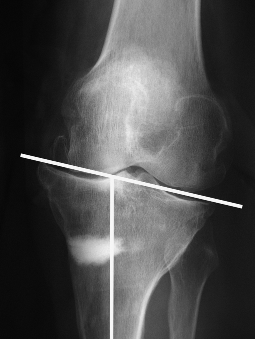
Medial knee osteoarthritis is not uncommon and high tibial osteotomy (HTO) was described for the first time more than 50 years ago [7, 9, 13]. Nowadays, HTO remains a good option [3–5, 8, 11, 17, 24, 27] despite the large expansion of total knee replacement (TKR) or the revival of unicompartmental knee prosthesis boosted by the less-invasive surgery concept. It is well indicated for "young" and active people (less than 65 years of age) with moderate arthrosis (narrowing joint line up to 100% without any bone wear or instability). Nevertheless, it is a demanding surgery with the risk of excessive over or under correction which can quickly lead to failure [8, 24, 26] or an oblique joint line leading to more difficulties in performing TKR (Fig. 1). This oblique joint line corresponds to an excessive valgus of the tibial mechanical axis [1]. It is all the more frequent when varus is substantial to have to decide whether to have to perform a femoral or a femoral and tibial correction. The desirable overcorrection to achieve a good clinical result (3–6°) increases the oblique joint line even more. When it reaches 10° of valgus one must often perform an osteotomy to set the tibial mechanical axis back to 90° [14] before implanting the prosthesis.
Fig. 1.
Severe oblique joint line after high tibial osteotomy. Notice the extreme tibial valgus
We thought for a long time that combined femoral and tibial osteotomy was a suitable procedure to avoid this drawback, but, because of the difficulty in obtaining an accurate lower leg axis without any reproducible assistance, we had performed it in only a few cases. Drawing on our experience with TKR and HTO navigation [15, 19, 20], we used the principles of computer-assisted surgery for double level osteotomy (DLO) hoping to increase the accuracy of this difficult procedure. Our experience is based on 42 DLO performed between August 2001 and June 2010 from 370 personal computer-assisted knee osteotomies for genu varum deformities (11.3%).
The objective of this article was to present the clinical and radiological results of these patients at a mean follow-up of 46 ± 27 months.
Material and methods
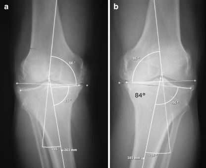
The series was composed of 38 patients (four bilateral), with nine females and 29 males aged from 39 to 64 years (mean age, 50.9 ± 7.1 years). We operated on 22 right knees and 20 left ones. The mean BMI was 29.3 ± 4.3 for a mean height of 171 cm and a mean weight of 85.8 kg. For functional assessment, we used the Lysholm-Tegner score [25] to evaluate patients, both pre-operatively and post-operatively. We felt this scoring system was better adapted than the IKS score which is usually used to evaluate surgical treatment for knee osteoarthritis. The mean score was 41.2 ± 8.9 points (22–69). According to the modified Ahlbäck criteria [21], we operated on nine stage 2, 25 stage 3, seven stage 4 and one stage 5. We measured HKA (hip-knee-ankle) angle using Ramadier’s protocol [16] and we also measured the medial distal femoral mechanical axis (MDFMA) and the medial proximal tibial mechanical axis (MPTMA) to ensure the right indication [23]. These measures were respectively: 167.7° ± 3.5° (159–172°), 87.28° ± 1.41° (83–90°) for the MDFMA and 83.51° ± 2.7° (78–88°) for the MPTMA.
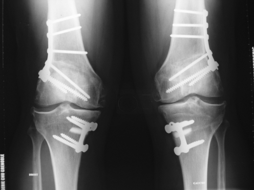
The inclusion criteria were a patient younger than 65 years old with a severe varus deformity (more than 8°; HKA angle ≤ to 172°) and a MDFMA at 91° or less (Fig. 2a, b). All the osteotomies were navigated using the Orthopilot® device (B-Braun-Aesculap, Tuttlingen, Germany). The procedure was performed as described previously [23]; after inserting the rigid-bodies and calibrating the lower leg, we first made the femoral closing wedge osteotomy (from 4 to 7 mm) which was fixed by an AO T-Plate (Fig. 3a, b), and then, after checking the residual varus, the tibial opening wedge osteotomy was made using a Biosorb® wedge (β Tricalcium phosphate, SBM, Lourdes, France) and a plate (AO T-plate or C-plate). The goals of the osteotomy were to achieve an HKA angle of 182° ± 2° and a MPTMA angle of 90° ± 2°. The functional results were evaluated not only according to the Lyshölm-Tegner score [25] but also to the KOOS score [18]. The patients answered the questionnaire at revision or by phone, and the radiological results were assessed by plain radiographs and standing long leg X-rays between three and six months post-operatively.
Fig. 2.
Bilateral severe genu varum. a Measurements of the right knee: HKA angle, MDFMA and MPTMA. b Measurements of the left knee
Fig. 3.
Bilateral DLO of the case of Fig. 2 (one year follow-up)
Results
We had no complications in this series but one case of recurrence of the deformity related to an impaction of the femoral osteotomy on the medial side (heavy patient). Two patients were lost to follow-up after removal of the plates (24 months) but were included in the results because the file was complete at that date. All the patients were assessed at a mean follow-up of 46 ± 27 months (12–108).
The mean Lyshölm-Tegner score was 83.3 ± 7.5 points (62–91) and the mean KOOS score was 95.1 ± 3.2 points (89–100). Forty patients were satisfied (22) or very satisfied (18) of the result. Only two were poorly satisfied.
Regarding the radiological results, if we exclude the patient who had a loss of correction not related to navigation, the goals were reached in 39 cases (92.7%) for the HKA angle and in 36 cases (88.1%) for the MPTMA with only one case at 93°. The mean angles were 181.83° ± 1.80° (177–185°) for HKA, 89.71° ± 1.72° (85–93°) for MPTMA and 92.76° ± 2.02° (89–97°) for MDFMA.
At that mid-term follow-up no patient had revision to a total knee arthroplasty.
Discussion
Combined distal femoral and proximal tibial osteotomy in the treatment of genu varum is technically difficult. Little has been said about this technique in the literature and we could find only one paper reporting on it [1]. It seems that this technique was first described by Benjamin [2] in 1969 for the treatment of rheumatoid arthritis of the knee, but at the time, he did not mention any HKA angle or joint line obliquity. In their paper, Babis et al. [1] reported on 24 patients (29 knees) operated on with a conventional technique (two closing wedge osteotomies). The mean pre-operative HKA angle was 193.3° (that is 13.3° of varus), and they used a computer-aided analysis of the mechanical status of the knee for pre-operative planning. This was limited to pre-operative evaluation, and the reliability of the pre-operative radiographic evaluation was not assessed. The results showed a mean post-operative HKA angle of 176.9° (169.4–184.9°). They had a residual varus in two cases (4.6–4.9°) and an over correction of more than 4° in ten cases and more than 6° in five. One knows that an under correction may lead to failure of the operative procedure and a too much overcorrection to cosmetic discomfort.
The difficulty of the technique comes from the fact that once the first osteotomy is performed, whether femoral or tibial, landmarks change and the ability to achieve a satisfactory alignment with the second osteotomy becomes challenging in the absence of reliable intra-operative landmarks. Martres et al. [12] suggested performing this operation in two different stages to improve its accuracy and reproducibility. It is also justified to consider that complication occurring at both osteotomy sites could lead to disastrous results. In our series we had no non-union and only one mal-union related to a secondary medial impaction of the femoral osteotomy in a heavy patient. Currently, we use a locking plate in spite of an AO T-plate, which could avoid this complication. On the other hand, every surgeon operating osteoarthritic knees should be aware of the risk of mal-union in the proximal tibia, for a procedure that is often considered temporary. In fact every osteotomy in a young adult is susceptible to lead subsequently to a TKR, and thus it is essential to plan ahead for the iterative surgery called revision.
Computer-assistance allows controlling the femoro-tibial axis (HKA angle) at every step of the procedure and thus makes it more accurate. Our present results are not far from a previous preliminary series [22] and argue in favour of a high reproducibility of this procedure. From a clinical point of view, the mean Lyshölm-Tegner score improved from 41.2 ± 8.9 points to 83.3 ± 7.5 points and the mean KOOS score was of 95.1 ± 3.2 points. These clinical results are remarkable and the satisfaction of the patients is very high (95% of the patients satisfied or very satisfied). At mid-term follow-up no patient was revised to TKR or to another osteotomy. This issue could be related not only to the accurate correction—good over correction and no oblique joint line—but also to the vascular effect of double osteotomy at each side of the joint line.
When should double level osteotomy be performed? If we consider the "normal" mechanical axis of the lower limb as described by Kapandji [10] and later taken up by Hungerford and Krackow [6] it should be 180° with an MDFMA of 93° and an MPTMA of 87° resulting in a joint line perfectly parallel to the ground. However, this assumption is not confirmed in cases of osteoarthritis with varus misalignment, because, in a personal unpublished series of 89 TKR, we found an MDFMA of 93° in only 43.8% of cases, at 90° in 33.7% of the cases, below 90° in 13.5% and above 93° in 9%.
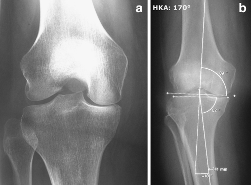
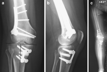
Thus, before performing high tibial osteotomy, it is crucial to have high quality and reproducible full-length AP radiographs of the lower limb, according to a specific protocol [23]. The HKA angle, the MDFMA and the MPTMA should be determined on this goniometry (Fig. 4a, b). Lateral instability testing has become less important than it once was, since the indications for osteotomy in this setting have become rare. In cases of femoral valgus (MDFMA > 90–91°), it is illogical to perform a femoral osteotomy because we do not want to create in the femur, the error, we are trying to avoid in the tibia. If the femur is in varus or at 90°, we believe that we should proceed with a femoral osteotomy to achieve an MDFMA of around 93° (93° ± 2°), and then complete it with a tibial osteotomy to achieve an HKA angle of 182° ± 2°. In our experience, it is useless to overcorrect more than this to obtain satisfactory results (Fig. 5a, b, c). Overcorrection, whether femoral or tibial, can distort the anatomy and lead to a much more complicated revision TKR. Our mid-term results have a trend to confirm this assertion. However, we think that a longer follow-up is needed to prove that overcorrection by ±2° is enough for a lasting good result. If the tibia is not in varus (MPTMA over 88°), we should perform a femoral osteotomy especially if the femur is at 90° or in varus, or contraindicate any osteotomy if it leads to joint line obliquity of more than 5°. If we stick strictly to these criteria, indications for double level osteotomy will likely increase with the development of navigation systems, especially since, as we said before, femurs in varus are not rare, and more so, those at 90°.
Fig. 4.
A 48-year-old woman with severe genu varum deformity (HKA angle = 170°) associated with a stage 2 medial femoro tibial osteoarthritis. a AP Rosenberg view. b Standing long leg X-rays according to Ramadier’s protocol
Fig. 5.
Radiological result at one year of the case of Fig. 4. AP (a) and lateral (b) X-rays. To notice the medial tibial mechanical axis and the absence of oblique joint line. c Standing long-leg X-ray. Note HKA angle (183°)
Finally, despite our trust in opening wedge osteotomies [24], we think that, at the femoral level, one should perform a closing wedge osteotomy to avoid excessive lengthening of the limb (double opening).
Conclusion
According to these results, computer-assisted double level osteotomy in severe genu varum is a reliable, reproducible, and accurate technique. This procedure, which is very delicate, especially in reaching pre-operative objectives, is simplified by computer-assistance. The functional results are satisfying and the satisfaction of the patients is very high. Despite the difficulty of the procedure, complications are, in our hands, very rare. We recommend DLO for severe genu varum deformity in young patients to avoid oblique joint line, which will be difficult to revise to TKR.
References
- 1.Babis GC, An KN, Chao EYS, et al. Double level osteotomy of the knee: a method to retain joint-line obliquity. J Bone J Surg Am. 2002;84:1380–1388. doi: 10.2106/00004623-200208000-00013. [DOI] [PubMed] [Google Scholar]
- 2.Benjamin A. Double osteotomy for the painful knee in rheumatoid arthritis and osteoarthritis. J Bone J Surg Br. 1969;51:694–699. [PubMed] [Google Scholar]
- 3.Coventry MB, Ilstrup DM, Wallrichs SL. Proximal tibial osteotomy: a critical long-term study of eighty-seven cases. J Bone Joint Surg Am. 1993;75:196–201. doi: 10.2106/00004623-199302000-00006. [DOI] [PubMed] [Google Scholar]
- 4.Flecher X, Parratte S, Aubaniac JM, Argenson JN. A 12–28-year follow-up study of closing wedge high tibial osteotomy. Clin Orthop Relat Res. 2006;452:91–96. doi: 10.1097/01.blo.0000229362.12244.f6. [DOI] [PubMed] [Google Scholar]
- 5.Hernigou Ph, Medevielle D, Debeyre J, et al. Proximal tibial osteotomy for osteoarthritis with varus deformity: a ten to thirteen-year follow-up study. J Bone Joint Surg Am. 1987;69:332–354. [PubMed] [Google Scholar]
- 6.Hungerford DS, Krackow KA. Total joint arthroplasty of the knee. Clin Orthop Relat Res. 1985;192:23–30. [PubMed] [Google Scholar]
- 7.Jackson JP, Waugh W. Tibial osteotomy for osteoarthritis of the knee. J Bone Joint Surg Br. 1961;43:746–751. doi: 10.1302/0301-620X.43B4.746. [DOI] [PubMed] [Google Scholar]
- 8.Jenny JY, Tavan A, Jenny G, et al. Taux de survie à long terme des ostéotomies tibiales de valgisation pour gonarthrose. Rev Chir Orthop. 1998;84:350–357. [PubMed] [Google Scholar]
- 9.Judet R, Dupuis JF, Honnard F, et al. Désaxations et arthroses du genou. Le genu varum de l’adulte. Indications thérapeutiques, résultats. Rev Chir Orthop. 1964;13:1–28. [Google Scholar]
- 10.Kapandji IA. Physiologie articulaire. Fascicule II quatrième édition: membre inférieur. Paris: Maloine SA; 1974. p. 104. [Google Scholar]
- 11.Lootvoet L, Massinon A, Rossillon R, et al. Ostéotomie tibiale haute de valgisation pour gonarthrose sur genu varum: à propos d’une série de 193 cas revus après 6 à 10 ans de recul. Rev Chir Orthop. 1993;79:375–384. [PubMed] [Google Scholar]
- 12.Martres S, Servien E, Aït Si Selmi T, et al. Double ostéotomie: indication dans la gonarthrose. Rev Chir Orthop. 2004;90(suppl n°6):2S137–2S138. [Google Scholar]
- 13.Merle d’Aubigné R, Ramadier JO. Arthrose du genou et surcharge articulaire. Acta Orthop Belg. 1961;27:365–375. [Google Scholar]
- 14.Neyret Ph, Deroche Ph, Deschamps G, et al. Prothèse totale du genou après ostéotomie tibiale de valgisation. Problèmes techniques. Rev Chir Orthop. 1992;77:438–448. [PubMed] [Google Scholar]
- 15.Picard F, Leitner F, Raoult, Saragaglia D. Computer assisted knee arthroplasty. In: Jerosch J, Nichol K, Peikenkam K, editors. Reschnergestützte Verfahren in Orthopädie und Unfallchirurgie. Darmstadt, Germany: Steinkopff Verlag; 1999. pp. 461–471. [Google Scholar]
- 16.Ramadier JO, Buard JE, Lortat-Jacob A, et al. Mesure radiologique des déformations frontales du genou. Procédé du profil vrai radiologique. Rev Chir Orthop. 1982;68:75–78. [PubMed] [Google Scholar]
- 17.Rinonapoli E, Mancini GB, Corvaglia A, et al. Tibial osteotomy for varus gonarthrosis. A 0 to 21-year follow-up study. Clin Orthop Relat Res. 1998;353:185–193. doi: 10.1097/00003086-199808000-00021. [DOI] [PubMed] [Google Scholar]
- 18.Roos EM, Roos HP, Ekdahl C, Lohmander LS. Knee injury and osteoarthritis outcome score (KOOS): validation of a Swedish version. Scand J Med Sci Sports. 1998;8:439–448. doi: 10.1111/j.1600-0838.1998.tb00465.x. [DOI] [PubMed] [Google Scholar]
- 19.Saragaglia D, Picard F, Chaussard C, et al. Mise en place des prothèses totales du genou assistée par ordinateur: comparaison avec la technique conventionnelle. Résultats d’une étude prospective randomisée de 50 cas. Rev Chir Orthop. 2001;87:18–28. [PubMed] [Google Scholar]
- 20.Saragaglia D, Pradel Ph, Picard F (2004) L’ostéotomie de valgisation assistée par ordinateur dans le genu varum arthrosique: résultats radiologiques d’une étude cas-témoin de 56 cas. E-mémoires de l’Académie Nationale de Chirurgie 3:21–25. Available at: http://www.bium.univ-paris5.fr/acad-chirurgie
- 21.Saragaglia D, Roberts J. Navigated osteotomies around the knee in 170 patients with osteoarthritis secondary to genu varum. Orthopaedics. 2005;28(Suppl 10):S1269–S1274. doi: 10.3928/0147-7447-20051002-13. [DOI] [PubMed] [Google Scholar]
- 22.Saragaglia D, Rubens-Duval B, Chaussard C. Double ostéotomie assistée par ordinateur dans les grands genu varum. Résultats préliminaires à propos de 16 cas. Rev Chir Orthop. 2007;93:351–356. doi: 10.1016/s0035-1040(07)90276-7. [DOI] [PubMed] [Google Scholar]
- 23.Saragaglia D, Mercier N, Colle PE. Computer-assisted osteotomies for genu varum deformity: which osteotomy for which varus? Int Orthop. 2010;34:185–190. doi: 10.1007/s00264-009-0757-6. [DOI] [PMC free article] [PubMed] [Google Scholar]
- 24.Saragaglia D, Blaysat M, Inman D, Mercier N. Outcome of opening wedge high tibial osteotomy augmented with a Biosorb® wedge and fixed with a plate and screws in 124 patients with a mean of ten years follow-up. Int Orthop. 2011;35:1151–1156. doi: 10.1007/s00264-010-1102-9. [DOI] [PMC free article] [PubMed] [Google Scholar]
- 25.Tegner Y, Lysholm J, Lysholm M, Gillquist J. A performance test to monitor rehabilitation and evaluate anterior cruciate ligament injuries. Am J Sports Med. 1986;14:156–159. doi: 10.1177/036354658601400212. [DOI] [PubMed] [Google Scholar]
- 26.Tunggal JAW, Higgins GA, Waddell JP. Complications of closing wedge high tibial osteotomy. Int Orthop. 2010;34:255–261. doi: 10.1007/s00264-009-0819-9. [DOI] [PMC free article] [PubMed] [Google Scholar]
- 27.Yasuda K, Majima T, Tsuchida T, et al. A ten to 15 year follow-up observation of high tibial osteotomy in medial compartment osteoarthrosis. Clin Orthop Relat Res. 1992;282:186–195. [PubMed] [Google Scholar]


