Abstract
Epizootic ulcerative syndrome (EUS) is a fish disease of international significance and reportable to the Office International des Epizootics. In June 2010, bony herring Nematalosa erebi, golden perch Macquaria ambigua, Murray cod Maccullochella peelii and spangled perch Leiopotherapon unicolor with severe ulcers were sampled from the Murray-Darling River System (MDRS) between Bourke and Brewarrina, New South Wales Australia. Histopathology and polymerase chain reaction identified the fungus-like oomycete Aphanomyces invadans, the causative agent of EUS. Apart from one previous record in N. erebi, EUS has been recorded in the wild only from coastal drainages in Australia. This study is the first published account of A. invadans in the wild fish populations of the MDRS, and is the first confirmed record of EUS in M. ambigua, M. peelii and L. unicolor. Ulcerated carp Cyprinus carpio collected at the time of the same epizootic were not found to be infected by EUS, supporting previous accounts of resistance against the disease by this species. The lack of previous clinical evidence, the large number of new hosts (n = 3), the geographic extent (200 km) of this epizootic, the severity of ulceration and apparent high pathogenicity suggest a relatively recent invasion by A. invadans. The epizootic and associated environmental factors are documented and discussed within the context of possible vectors for its entry into the MDRS and recommendations regarding continued surveillance, research and biosecurity are made.
Introduction
The emergence and spread of aquatic freshwater diseases are a major conservation concern [1]. One aquatic disease implicated in mass mortalities of cultured and wild fish in many countries is epizootic ulcerative syndrome (EUS) [2]. Also known as mycotic granulomatosis, red spot disease and ulcerative mycosis, EUS is caused by the fungus-like oomycete Aphanomyces invadans ( = A. piscicida) and can cause significant ulceration of the skin, necrosis of muscle with extension to subjacent structures including abdominal cavity and cranium, and leading to mortality in many cases [2]–[4]. Originally described in cultured ayu Plecoglossus altivelis in Japan [5], within three decades EUS had been reported in more than 100 fish species [6] in both freshwater and estuarine environments throughout south, south-eastern and western Asia [7]–[9], the east coast of North America [10], [11], in distinct regions of Australia: including New South Wales (NSW), Northern Territory, Queensland and Western Australia [3] and recently in Africa [12]. Due to concern over its potential impact on cultured and wild fisheries, EUS is officially recognised as a reportable disease by the Network of Aquaculture Centres in Asia-Pacific (NACA) and internationally by the World Organisation for Animal Health (Office International des Epizooties or OIE) [13].
Little is known about the infectious diseases of native fish in the Murray-Darling River System (MDRS), which drains inland catchments, west of the Great Dividing Range in south-eastern Australia. Until recently (2001 in an aquaculture facility), EUS had not been reported from the MDRS, and within Australia was generally considered endemic only to coastal drainages. Originally referred to as Bundaberg Fish Disease, EUS was first reported in Australia in 1972 [14], and subsequently there have been numerous outbreaks reported in wild freshwater and estuarine fishes in the eastern, northern and western coastal drainages [15]–[18].
EUS has been reported in silver perch Bidyanus bidyanus (Mitchell, 1838), a species endemic to the MDRS, being farmed in coastal drainages in northern NSW and south-eastern Queensland [19]. More recently, there have been reports of the presence of EUS within the MDRS, although little is known of its pathogenicity, distribution or susceptibility of species to infection in wild populations in this river system. In May 2008, A. invadans was isolated from bony herring Nematalosa erebi (Günther, 1868) immediately upstream of Bourke town weir on the Barwon-Darling River, representing the first confirmed, reported diagnosis of EUS in the MDRS, and significantly extending the known range of this pathogen within Australia (J. Go, Elizabeth Macarthur Agricultural Institute, unpubl. data).
During routine sampling of fish in the same section of the Barwon-Darling River in June 2010, dermal lesions and ulcers were observed in six species of fish. Many of the lesions and ulcers appeared to be characteristic of EUS, raising the concern of an epizootic of this reportable disease. As a member country of the OIE, Australia is obliged to report any significant new incursions of EUS, whether it is in a new species or a new area. This paper is the first published account of EUS in four native species: bony herring, golden perch Macquaria ambigua (Richardson, 1845), Murray cod M. peelii and spangled perch Leiopotherapon unicolor (Günther, 1859). The objectives are three-fold. Firstly to document the 2010 epizootic and report its prevalence and extent compared to the time of its first report in N. erebi in 2008. Secondly, to document some of the environmental factors associated with the epizootic. Finally, potential explanations of the route of introduction of A. invadans into the MDRS are suggested and discussed, with recommendations made for future surveillance, research and biosecurity.
Results
Diagnosis of EUS
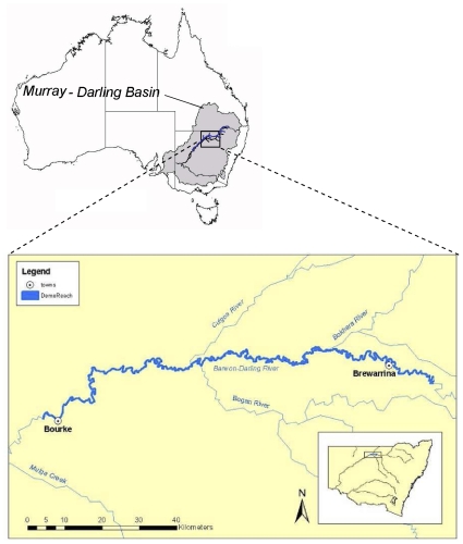
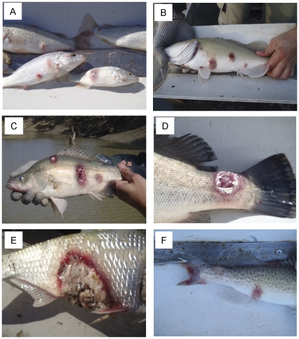
In June 2010, N. erebi, M. ambigua, M. peelii, L. unicolor, carp C. carpio and goldfish C. auratus caught from a 200 km section of the Barwon-Darling River between Bourke and Brewarrina weirs (Figure 1) had raised lesions and mild to severe ulcers characteristic of the invasive, tissue-destructive stages of EUS (Figure 2A–F). In some fish the caudal peduncle, caudal fin or dorsal fins were severely eroded (Figure 2D,F), and in others, deep ulcers penetrated into and exposed the peritoneal cavity (Figure 2E).
Figure 1. Section of the Barwon-Darling River between Bourke and Brewarrina weirs where fish with EUS were collected.
Figure 2. Diseased fish collected from the Barwon-Darling River between Bourke and Brewarrina weirs in June 2010.
Panels show A) L. unicolor, B) M. peelii with raised lesions, C,D) M. ambigua and F) M. peelii with deep ulceration and muscle or fin necrosis, and E) N erebi showing severe ulceration and tissue necrosis exposing the peritoneal cavity and internal organs.
Aphanomyces invadans was detect as being present in N. erebi, M. ambigua, M. peelii and L. unicolor using histopathology, and the diagnosis was further confirmed in N. erebi and M. ambigua using PCR. Although a small number of C. carpio and C. auratus were sampled with distinct haemorrhagic, dermal lesions, histopathology and PCR (PCR performed on C. carpio only) confirmed that these were not consistent with the case definition of A. invadans [2], [6], [20]. PCR was not performed on L. unicolor and M. peelii.
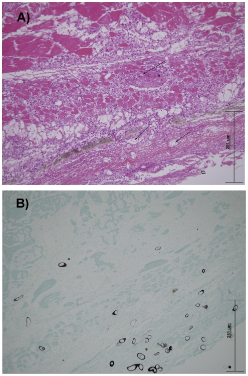
Gross observations were consistent in all of the confirmed cases, with focal to multifocal cutaneous ulceration of varying degrees of severity and distribution. Histologically, there was extensive necrosis and ulceration of the epidermis with adjacent epithelial hyperplasia. Subjacent myofibres were, in most cases, severely necrotic, with extensive myofibrillar liquefaction. The endomysium was infiltrated with moderate numbers of histiocytes, lymphocytes and plasma cells, with lesser granulocytes. Distinct, thin sheaths of macrophages (linear granulomas) were frequently observed, surrounded by a narrow band of lymphocytes and plasma cells (Figure 3A). In all cases, myriad fungal hyphae were associated with the ulcers, infiltrating into the myofibres and surrounding connective tissue. Cross sectioned hyphae varied from 10–35 µm in width, non-septate, thick-walled, with occasional branching. The morphology of hyphae was accentuated with GMS staining (Figure 3B). Typically, abundant fibrinocellular debris was associated with the eroded surfaces. In some L. unicolor and N. erebi, there were multiple infiltrative granulomas associated with fungal hyphae within internal organs, including kidneys, abdominal adipose tissue, ovary and swim bladder.
Figure 3.
A) N. erebi, skin and underlying muscle. Photo micrograph of developing linear granulomas (thin arrow) surrounding faintly eosinophilic fungal hyphae (*). The overlying epithelium is ulcerated ( ) H & E. (X200). B) N. erebri, skin and underlying muscle. Photo micrograph of black staining longitudinal and cross sectional fungal hyphae (*) against green stained tissue. GMS. (X200).
Nucleic acid detection of A. invadans
PCR performed on tissue samples detected A. invadans DNA in three of three N. erebi and three of four M. ambigua samples. A negative PCR result was obtained for one C. carpio analysed.
Difference in the prevalence of ulcerated fish between 2008 and 2010
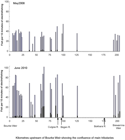
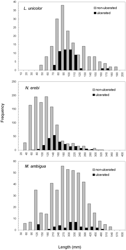
In 2008, ulcerated fish representing four species were sampled at 13 of the 30 locations over 200 km from Bourke Weir to Brewarrina Weir, although only N. erebi were submitted for histopathology testing and subsequently confirmed to have EUS (Jeffery Go, unpublished data). By comparison, in 2010 six species with ulcers were sampled from 29 of the 30 locations (Table 1; Figure 4). The prevalence of cutaneous lesions and/or ulcers was 2% (of all fish sampled) in 2008, but 10% in 2010 (Table 1). No N. erebi, L. unicolor and M. ambigua <60 mm had lesions or ulcers (Figure 5). Ulcerated L. unicolor ranged in length from 70–170 mm (n = 50), N. erebi 60–320 mm (n = 216), and M. ambigua 120–480 mm (n = 40). Most ulcerated L. unicolor were in the range 70–130 mm, and a majority of N. erebi over 140 mm were ulcerated (Figure 5).
Table 1. Number of ulcerated fish from the assemblage of species collected from 30 locations in the Barwon-Darling River between Bourke and Brewarrina weirs in May 2008 and June 2010.
| Common name | Scientific name | May 2008 | June 2010 |
| Olive perchlet | Ambassis agassizii (Steindachner, 1866) | - | - |
| Silver perch | Bidyanus bidyanus | - | - |
| goldfish | Carassius auratus | - | 2 (<1%) |
| Un-specked hardyhead | Craterocephalus stercusmuscarum fulvus (Ivantsoff, Crowley & Allen, 1987) | - | - |
| Carp | Cyprinus carpio | 7 (3%) | 7 (<1%) |
| Mosquito fish | Gambusia holbrooki (Girard, 1859) | - | - |
| Carp gudgeon | Hypseleotris spp. | - | - |
| Spangled perch | Leiopotherapon unicolor | 3 (8%) | 50 (21%) |
| Golden perch | Macquaria ambigua | 3 (2%) | 40 (8%) |
| Murray cod | Maccullochella peelii | - | 4 (5%) |
| Murray-Darling rainbowfish | Melanotaenia fluviatilis (Castelnau, 1878) | - | - |
| Bony herring | Nematalosa erebi | 28 (<1%) | 216 (16%) |
| Hyrtl's tandan | Neosiluris hyrtlii (Steindachner, 1867) | - | - |
| Australian smelt | Retropinna semoni (Weber, 1895) | - | - |
| Total number individuals ulcerated | 41 (2%) | 319 (10%) | |
| Total number of species ulcerated | 4 (29%) | 6 (32%) | |
Data in parentheses are the proportion of sampled individuals of each species that were ulcerated. Bold type identifies species in which EUS was confirmed using histopathology. No data means that the species was caught but no specimens were ulcerated.
Figure 4. Distribution of fish in the Barwon-Darling River between Bourke and Brewarrina weirs with ulcers (black) and without ulcers (grey) in 2008 and 2010.
Figure 5. Length frequency of ulcerated and non-ulcerated fish collected from the Barwon-Darling River between Bourke and Brewarrina weirs in June 2010.
Environmental conditions
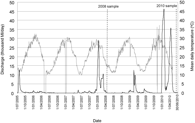
The epizootics occurred in autumn (2008) and winter (2010) with water temperatures below 16°C and decreasing (Figure 6). In both years, detection of ulcerated fish occurred within two months of significant within-channel flow events (equivalent to between four and eight percentile flows at the Bourke gauge), after an extended period of low-flow conditions (Figure 6). Water quality variables monitored at the time of fish sampling (i.e. once lesions/ulcers were already established) were: temperature 12.7–16.3°C; pH 7.3–8.7; electrical conductivity 350–600 µs.cm−1 (electrical conductivity was not significantly different from the five year average obtained from the Bourke gauge; Student t-test, d.f. 79, p = 0.593); and dissolved oxygen 4.2–10.6 mg.L−1. Dissolved oxygen concentrations were not below acceptable trigger values for aquatic ecosystems for lowland rivers in south eastern Austrlaia [21].
Figure 6. Daily discharge (black line) and mean daily water temperature (grey line) recorded at the Bourke gauge (425003) between July 2005 and July 2010; dotted lines show when the 2008 and 2010 fish samples were collected.
Source: NSW Office of Water.
Discussion
Hosts and distribution
Gross observations and histopathology identified EUS as the cause of the recent epizootic in the Barwon-Darling River in 2010. This is the first published account of EUS in N. erebi and first confirmed case of the disease in the native species M. ambigua, M. peelii and L unicolor in the wild. The findings are consistent with the earlier unpublished reports of EUS in N. erebi in the Barwon-Darling River in 2008 (Jeffery Go, unpublished data). This increases the number of native fish in the MDRS known to be susceptible to EUS to five, with B. bidyanus previously being shown to carry the disease under some culture conditions [19]. Additionally, there has recently (January 2010) been unpublished confirmation of EUS in farmed M. peelii at a facility on the Murray River (Brett Ingram and Tracey Bradley, pers. comm). Prior to this, the disease had never been reported in wild or cultured M. peelii [22].
Carasius auratus are known to be susceptible to EUS [23], however, we found no histopathological evidence that A. invadans was the causative pathogen in the two ulcerated C. auratus sampled during the recent epizootic. Similarly, despite the abundance of C. carpio (n = 953) sampled in the study area during the epizootic, and the observation of seven ulcerated individuals, there was similarly no evidence that A. invadans was the causative pathogen for the individuals examined. This is consistent with current evidence through Asia and Europe that C. carpio is resistant to EUS [6], [24], [25].
Prior to 2008, all previous reports of EUS in Australia were from coastal drainages from Bundaberg in Queensland to the Hawkesbury River in NSW [6], [14], [16], [18], [19], [26]. Following the first report of red spot disease in Australia near Bundaberg in Queensland in 1972 [14], numerous outbreaks followed during the 1980s, and the distribution of the disease expanded south to include the Clarence and Richmond rivers in NSW [16], [18], [26]. Elsewhere in the world, EUS has spread rapidly throughout south-east Asia and extended deep into the Indian sub-continent since the mid 1980s [27]. These and more recent reports from Africa, Zambia and the USA [12], [28]–[30] demonstrate the extent and ease at/with which this disease can spread.
EUS is a very invasive disease, and when it first occurs in an area, there are high levels of mortality over a very short time, many susceptible species are affected, and individual fish can have numerous lesions and ulcers [27], [31]. The severity and prevalence of this recent epizootic and lack of previous reporting suggests that EUS may be a recent incursion into the MDRS. More than 100 fish species have been reported to be affected by EUS [6], and susceptibility varies between species, with some, including tilapia Oreochromis spp. (Castelnau, 1861) resistant to infection by A. invadans [24], [25], [32], [33]. Resistance to EUS in tilapia is of significance to the Murray-Darling River System as this species currently poses a significant invasion risk in northern catchments [34]. The severity of ulceration that we observed in N. erebi, M. ambigua and M. peelii suggest that these species are very sensitive to infection by A. invadans.
The current geographic distribution of EUS in the MDRS remains largely unclear. The epizootic reported in this study covers a 200 km section of the Barwon-Darling River in the northern region of the Murray-Darling Basin. During publication of this paper, EUS has also been confirmed (histopathologically) in two M. ambigua sampled (in July 2011) below Lock 7 in the Murray River, a site downstream of the confluence with the Darling River (M. Gabor and D. Gilligan, NSW Department of Primary Industries, unpubl. data). This, and the previously mentioned report of infected M. peelii on a fish farm near the Murray River, confirms that this disease is now widespread in the Murray-Darling River system.
Predisposing environmental factors
Exposure to A. invadans spores is a key factor causing EUS [2], and the incidence and transmission of this pathogen throughout the MDRS will largely determine the potential range this disease. Once a pathogen is in an area, subsequent outbreaks of infectious diseases in fishes are closely linked to environmental conditions, particularly temperature and other water quality variables through their effects on stress and the immune system [35]–[37]. The prevalence of EUS in four species in the Barwon-Darling River in 2010 suggests that conditions were conducive to initial infection and transmission of the pathogen within and between species [2]. Caution must be exercised when interpreting the significance of the water quality measurements taken during this study, as they were taken only at the time of sampling, when fish were ulcerated and the epizootic well advanced. Nevertheless, it is prudent to document the environmental variables associated with all epizootics, to facilitate the development of causal links should more data become available from future outbreaks.
Both outbreaks in 2008 and 2010 were detected at relatively low water temperatures (<16.3°C) following periods of very high flow and flooding. This is consistent with most outbreaks of EUS, which tend to be associated with low and declining water temperatures and high rainfall [6], [9], [25], [33]. Virgona [16] reported significant correlation between rainfall and the prevalence of early stage lesions in sea mullet Mugil cephalus (L., 1758), and found that progression to later stage ulcers occurred after the high flows. Outbreaks of EUS in cultured B. bidyanus at the Grafton Aquaculture Centre occur only when water is pumped from the Clarence River during high flows or floods, and when fish in the river are known to have EUS [38].
Temperature is a critical factor determining the severity of EUS outbreaks and most mortalities occur when water temperatures are relatively low [39]. The findings in this paper suggest that high flows and low temperatures may have been predisposing factors to the outbreaks in 2008 and 2010. Low water temperatures (<16°C) and rapid decreases in temperature are immunosuppressive and induce changes to the epidermis, including loss of mucus that predispose fish to infection [37], [40]. Outbreaks of the fungal disease saprolegniosis during winter can cause significant mortalities in many species of freshwater fish in the wild and under culture conditions, including N. erebi and B. bidyanus [38], [41].
Other water quality variables including low pH, low dissolved oxygen, decreasing alkalinity, hardness and conductivity have been implicated in outbreaks of EUS [6], [9], [33]. Although low pH (<6) has been associated with some EUS outbreaks [12], [17], Rowland [42] reported an outbreak of EUS in B. bidyanus in earthen ponds following a bloom of the blue-green algae Microcystis and a rapid rise in afternoon pH values to 9.4 and unionised ammonia to 0.39 mg/L. These findings suggest that rapid changes in pH and possibly other water quality variables, and not necessarily absolute values, may initiate changes to the skin which allow attachment of A. invadans spores and subsequent invasion of underlying tissue as suggested by Callinan et al. [18]. Outbreaks of EUS in the Richmond River in Australia, as well as in the Philippines, Bangladesh and Zambia have been attributed to exposure to acidic runoff draining from acid sulphate soils following heavy rainfall [9], [12], [43], [44]. Sulphidic sediments are not uncommon in floodplain wetlands of the MDRS, potentially being caused by hydrological change brought about by river regulation [45]. Although the risk of wetland acidification appears to be lowest in the Darling River within the vicinity of the recent EUS outbreak, areas of the Murray River appear to contain sulfidic sediments at concentrations which could pose an acidification risk [45]. It is plausible that recent record low flows in the MDRS, combined with the reinstatement of wetting and drying regimes to wetlands may alter pH and provide conditions that predispose fish to infection by A. invadans. Extensive cyanobacterial blooms are known to occur in the Barwon-Darling River [46], but there were no reports coinciding with either of the epizootics.
Possible vectors for introduction of EUS into the MDRS
Controlling the spread of infectious diseases through cultured fish has been a serious problem in many countries, including Australia [47], [48]. It is unclear how A. invadans has entered the MDRS, but the translocation of cultured B. bidyanus, M. peelii and M. ambigua may have provided a vector for its introduction. In the past, fingerlings have been translocated from Government and commercial native fish farms in eastern drainages in southern Queensland and north-eastern NSW to the western drainage for stock enhancement in impoundments and for commercial aquaculture. In 2001, B. bidyanus with EUS were translocated from a commercial hatchery in north-eastern NSW to a fish farm on the Murray River, and although quarantine procedures were implemented, it is unsure if pathogens escaped from the farm (R. Callinan and S. Rowland, unpubl. data). A hatchery quality assurance program and biosecurity measures in NSW now prevent the translocation of native fish fingerlings from eastern drainages to the MDRS, and all fingerlings leaving each hatchery must be free of pathogens and signs of diseases [49]H. Such measures should now be considered for all within-MDRS translocations, including the interstate movement of fish. Until the aspects of distribution and hosts of EUS are better known, no fish should be transported from the Barwon-Darling River to hatcheries for use in breeding programs unless they can be certified free of pathogens.
The extensive, east to west migration of waterbirds from coastal drainages may be a potential vector for the translocation of A. invadans to the MDRS that warrants further investigation. Waterbirds are known to disperse a range of aquatic organisms [50]H, and fish-eating birds have been implicated in the spread of some infectious diseases in fish [51]–[53]. Cormorants Phalacrocorax spp., pelicans Pelecanus conspicillatus, ibis Threskiornis spp., and various species of ducks, including grey teal Anas gracilis are commonly found in both coastal and inland drainages, at times aggregating in large numbers on B. bidyanus farms where they can introduce pathogens from the wild and move pathogens from pond to pond [38], [42]; Jeff Guy, pers. comm.).
It is plausible that A. invadans may have been carried into the MDRS from coastal drainages in boats or other equipment. Boats and fishing equipment have been implicated in the transportation of larval and adult stages of some aquatic organisms (in live wells, bilges, bait buckets and engines and the transmission of whirling disease in trout in the USA [54]–[56]. Whilst there is no evidence that this is responsible for the movement of A invadans into the MDRS, it cannot be discounted because is not uncommon for research boats involved in the State and Basin-wide monitoring programs to frequently move between coastal and inland drainages. It would therefore be prudent to ‘disinfect’ boats and equipment moving between different aquatic environments and this warrants further consideration by biosecurity agencies.
Conclusions
The five known host native fish species, N. erebi, M. ambigua, M. peelii, L. unicolor and B. bidyanus appear very susceptible to infection by A. invadans. Given the invasive nature of EUS, it can be expected to spread to other parts of the MDRS, and the recent confirmation of EUS in farmed and wild fish in the Murray River demonstrates that the disease in not restricted to the Barwon-Darling River. Although no attempt was made to estimate the level of mortality in our study, EUS is known to cause losses of 100% in susceptible species in captivity [29], [57]. Oomycete infections cause significant problems in aquaculture and have been implicated in the decline of some wild fish stocks around the world [58]. In B. bidyanus culture, the prevalence of EUS can be as high as 90%, with mortality rates >50% in tanks, and winter saprolegniosis can cause total mortality of B. bidyanus in earthen ponds [38], [59]. The invasive nature of EUS and apparent pathogenicity to at least five endemic species suggest that it poses a significant threat to the fishes of the MDRS warranting surveillance, more vigilant reporting, pathology testing of suspected outbreaks and further research into its potential spread and impact on wild an cultured fish. This will assist in the development of sound biosecurity and fisheries management actions to combat this emerging disease.
Materials and Methods
Ethics statement
All field studies outlined in this paper were authorised under a scientific research permit (permit No: P01/0059) issued by the NSW Department of Primary Industries under section 37 of the Fisheries Management Act 1994. This permit authorises the collection of fish in all waters of NSW. The river sites sampled were not privately owned or protected and no endangered or protected species were involved in this study. All fish collection was carried out in an ethical manner and any fish euthanased were done so in accordance with the Australian code of practice for the care and use of animals for scientific purposes (2004) and NSW Primary Industries (Fisheries) Animal Research Authority 98/14.
Fish and water sampling
Fish were sampled from 30 locations in the Barwon-Darling River over approximately 200 km of river channel between the town weirs of Bourke (30°05′12.77″S, 145°53′39.41″E) and Brewarrina (29°57′27.63″S, 146°51″24.73″E) in north-western NSW (Figure 1). At each location, fish were immobilised using a total of 1080 seconds (‘power-on’) of electric fishing (boat-mounted 7.5 kW Smith-Root Model GPP 7.5 H.L−1). All fish were collected using a 3 mm mesh dip net and placed in a live-well before being identified, measured (fork length (FL) for fork-tailed species and total length (TL) for others) and assessed for disease or abnormalities. The total catch and the number of individuals and species with lesions or ulcers were compared to data obtained using equivalent methods and fishing effort from the same 30 locations in 2008 (obtained from the Department of Industry & Investment NSW, Freshwater Fish Research Database). Dissolved oxygen, pH, temperature and conductivity were recorded at the time of sampling using a Horiba U-10 water quality meter near the surface (<1 m) for each location in 2010.
Histopathology
Diagnostic tests for EUS were carried out on a sub-sample of ulcerated fish (haphazardly selected). After capture, these fish were immediately sealed in bags (one fish per bag) and placed in an ice slurry to induce euthanasia and to preserve specimens until a fixative could be added. Specimens (n = 23) sent for laboratory examination were L. unicolor (n = 2), N. erebi (n = 7), M. ambigua (9), M. peelii (n = 1), carp Cyprinus carpio (Linnaeus, 1758) (n = 2) and goldfish Carassius auratus, (L., 1758) (n = 2).
We used a case definition currently accepted by OIE to confirm the presence of EUS by histopathology. This involved identifying mycotic granulomas in histological sections, with further isolation of A. invadans from internal tissues in a subset of cases (Level II diagnosis: [2], [6], [20]. Within 24 to 48 hours, necropsy examination and sample fixing was carried out on all submitted fish. Samples of skin, underlying muscle tissue and major internal organs were removed and fixed in 10% neutral buffered formalin, and processed for histological evaluation in a standard manner. Slides were stained with Haematoxylin and Eosin (H&E) and Gomoris Hexamine Silver (GMS: [60]). Additionally, samples of tissues underlying ulcers were fixed in 95% ethanol prior to molecular examination.
Nucleic acid detection of A. invadans
DNA extraction from ulcerated lesion of examined fish and EUS PCR was performed as described by Buller et al. [61] and the manufacturers recommendations for DNAzol. Briefly, 25–50 mg tissue (about 5 mm) was homogenised in 700 µL of DNAzol reagent (DNAzol® Genomic DNA Isolation Reagent, Molecular Research Centre Inc., Cat. No. DN 127). The homogenate was allowed to stand at room temperature for five to 10 minutes and then centrifuged for 10 minutes at 16,060×g. The supernatant was transferred to a fresh tube and the DNA was precipitated by adding 400 µL of 100% ethanol and mixing by inversion. This was allowed to stand at room temperature for 1 minute and then centrifuged for five minutes at 3421×g. The supernatant was removed and the pellet was washed twice with 600 µL of 75% ethanol by inverting the tube three to six times to re-suspend the DNA then centrifuging for three minutes at 3421×g to collect the DNA. The remaining ethanol was removed by pipette and the DNA was air dried for five to 15 seconds. The DNA pellet was dissolved by 8 mM NaOH (pellet <2 mm diameter: 50 to100 µL, pellet >2 mm diameter: 150 µL) and then stored at 4°C for immediate use or −20°C for long term storage.
A specific PCR for the direct detection of A. invadans in tissue, that targets a 554 bp region of the internal transcribed spacer (ITS) regions [61], [62], was undertaken in a 25 µL reaction including: 12.5 µL of Promega PCR Master Mix (Promega Cat. No. M7502), 0.5 µL (800 nM) of each primer (AIFP10, ATTACACTATCTCACTCCGC and AIFP 14, CTGACTCACACTCGGCTAGC), 2.0 µL of template DNA and purified water. Amplification was then performed in a 96-place thermal cycler (Corbett Research, Sydney, Australia) using the following conditions: one cycle of denaturation at 94°C for five minutes followed by 35 cycles of denaturation at 94°C for one minute, annealing at 55°C for 30 seconds, extension at 72°C for 30 seconds and a final extension of one cycle at 72°C for five minutes. PCR results were assessed by electrophoresis in 2% agarose gels stained with ethidium bromide. A sample was considered positive when a 554 bp product was produced corresponding to the A. invadans positive control.
Acknowledgments
We thank T. Fowler and B. Rampano for assistance with the collection of fish and D. Ballagh for assistance with preparing and fixing samples. Various staff from the Elizabeth Macarthur Agricultural Institute assisted with histopathology and Vanessa Saunders assisted in all PCR analyses. Thanks to J. Humphrey, B. Ingram, T. Bradley and T. Hawkesford for records and information on EUS. The authors would like to thank N. Otway, A. Boulton and anonymous reviewers whose comments improved this manuscript.
Footnotes
Competing Interests: The authors have declared that no competing interests exist.
Funding: Fish were collected during routine sampling carried out as part of the Bourke to Brewarrina Demonstration Reach Project, funded under the Native Fish Strategy Program of the Murray-Darling Basin Authority. The funders had no role in study design, data collection and analysis, decision to publish, or preparation of the manuscript.
References
- 1.Johnson PTJ, Paull SH. The ecology and emergence of diseases in fresh waters. Freshwater Biology. 2011;56:638–657. [Google Scholar]
- 2.Baldock FC, Blazer V, Callinan R, Hatai K, Karunasagar I, et al. Outcomes of a short expert consultation on epizootic ulcerative syndrome (EUS): Re-examination of causal factors, case definition and nomenclature. In: Walker P, Laster R, Bondad-Reantaso MG, editors. Diseases in Asian Aquaculture V. Manila, Philippines: Fish Health Section, Asian Fisheries Society; 2005. pp. 555–585. [Google Scholar]
- 3.Callinan RB, Paclibare JO, Bondad-Reantaso MG, Chin JC, Gogolewski RP. Aphanomyces species associated with epizootic ulcerative syndrome (EUS) in the Philippines and red spot disease (RSD) in Australia: Preliminary comparative studies. Diseases of Aquatic Organisms. 1995;21:233–238. [Google Scholar]
- 4.Lilley JH, Roberts RJ. Pathogenicity and culture studies comparing the Aphanomyces involved in epizootic ulcerative syndrome (EUS) with other similar fungi. Journal of Fish Diseases. 1997;20:135–144. [Google Scholar]
- 5.Egusa S, Masuda N. A new fungal disease of Plecoglossus altivelis. Fish Pathology. 1971;6:41–46. [Google Scholar]
- 6.Lilley JH, Callinan RB, Chinabut S, Kanchanakhan S, MacRae IH, et al. Epizootic ulcerative syndrome (EUS) technical handbook. 1998. 88 Aquatic Animal Health Research Institute, Bangkok.
- 7.Lilley JH, Hart D, Richards RH, Roberts RJ, Cerenius L, et al. Pan-Asian spread of single fungal clone results in large scale fish kills. Veterinary Record. 1997;140:653–654. doi: 10.1136/vr.140.25.653. [DOI] [PubMed] [Google Scholar]
- 8.Callinan RB, Chinabut S, Kanchanakhan S, Lilley JH, Phillips MJ. Epizootic ulcerative syndrome (EUS) of fishes in Pakistan. 1997. A report of the findings of a mission to Pakistann, 9–19 March 1997. Prepared by collaboration between ACIAR, AAHRI, NACA, ODA, NAWFisheries and Stirling University, UK.
- 9.Sanaullah M, Hjeltnes B, Ahmed ATA. The relationship of some environmental factors and the Epizootic Ulceration Syndrome outbreaks in Beel Mahmoodpur, Faridpur, Bangladesh. Asian Fisheries Science. 2001;14:301–315. [Google Scholar]
- 10.Blazer VS, Lilley JH, Schill WB, Kiryu Y, Densmore CL, et al. Aphanomyces invadans in Atlantic menhaden along the east coast of the United States. Journal of Aquatic Animal Health. 2002;14:1–10. [Google Scholar]
- 11.Blazer VS, Vogelbein WK, Densmore CL, May EB, Lilley JH, et al. Aphanomyces as a cause of ulcerative skin lesions of menhaden from Chesapeake Bay tributaries. Journal of Aquatic Animal Health. 1999;11:340–349. [Google Scholar]
- 12.Choongo K, Hang'ombe B, Samui KL, Syachaba M, Phiri H, et al. Environmental and climatic factors associated with epizootic ulcerative syndrome (EUS) in fish from the Zambezi floodplains, Zambia. Bulletin of Environmental Contamination and Toxicology. 2009;83:474–478. doi: 10.1007/s00128-009-9799-0. [DOI] [PubMed] [Google Scholar]
- 13.AFFA. AQUAVETPLAN. 2000. Agriculture, Fisheries and Forestry - Australia, Canberra.
- 14.MacKenzie RA, Hall WTK. Dermal ulceration of mullet (Mugil cephalus). Australian Veterinary Journal. 1976;52:230–231. doi: 10.1111/j.1751-0813.1976.tb00076.x. [DOI] [PubMed] [Google Scholar]
- 15.Rogers LJ, Burke JB. Seasonal variation in the prevalence of ‘red spot’ disease in estuarine fish with particular reference to the sea mullet, Mugil cephalus L. Journal of Fish Diseases. 1981;4:297–307. [Google Scholar]
- 16.Virgona JL. Environmental factors influencing the prevalence of cutaneous ulcerative disease (red spot) in the sea mullet, Mugil cephalus L., in the Clarence River, New South Wales, Australia. Journal of Fish Diseases. 1992;15:363–387. [Google Scholar]
- 17.Callinan RB, Sammut J, Fraser GC. Dermatitis, branchitis and mortality in empire gudgeon Hypseleotris compressa exposed naturally to runoff from acid sulphate soils. Diseases of Aquatic Organisms. 2005;63:247–253. doi: 10.3354/dao063247. [DOI] [PubMed] [Google Scholar]
- 18.Callinan RB, Fraser GC, Virgona JL. Pathology of red spot disease in sea mullet, Mugil cephalus L., from eastern Australia. Journal of Fish Diseases. 1989;12:467–479. [Google Scholar]
- 19.Callinan RB, Rowland SJ. Diseases of silver perch. In: Rowland SJ, Bryant C, editors. Silver Perch Culture Proceedings of Silver Perch Workshops, Grafton & Narrandera, NSW, Australia, April 1994. Sandy Bay: Austasia Aquaculture; 1995. pp. 67–76. [Google Scholar]
- 20.OIE. Manual of diagnostic tests for aquatic animals. 2003. Office International des Epizooties, Paris, France.
- 21.ANZECC. Australian and New Zealand guidelines for fresh and marine water quality. 2000. Volume 1, The guidelines. Australian and New Zealand Environment and Conservation Council, Agriculture and Resource Management Council of Australia and New Zealand, Canberra.
- 22.Ingram BA, Gavine FM, Lawson P. Fish health management guidelines for farmed Murray cod. 2005. Fisheries Victoria, Research Report Series No. 32, Snobs Creek.
- 23.Hatai K, Egusa S, Takahashi S, Ooe K. Study on the pathogenic fungus of mycotic granulomatosis. I: Isolation and pathogenicity of the fungus from cultured-ayu infected with the disease. Fish Pathology. 1977;12:129–133. [Google Scholar]
- 24.Ahmed M, Rab MA. Factors affecting outbreaks of epizootic ulcerative syndrome in farmed fish in Bangladesh. Journal of Fish Diseases. 1995;18:263–272. [Google Scholar]
- 25.Ahmed GU, Hoque MA. Mycotic involvement in epizootic ulcerative syndrome of freshwater fishes of Bangladesh: A histopathological study. Asian Fisheries Science. 1999;12:381–390. [Google Scholar]
- 26.Fraser GC, Callinan RB, Calder LM. Aphanomyces species associated with red spot disease: an ulcerative disease of estuarine fish from eastern Australia. Journal of Fish Diseases. 1992;15:173–181. [Google Scholar]
- 27.Roberts RJ, Willoughby LG, Chinabut S. Mycotic aspects of epizootic ulcerative syndrome (EUS) of Asian fishes. Journal of Fish Diseases. 1993;16:169–183. [Google Scholar]
- 28.Sosa ER, Landsberg JH, Stephenson CM, Forstchen AB, Vandersea MW, et al. Aphanomyces invadans and ulcerative mycosis in estuarine and freshwater fish in Florida. Journal of Aquatic Animal Health. 2007;19:14–26. doi: 10.1577/H06-012.1. [DOI] [PubMed] [Google Scholar]
- 29.Saylor RK, Miller DL, Vandersea MW, Bevelhimer MS, Schofield PJ, et al. Epizootic ulcerative syndrome caused by Aphanomyces invadans in captive bullseye snakehead Channa marulius collected from south Florida, USA. Diseases of Aquatic Organisms. 2010;88:169–175. doi: 10.3354/dao02158. [DOI] [PubMed] [Google Scholar]
- 30.Hawke JP, Grooters AM, Camus AC. Ulcerative mycosis caused by Aphanomyces invadans in channel catfish, black bullhead, and bluegill from southeastern Louisiana. Journal of Aquatic Animal Health. 2003;15:120–127. [Google Scholar]
- 31.Vishwanath TS, Mohan CV, Shankar KM. Epizootic Ulcerative Syndrome (EUS), associated with fungal pathogen, in Indian fisheries: histopathology – ‘a cause for invasiveness’. Aquaculture. 1998;165:1–9. [Google Scholar]
- 32.Khan MH, Marshall L, Thompson KD, Campbell RE, Lilley JH. Susceptibility of five fish species (Nile tilapia, rosy barb, rainbow trout, stickleback and roach) to intramuscular injection with the oomycete fish pathogen, Aphanomyces invadans. Bulletin of the European Association of Fish Pathologists (United Kingdom) 1998;18:192–197. [Google Scholar]
- 33.Pathiratne A, Jayasinghe RPPK. Environmental influence on the occurrence of epizootic ulcerative syndrome (EUS) in freshwater fish in the Bellanwila–Attidiya wetlands, Sri Lanka. Journal of Applied Ichthyology. 2001;17:30–34. [Google Scholar]
- 34.Hopley D, Smithers S, Parnell K. Thirty years later, should we be more concerned for the ongoing invasion of Mozambique Tilapia in Australia? Science. 2008;57:1359–1368. [Google Scholar]
- 35.Wedemeyer GA. Physiology of Fish in Intensive Culture Systems. Melbourne: Chapman and Hall; 1996. [Google Scholar]
- 36.Hrubec TC, Robertson JL, Smith SA, Tinker MK. The effect of temperature and water quality on antibody response to Aeromonas salmonicida in sunshine bass (Morone chrysops×Morone saxatilis). Veterinary immunology and immunopathology. 1996;50:157–166. doi: 10.1016/0165-2427(95)05491-x. [DOI] [PubMed] [Google Scholar]
- 37.Bly JE, Clem LW. Temperature and teleost immune functions. Fish & Shellfish Immunology. 1992;2:159–171. [Google Scholar]
- 38.Rowland SJ, Landos M, Callinan RB, Allan GL, Read P, et al. Development of a health management strategy for the silver perch aquaculture industry. 2007. 219 Report to Fisheries Research and Development Corporation on Projects 2000/267 and 2004/089. NSW Department of Primary Industries, Fisheries Final Report Series No. 93. NSW Department of Primary Industries, Cronulla.
- 39.Chinabut S, Roberts RJ, Willoughby GR, Pearson MD. Histopathology of snakehead, Channa striatus (Bloch), experimentally infected with the specific Aphanomyces fungus associated with epizootic ulcerative syndrome (EUS) at different temperatures. Journal of Fish Diseases. 1995;18:41–47. [Google Scholar]
- 40.Quiniou SMA, Bigler S, Clem LW, Bly JE. Effects of water temperature on mucous cell distribution in channel catfish epidermis: a factor in winter saprolegniasis. Fish & Shellfish Immunology. 1998;8:1–11. [Google Scholar]
- 41.Puckridge JT, Walker KF, Langdon JS, Daley C, Beakes GW. Mycotic dermatitis in a freshwater gizzard shad, the bony bream, Nematalosa erebi (Günther), in the River Murray, South Australia. Journal of Fish Diseases. 1989;12:205–221. [Google Scholar]
- 42.Rowland S. High density pond culture of silver perch, Bidyanus bidyanus. Asian fisheries science. 1995;8:73–79. [Google Scholar]
- 43.Sammut J, Melville MD, Callinan RB, Fraser GC. Estuarine acidification: impacts on aquatic biota of draining acid sulphate soils. Australian Geographical Studies. 1995;33:89–100. [Google Scholar]
- 44.Sammut J, White I, Melville D. Acidification of an estuarine tributary in eastern Australia due to drainage of acid sulphate soils. Marine and Freshwater Research. 1996;47:669–684. [Google Scholar]
- 45.Hall KC, Baldwin DS, Rees GN, Richardson AJ. Distribution of inland wetlands with sulfidic sediments in the Murray-Darling Basin, Australia. Science of The Total Environment. 2006;370:235–244. doi: 10.1016/j.scitotenv.2006.07.019. [DOI] [PubMed] [Google Scholar]
- 46.Bowling LC, Baker PD. Major cyanobacteria bloom in the Barwon-Darling River, Australia, in 1991, and underlying limnological conditions. Marine and Freshwater Research. 1996;47:643–657. [Google Scholar]
- 47.Callinan RB. Diseases of Australian native fishes. In: Bryden DI, editor. Fish Diseases Refresher Course for Veterinarians Proceedings 106, May 1988. Sydney: University of Sydney; 1988. pp. 459–472. [Google Scholar]
- 48.Paperna I. Diseases caused by parasites in the aquaculture of warm water fish. Annual Review of Fish Diseases. 1991;1:155–194. [Google Scholar]
- 49.Rowland S, Tully P. Hatchery quality assurance program for Murray cod (Maccullochella peelii peelii), golden perch (Macquaria ambigua) and silver perch (Bidyanus bidyanus). 2004. NSW Department of Primary Industries, Sydney.
- 50.Charalambidou I, Santamaria L. Field evidence for the potential of waterbirds as dispersers of aquatic organisms. Wetlands. 2005;25:252–258. [Google Scholar]
- 51.Willumsen B. Birds and wild fish as potential vectors of Yersinia ruckeri. Journal of Fish Diseases. 1998;12:275–277. [Google Scholar]
- 52.Taylor PW. Fish-eating birds as potential vectors of Edwardsiella ictaluri. Journal of Aquatic Animal Health. 1992;4:240–243. [Google Scholar]
- 53.Koel TM, Kerans BL, Barras SC, Hanson KC, Wood JS. Avian piscivores as vectors for Myxobolus cerebralis in the Greater Yellowstone Ecosystem. Transactions of the American Fisheries Society. 2010;139:976–988. [Google Scholar]
- 54.Meyers TU, Scala J, Simmons E. Modes of transmission of whirling disease of trout. Nature. 1970;227:622–623. doi: 10.1038/227622b0. [DOI] [PubMed] [Google Scholar]
- 55.Johnson LE, Ricciardi A, Carlton JT. Overland dispersal of aquatic invasive species: a risk assessment of transient recreational boating. Ecological Applications. 2001;11:1789–1799. [Google Scholar]
- 56.Gates KK, Guy CS, Zale AV. Adherence of Myxobolus cerebralis to waders: implications for disease dissemination. North American Journal of Fisheries Management. 2008;28:1453–1458. [Google Scholar]
- 57.Pradhan P, Mohan C, Shankar K, Kumar B. Infection Experiments with Aphanomyces invadans in Advanced Fingerlings of Four Different Carp Species. In: Bondad-Reantaso M, Mohan C, Crumlish M, Subasinghe R, editors. Diseases in Asian Aquaculture VI. Manila: Fish Health Section, Asian Fisheries Society; 2008. pp. 105–114. [Google Scholar]
- 58.van West P. Saprolegnia parasitica, an oomycete pathogen with a fishy appetite: new challenges for an old problem. Mycologist. 2006;20:99–104. [Google Scholar]
- 59.Callinan R, Rowland S, Jiang L, Mifsud C. Outbreaks of epizootic ulcerative syndrome (EUS) in farmed silver perch Bidyanus bidyanus in New South Wales, Australia; 26 April–2 May 1999; Sydney. 1999. 127 World Aquaculture Society, Baton Rouge.
- 60.Brown GG. Micro-organisms, fungi, virus inclusion bodies, and parasites. An introduction to histotechnology 1st edn. New York: Appleton-Century-Crofts; 1978. [Google Scholar]
- 61.Buller N. Molecular diagnostic tests to detect epizootic ulcerative syndrome (Aphanomyces invadans) and crayfish plague (Aphanomyces astaci). 2004. 99 Fisheries Research and Development Corporation, Canberra.
- 62.Buller N. Further research and laboratory trials for diagnostic tests for the detection of Aphanomyces invadans) and A. astaci (Crayfish plague). 2007. 130 Fisheries Research and Development Corporation, Canberra.


