Abstract
There are several human atavisms that reflect our common genetic heritage with other mammals. One of the most striking is the existence of the rare ‘true human tail’. It is a rare event with fewer than 40 cases reported in the literature. The authors report a case of an infant born with the true tail. A 3-month-old baby girl, presented with an 11 cm long tail, which was successfully surgically removed. Human embryos normally have a prenatal tail, which disappears in the course of embryogenesis by programmed cell death. Recent advances in genetic research reveal that ‘of those organs lost, in evolution, most species carry ‘genetic blue prints’. Thus, rarely the appearance of ancient organs like tail may be the result of re-expression of these switched off gene.
Background
Rarity of the case and its interesting presentation led us to report this case.
Case presentation
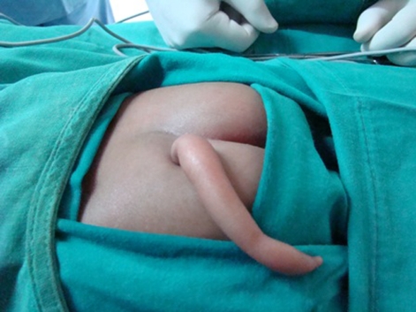
A human baby having caudal appendage resembling a tail generates an unusual amount of interest, excitement and anxiety.1 True human tail is a rare event with fewer than 40 cases reported in the literature (figure 1).2 Here we present a case report of an infant born with a true tail. A 3-month-old baby girl was brought to paediatric surgery outpatient department, with the complaint of having an 11 cm long tail. At birth it was approximately 7 cm long which grew to 11 cm in 3 months. Out of superstition, the parents did not get it excised but finally decided to take medical advice due to its alarming growth.
Figure 1.
Preoperative photograph of the baby with the caudal appendage (tail).
On examination, there was an elongated soft, non-tender mass, with normal skin (colour, texture and temperature) covering, located 1 cm to the left of natal cleft in the sacrococcygeal region. It was well circumscribed measuring 11 cm with 3.5 cm diameter at the base, tapering towards the tip. No voluntary movement was observed in the mass but it could be freely moved in all the directions. On palpation no bony or cartilaginous element was palpable. Sensation over the skin of the appendage was intact. There was normal anal tone. The infant was born after an uneventful pregnancy with no antenatal history of illness, exposure to radiation, or intake of any drug. Except for the presence of the tail the child was otherwise clinically normal. There was no family history of such or any other congenital anomalies. After complete preoperative investigation and preanaesthetic check-up the child was taken up for surgery.
Investigations
All routine investigations were within the normal limits.
X-ray spines showed spina bifida in sacral vertebrae. MRI revealed spina bifida in the sacral vertebrae but no herniation of meninges in posterior subcutaneous region. Few fibrous tissue bands were seen in subcutaneous coccygeal region extending up to the tail.
Differential diagnosis
-
▶
True tail.
-
▶
Pseudo tail (lipoma, teratoma, myelomeningocoele or parasitic fetus etc.).
Treatment
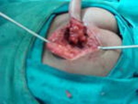
Surgery: elliptical incision was made at the base of the tail. Incision was deepened through the layers reaching up to dorsal lumbosacral fascia. Few fibrous bands were found to attach the tail tissue to lumbosacral fascia (figure 2).
Figure 2.
Intraoperative photograph showing dissection of the base of the tail.
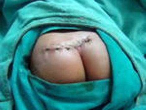
Even on careful dissection lumbosacral fascia was found to be intact and no subfascial extension of the tail structure was found. The tail was removed enbloc. Closure was done in layers (figure 3).
Figure 3.
Postoperative photograph of the surgical site after removal of the tail.
Histopathological examination of the excised tail showed skin covering a core of adipose tissue, collagen fibres and skeletal muscle fibres. No bony or cartilaginous element was present.
Outcome and follow-up
The infant recovered uneventfully in the postoperative period. On follow-up, she was all right without any neurological deficit with normal bowel and bladder habits.
Discussion
Human tail is a caudal, vestigial, midline protrusion with skin covering a combination of muscle and adipose tissue. It may be a ‘true tail’ or a ‘pseudo-tail’. Pseudo-tails are lesions of various types coincidentally found in the caudal region of newborns, often associated with the spinal column and coccygeal malformations. The true human tail is characterised by a complex arrangement of adipose and connective tissue, central bundles of longitudinally arranged striated muscle in the core, blood vessels, nerve fibres, nerve ganglion cells and specialised pressure sensing nerve organs (Vater–Pacini corpuscles). It is covered by normal skin, replete with hair follicles, sweat glands and sebaceous glands. Rarely, voluntary contractions in response to emotional states have been documented.3 4
The dorsal cutaneous appendage, or so-called human tail, is considered to be a marker of underlying intraspinal pathology of occult spinal dysraphism.5 Chunquan Cai et al have reported a case of human tail coexisting with type I split cord malformations.6 Donovan et al have reported child with a tail and intraspinal lipoma that were not contiguous with each other, and were separated by an intact layer of lumbosacral fascia, in our case also there were no subfascial extension of the tail structure.7
True human tails are rarely inherited, though familial cases have been reported. In one case the tail has been inherited through three generations of females.8 Human tails may be associated with other congenital anomalies in 29% of cases,9 commonest is spina bifida. Cleft palate was reported once.4
The true atavistic tail of humans results from incomplete regression of the most distal end of the normal embryonic tail found in the developing human fetus. Human embryos normally have a prenatal tail that measures about one-sixth of the size of the embryo itself.1 At between 4 and 5 weeks of age, the normal human embryo has 10–12 developing tail vertebrae. By the 8th week of gestation, the sixth to twelfth tail vertebrae have disappeared via cell death, and likewise, the associated tail tissues also undergo cell death and regress.
Recent advances in genetic research reveal that ‘of those organs lost’, most species carry ‘genetic blue prints’ which are ‘switched off’ but remain there as genetic storage. The rare re-appearance of these ancient organs are result of re-expression of their switched off genes.10 As with other atavistic structures, human tails are most likely the result of a somatic mutation, a germ line mutation or an environmental influence that reactivates an underlying developmental pathway which has been retained, in the human genome.3 11 12 The genes that control the development of tails in mice and other vertebrates have been identified (the Wnt-3a and Cdx1 genes).13–16 Apoptosis (programmed cell death) plays a significant role in removing the tail of a human embryo after it has formed. It is now known that downregulation of the Wnt-3a gene induces apoptosis of tail cells during mouse development.13 16 Thus, any mutation resulting in increased dosage of the Wnt-3a gene would reduce apoptosis of the human tail during development and would result in its retention in the newborn.
Learning points.
-
▶
Mutations in genes involved in apoptosis may manifest with appearance of vestigial organs in body. The causative factors require further research.
Footnotes
Competing interests None.
Patient consent Obtained.
References
- 1.Ledley FD. Evolution and the human caudal appendage: a case report. N Engl J Med 1982;306:1212–5 [DOI] [PubMed] [Google Scholar]
- 2.Alashari M, Torakawa J. True tail in a newborn. Pediatr Dermatol 1995;12:263–6 [DOI] [PubMed] [Google Scholar]
- 3.Dao AH, Netsky MG. Human tails and pseudotails. Hum Pathol 1984;15:449–53 [DOI] [PubMed] [Google Scholar]
- 4.Lundberg GD, Parsons RW. A case of a human tail. Am J Dis Child 1962;104:72–3 [DOI] [PubMed] [Google Scholar]
- 5.Humphreys RP. Clinical evaluation of cutaneous lesions of the back: spinal signatures that do not go away. Clin Neurosurg 1996;43:175–87 [PubMed] [Google Scholar]
- 6.Cai C, Shi O, Shen C. Surgical treatment of a patient with human tail and multiple abnormalities of the spinal cord and column. Advances in orthopedics 2011:2011:153797. [DOI] [PMC free article] [PubMed] [Google Scholar]
- 7.Donovan DJ, Pedersen RC. Human tail with noncontiguous intraspinal lipoma and spinal cord tethering: case report and embryologic discussion. Pediatr Neurosurg 2005;41:35–40 [DOI] [PubMed] [Google Scholar]
- 8.Standfast AL. The human tail. N Y State J Med 1992;92:116. [PubMed] [Google Scholar]
- 9.Parsons RW. Human tails. Plast Reconstr Surg Transplant Bull 1960;25:618–21 [DOI] [PubMed] [Google Scholar]
- 10.Bakker RT. The Dinosaur Heresis. New York: William Morrow; 1986;1:314–16 [Google Scholar]
- 11.Hall BK. Development mechanisms underlying the formation of atavisms. Biol Rev Camb Philos Soc 1984;59:89–124 [DOI] [PubMed] [Google Scholar]
- 12.Hall BK. Atavisms and atavistic mutations. Nat Genet 1995;10:126–7 [DOI] [PubMed] [Google Scholar]
- 13.Greco TL, Takada S, Newhouse MM, et al. Analysis of the vestigial tail mutation demonstrates that Wnt-3a gene dosage regulates mouse axial development. Genes Dev 1996;10:313–24 [DOI] [PubMed] [Google Scholar]
- 14.Prinos P, Joseph S, Oh K, et al. Multiple pathways governing Cdx1 expression during murine development. Dev Biol 2001;239:257–69 [DOI] [PubMed] [Google Scholar]
- 15.Schubert M, Holland LZ, Stokes MD, et al. Three amphioxus Wnt genes (AmphiWnt3, AmphiWnt5, and AmphiWnt6) associated with the tail bud: the evolution of somitogenesis in chordates. Dev Biol 2001;240:262–73 [DOI] [PubMed] [Google Scholar]
- 16.Takada S, Stark KL, Shea MJ, et al. Wnt-3a regulates somite and tailbud formation in the mouse embryo. Genes Dev 1994;8:174–89 [DOI] [PubMed] [Google Scholar]


