Abstract
The field of mouse phenotyping with magnetic resonance imaging (MRI) is rapidly growing, motivated by the need for improved tools for characterizing and evaluating mouse models of human disease. MRI is an excellent modality for investigating genetically altered animals. It is capable of whole brain coverage, can be used in vivo, and provides multiple contrast mechanisms for investigating different aspects of neuranatomy and physiology. The advent of high-field scanners along with the ability to scan multiple mice simultaneously allows for rapid phenotyping of novel mutations.
Effective mouse MRI studies require attention to many aspects of experiment design. In this article, we will describe general methods to acquire quality images for mouse phenotyping using a system that images mice concurrently in shielded transmit/receive radio frequency (RF) coils in a common magnet (Bock et al., 2003). We focus particularly on anatomical phenotyping, an important and accessible application that has shown a high potential for impact in many mouse models at our imaging centre. Before we can provide the detailed steps to acquire such images, there are important practical considerations for both in vivo brain imaging (Dazai et al., 2004) and ex vivo brain imaging (Spring et al., 2007) that should be noted. These are discussed below.
Keywords: Neuroscience, Issue 48, magnetic resonance imaging, mouse, phenotyping, mouse handling, monitoring, brain, multiple mouse imaging
Protocol
1. Multiple-mouse in vivo Brain Imaging:
When imaging live animals, several key features must be present throughout the imaging session: 1) a safe method of anesthesia, 2) environmental control and 3) physiological monitoring. Furthermore, when imaging multiple subjects simultaneously, there are added complexities regarding the ease and speed of preparation and the reproducibility of animal positions to facilitate image registration. Consequently, three major custom components were designed and fabricated: a loading system to insert the mice into RF coils within the MRI, an induction chamber to facilitate preparation and a platform with embedded monitoring leads to standardize position.
The Loading System:
The mouse loading system consists of two major parts: the 'mouse hive' and the 'loading array'. The mouse hive's main function is to position seven Millipede RF coils (Varian NMR Systems, Palo Alto, CA) in a hexagonal array inside the magnet bore. The loading array is designed to hold and transport multiple mice housed in 50 milliliter centrifuge tubes with holes drilled through their tips to allow entry of anesthetic gas. After the mice are anesthetized and interfaced to the monitoring equipment at a preparation area in the vicinity of the magnet, they are inserted into the modified centrifuge tubes and mounted onto the loading array. After mounting all mice, the loading array is transported and inserted into the magnet and positioned on a rail system. The rail system allows the array to couple with the mouse hive when pushed down the bore of the magnet. When fully inserted into the magnet, the centrifuge tubes dock onto the anesthetic delivery system within the RF coils. Isoflurane mixed with oxygen is supplied from the mouse hive end to the specimen through a tube along the axis of each individual coil. This anesthetic gas mixture flows into the tubes, past the mice and is collected by an active scavenging unit attached to the back of the loading array (Figure 1).
The Induction Chamber:
Since imaging times can take up to three hours, minimizing animal preparation time is crucial to limit the mouse's exposure to anesthesia. Hence, we have developed a custom induction chamber to streamline the preparation process (Figure 2). The custom induction chamber creates a single environment for both induction and handling of multiple mice. Constructed from clear acrylic, the induction chamber features selfclosing silicone iris ports to minimize anesthetic leakage and allows the user to access the internal environment without the need for special gloves. Compared to conventional mask and circuits for a single mouse, the induction chamber is large enough to house up to twenty mice and allows for free manipulation of the mice without the attachment of cumbersome tubes and masks. The unit is supplied with a constant flow of anesthetic gas which is collected using a passive scavenging system. Resistive heating elements are used to heat the floor of the chamber to maintain the animals' body temperature during preparation.
The Sled:
One of the most awkward and time-consuming aspects of preparing mice for the MRI are the application of electrocardiograph (ECG) electrodes and rectal temperature probes. In addition, many of the conventional electrodes, such as cuff and needle electrodes, were found to distort the animal's posture making it difficult to standardize positioning. Therefore, we devised a custom form-fitted positioning platform with embedded ECG, respiration and temperature probes called the 'sled' (U.S. Patent 7,146,936) (Figure 3). Motion restraints made from Velcro fasteners were used to limit movement of the head.
Multiple-mouse in vivo brain imaging steps:
All mouse research requires local ACC (Animal Care Committee), IACUC (Institutional Animal Care and Use Committee) or equivalent approval for mouse handling procedures.
All procedures such as identification and weighing of animals should be performed under a Biological Safety Cabinet (BSC) and in the MRI Unit. The animals are transferred into an autoclavable plastic container and transported to the MRI induction chamber.
Mice are anesthetized in a prewarmed induction chamber using 4% isoflurane and 4 L/min of oxygen. Animals are fully anesthetized once they fail to respond to paw pinch. Fur from the chest is removed using a hair remover (NAIR) if necessary to provide better contact with ECG and temperature monitoring devices that have been built into a custom sled. Eve salve (Tears Naturale P.M.) is applied to the eyes to prevent drying, and approximately 0.3 mL of saline is administered subcutaneously to maintain hydration.
Gd-DTPA-BMA (Omniscan) may be used if contrast enhancement is desired. If Gd-DTPA is to be used, it will be diluted in saline (final volume 300uL) and administered in a single dose of 1 mmol/kg prior to the MR session via IP.
Mice are then loaded into individual sleds, immobilized with head straps and slide into open-ended 50mL conical tube (Figure 3). Up to 7 live mice can be scanned at one time. Once all animals are loaded with physiological monitoring connected, set the isoflurane level in the magnet bore to 2% and the oxygen level to 8 L/min.
The conical tubes are mounted into a docking system (Figure 1) designed to position mice uniformly into each RF coil positioned in the centre of the magnet bore. Once loaded, isoflurane can be reduced to 0.9-2%. ECG and temperature are monitored on each animal throughout the course of the scan. Animals are kept warm during the course of the scan with warmed air.
The duration of each three dimensional scan is approximately 3 hours. The details are as follows: fast spin echo with TR of 2300 ms and a TEeff of 36 ms. Echo train length of 8 with 1 average. Resulting image resolution is 125 μm (Figure 4).
When the scan is complete, the animals are removed from the magnet and unloaded in a warm induction chamber filled with 100% oxygen. The animals are transferred into a plastic sealed containers and transported under a BSC. They are placed on a warm draft free cage and allowed to recover from anesthesia.
2. Multiple-mouse ex vivo Brain Imaging:
Unaffected by motion artifacts, fixed MR imaging achieves higher resolution than live imaging. High-resolution, Three-dimensional datasets provide maximal flexibility in extracting quantitative information and allow for automated image analysis. A custom RF coil array was developed for parallel acquisition of 16 high-resolution MR datasets of fixed in-skull mouse brains in overnight scanning sessions.
The 16-Coil ex vivo Brain Imaging Array:
A custom-built 16-coil solenoid array was created to image 16 samples concurrently. This design improves upon an earlier prototype used to image three samples concurrently in a 60mm insert gradient set. The 8-turn solenoid coils are over wound at the ends to provide uniform sensitivity to within 10% over a length of 26mm and are individually shielded within modular compartments (Idziak and Haeberlen, 1982). The 16 coil compartments are assembled to a frame (Figure 5), which positions the coils within the gradient and clamps it in place using a pneumatic bladder to minimize motion.
Multiple-mouse ex vivo brain imaging steps:
Open chest cavity and insert needle (or Safety Winged infusion set, 25G X 3⁄4) into the left ventricle of the heart of an anesthetized mouse (via intraperitoneal injection of ketamine (150 mg/kg) and xylazine (10 mg/kg). Cut the right auricle.
Transcardiac perfusion flush with 30 mL room temp 1 X PBS + 1 μL/ mL heparin (1000 USP units/ mL) + 2 mM ProHance at a flow rate of approximately 100 mL/hr.
Fixation pass with 30 mL 4% PFA (room temperature) + 2 mM ProHance at 100 mL/hr.
Decapitate and remove skin, lower jaw, ears, cartilaginous nose tip.
Place remaining skull structure in 4% PFA + 2 mM ProHance overnight at 4°C.
Transfer to 1X PBS + 0.02% sodium azide + 2 mM ProHance.
High resolution three dimensional MRI scanning at 7 Tesla occurs between 4 days and not longer than 2.5 months after perfusion. Brains are placed in a 16-channel solenoid coil array (Figure 5).
Imaging parameters are as follows: fast spin echo with TR 325 ms and a TEeff of 30 ms, echo train length of 6 with 4 averages. The final images have an isotropic resolution of 32 μm (Figure 6) and the scan duration is approximately 12 hours.
After scanning, place the skull in 10% Formalin + 2 mM ProHance for preservation.
3. Representative Results:
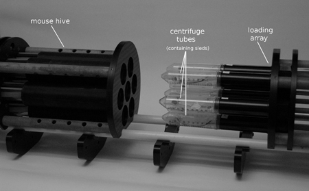 Figure 1. The mouse loading system. The "loading array" and "mouse hive" connected with a common fiberglass rail system.
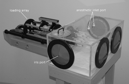 Figure 2. The induction chamber.
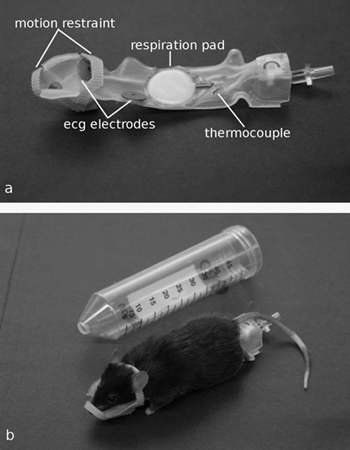 Figure 3. a) The sled, showing embedded monitoring sensors and head restraint. b) An anesthetized mouse on a sled with the head restraint attached. The sled assembly easily slides into the centrifuge tube.
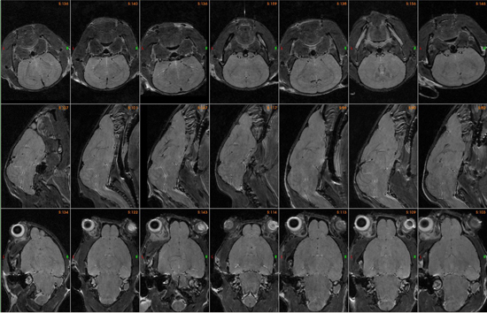 Figure 4. Representative in vivo multiple brain images from one 3 hour three dimensional scan.
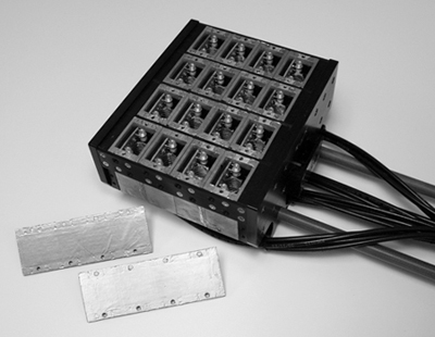 Figure 5. 16-channel solenoid coil array for scanning of 16 fixed brain specimens.
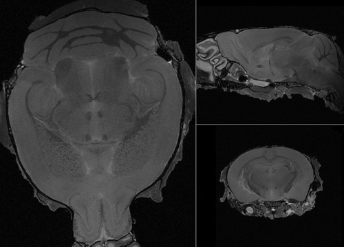 Figure 6. Representative ex-vivo brain image.
Discussion
Both the in vivo and ex vivo mouse imaging systems image multiple subjects at once to increase throughput of imaging studies considerably without increasing the imaging time. The brain images from both the in vivo and ex vivo multiple-mouse imaging techniques are of high quality and are suitable for phenotyping major and minor structures in the mouse brain respectively.
To minimize animal preparation time of multiple specimens, parallelization of processes is of utmost importance. For example, the development of the induction chamber allowed for the induction of multiple specimens simultaneously, and the sled has synchronized the application of the ECG and temperature probes while standardizing body positioning. Furthermore, our ex vivo imaging system enables us to acquire high resolution three dimensional images of 16 fixed whole brains at one time which is ideal for high-throughput phenotyping studies.
Possible limitations of our in vivo imaging system include the inability to individually control anesthetic and temperature for each mouse. If required individual anesthetic control can be implemented with the addition of exclusive anesthetic vaporizers for each mouse. Another limitation of the in vivo imaging system is that scanning is restricted to mice that are less than approximately 32 grams. However, there is a plan currently to increase coil size to accommodate larger animals.
Disclosures
No conflicts of interest declared.
Acknowledgments
This work is part of the Mouse Imaging Centre (MICe) at the Hospital for Sick Children and the University of Toronto.
References
- Bock NA, Konyer NB, Henkelman RM. Multiple-mouse MRI. Magn. Reson. Med. 2003;49:158–167. doi: 10.1002/mrm.10326. [DOI] [PubMed] [Google Scholar]
- Dazai J, Bock NA, Nieman BJ, Davidson LM, Henkelman RM, Chen XJ. Multiple mouse biological loading and monitoring system for MRI. Magn. Reson. Med. 2004;52:709–715. doi: 10.1002/mrm.20215. [DOI] [PubMed] [Google Scholar]
- Spring S, Lerch JP, Henkelman RM. Sexual dimorphism revealed in the structure of the mouse brain using three-dimensional magnetic resonance imaging. NeuroImage. 2007;35:1424–1433. doi: 10.1016/j.neuroimage.2007.02.023. [DOI] [PubMed] [Google Scholar]
- Idziak S, Haeberlen U. Design and construction of a high homogeneity rf coil for solid-state multiple-pulse NMR. J. Magn. Reson. 1982;50:281–288. [Google Scholar]


