Abstract
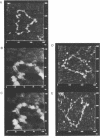
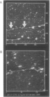
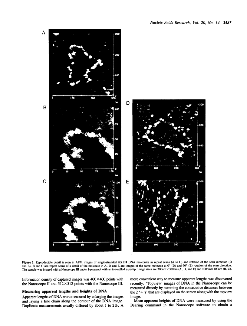
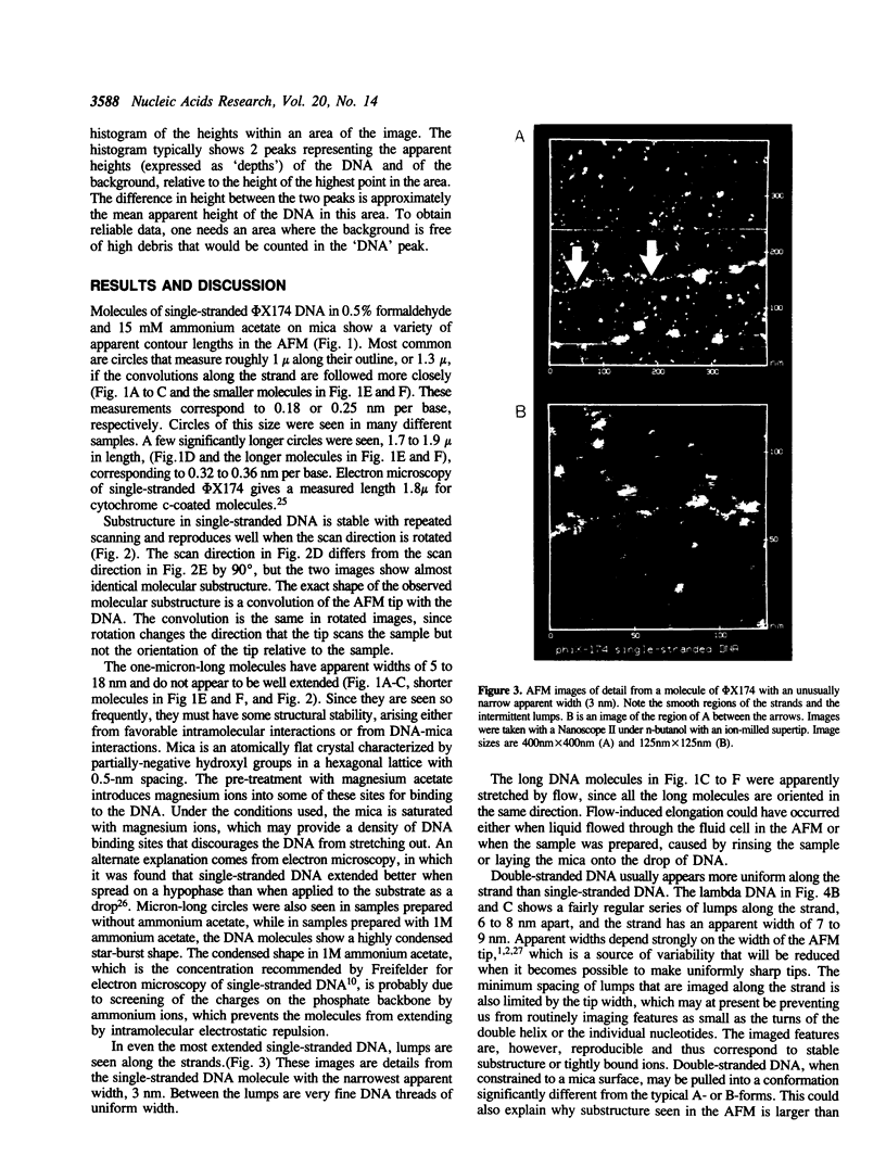
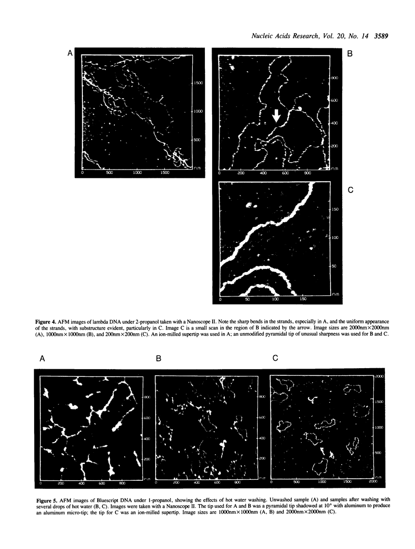
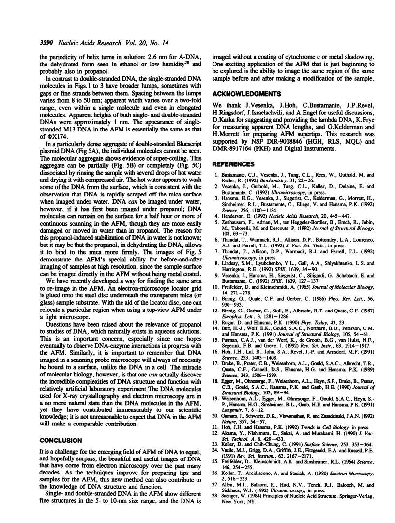
A method has been developed for imaging single-stranded DNA with the atomic force microscope (AFM). phi X174 single-stranded DNA in formaldehyde on mica can be imaged in the AFM under propanol or butanol or in air. Measured lengths of most molecules are on the order of 1 mu, although occasionally more extended molecules with lengths of 1.7 to 1.9 mu are seen. Single-stranded DNA in the AFM generally appears lumpier than double-stranded DNA, even when extended. Images of double-stranded lambda DNA in the AFM show more sharp kinks and bends than are typically observed in the electron microscope. Dense, aggregated fields of double-stranded plasmids can be converted by gentle rinsing with hot water to well spread fields.
Full text
PDF
Images in this article
Selected References
These references are in PubMed. This may not be the complete list of references from this article.
- Binnig G, Quate CF, Gerber C. Atomic force microscope. Phys Rev Lett. 1986 Mar 3;56(9):930–933. doi: 10.1103/PhysRevLett.56.930. [DOI] [PubMed] [Google Scholar]
- Bustamante C., Vesenka J., Tang C. L., Rees W., Guthold M., Keller R. Circular DNA molecules imaged in air by scanning force microscopy. Biochemistry. 1992 Jan 14;31(1):22–26. doi: 10.1021/bi00116a005. [DOI] [PubMed] [Google Scholar]
- Butt H. J., Wolff E. K., Gould S. A., Dixon Northern B., Peterson C. M., Hansma P. K. Imaging cells with the atomic force microscope. J Struct Biol. 1990 Oct-Dec;105(1-3):54–61. doi: 10.1016/1047-8477(90)90098-w. [DOI] [PubMed] [Google Scholar]
- Drake B., Prater C. B., Weisenhorn A. L., Gould S. A., Albrecht T. R., Quate C. F., Cannell D. S., Hansma H. G., Hansma P. K. Imaging crystals, polymers, and processes in water with the atomic force microscope. Science. 1989 Mar 24;243(4898):1586–1589. doi: 10.1126/science.2928794. [DOI] [PubMed] [Google Scholar]
- FREIFELDER D., KLEINSCHMIDT A. K., SINSHEIMER R. L. ELECTRON MICROSCOPY OF SINGLE-STRANDED DNA: CIRCULARITY OF DNA OF BACTERIOPHAGE PHI-X174. Science. 1964 Oct 9;146(3641):254–255. doi: 10.1126/science.146.3641.254. [DOI] [PubMed] [Google Scholar]
- Freifelder D., Kleinschmidt A. K. Single-strand breaks in duplex DNA of coliphage T7 as demonstrated by electron microscopy. J Mol Biol. 1965 Nov;14(1):271–278. doi: 10.1016/s0022-2836(65)80246-7. [DOI] [PubMed] [Google Scholar]
- Hansma H. G., Vesenka J., Siegerist C., Kelderman G., Morrett H., Sinsheimer R. L., Elings V., Bustamante C., Hansma P. K. Reproducible imaging and dissection of plasmid DNA under liquid with the atomic force microscope. Science. 1992 May 22;256(5060):1180–1184. doi: 10.1126/science.256.5060.1180. [DOI] [PubMed] [Google Scholar]
- Henderson E. Imaging and nanodissection of individual supercoiled plasmids by atomic force microscopy. Nucleic Acids Res. 1992 Feb 11;20(3):445–447. doi: 10.1093/nar/20.3.445. [DOI] [PMC free article] [PubMed] [Google Scholar]
- Hoh J. H., Lal R., John S. A., Revel J. P., Arnsdorf M. F. Atomic force microscopy and dissection of gap junctions. Science. 1991 Sep 20;253(5026):1405–1408. doi: 10.1126/science.1910206. [DOI] [PubMed] [Google Scholar]
- Zenhausern F., Adrian M., ten Heggeler-Bordier B., Emch R., Jobin M., Taborelli M., Descouts P. Imaging of DNA by scanning force microscopy. J Struct Biol. 1992 Jan-Feb;108(1):69–73. doi: 10.1016/1047-8477(92)90008-x. [DOI] [PubMed] [Google Scholar]


