Abstract
Avascular necrosis (AVN) of the femoral head is a debilitating disease of multifactorial genesis, predominately affects young patients, and often leads to the development of secondary osteoarthritis. The evolving field of regenerative medicine offers promising treatment strategies using cells, biomaterial scaffolds, and bioactive factors, which might improve clinical outcome. Early stages of AVN with preserved structural integrity of the subchondral plate are accessible to retrograde surgical procedures, such as core decompression to reduce the intraosseous pressure and to induce bone remodeling. The additive application of concentrated bone marrow aspirates, ex vivo expanded mesenchymal stem cells, and osteogenic or angiogenic growth factors (or both) holds great potential to improve bone regeneration. In contrast, advanced stages of AVN with collapsed subchondral bone require an osteochondral reconstruction to preserve the physiological joint function. Analogously to strategies for osteochondral reconstruction in the knee, anterograde surgical techniques, such as osteochondral transplantation (mosaicplasty), matrix-based autologous chondrocyte implantation, or the use of acellular scaffolds alone, might preserve joint function and reduce the need for hip replacement. This review summarizes recent experimental accomplishments and initial clinical findings in the field of regenerative medicine which apply cells, growth factors, and matrices to address the clinical problem of AVN.
Introduction
Osteonecrosis or avascular necrosis (AVN) is defined as a pathologic process that results from a critical reduction of blood supply to the bone and elevated intraosseous pressure. Although the pathogenic process is poorly understood, osteonecrosis is the final common pathway of traumatic and non-traumatic insults and compromises the already precarious circulation of the femoral head. Femoral head ischemia subsequently leads to the death of bone marrow and osteocytes and usually results in the collapse of the necrotic segment [1]. AVN is a debilitating disease that usually leads to osteoarthritis of the hip joint in young adults. The disease prevalence is unknown, but estimates indicate that 10,000 to 20,000 new cases are diagnosed in the US each year. Furthermore, it is estimated that 5% to 18% of the more than 500,000 total hip arthroplasties performed annually are related to advanced AVN [2]. Joint-preserving techniques such as core decompression, avascular or vascularized bone grafting, and various femoral osteotomies are most promising in early stages of AVN - Association Research Circulation Osseous (ARCO) stage I and II - with preserved structural integrity of the subchondral plate [2]. The structural collapse of the osteonecrotic segment (ARCO stage III and IV) indicates the progressive course of the disease, and total hip replacement is inevitable in most cases in the long term [1,3].
Cell-based strategies
To enhance osseous regeneration, the application of osteogenic or angiogenic precursor cells (or both) in combination with or without supporting growth factors is an appealing possibility. Among the various cell types, adult tissue-derived, multipotent mesenchymal stem cells (MSCs) represent a highly promising candidate for cell-based therapeutic approaches [4]. These somatic adult stem cells can be detected in specific tissue compartments of the human body and are considered to play a critical role in maintaining the integrity of different tissues such as skin, bone, and blood [5]. MSCs maintain the ability of mitotic multiplication without significant loss of their specific biomolecular characteristics over extensive population expansion and are capable of differentiating into multiple mesenchymal phenotypes, including osteoblasts, chondrocytes, and adipocytes [5]. MSCs have been shown to enhance tissue regeneration after transplantation in an experimental AVN model in dogs [6]. However, whether the observed effects resulted from the osteogenic differentiation of transplanted cells or are mediated via their trophic activities remains unclear [6,7].
Proper vascularization is essential for the viability and function of the majority of tissues in the body. Without sufficient blood supply, cells suffer from hypoxia, lack of nutrients, and the accumulation of waste products, and biochemical signaling mechanisms are disrupted, affecting tissue homeostasis and preventing tissue regeneration. Endothelial progenitor cells (EPCs) are spindle-shaped cells capable of differentiation into a mature endothelial phenotype. These cells can be isolated from bone marrow and peripheral blood [8,9]. Their role in angiogenesis and neovascularization has been studied extensively, and positive effects on blood vessel formation after transplantation have been reported [10]. So far, however, these cells have not been applied clinically for the treatment of AVN.
Interestingly, Feng and colleagues [9] recently reported significantly decreased numbers of circulating EPCs and colonyforming units (CFUs) in patients with diagnosed AVN in comparison with a healthy control group. Furthermore, EPCs of patients with AVN exhibited impaired migratory capacities and increased cellular senescence, resulting in reduced angiogenesis in vitro [9].
Application of concentrated bone marrow aspirates
Hernigou and colleagues [11-13] pioneered the clinical application of a cell-based strategy for the treatment of AVN, percutaneously injecting an autologous bone marrow concentrate into the necrotic area of femoral heads. The strategy is driven by the hypothesis that injected bone marrow cells can repopulate the trabecular bone structure and subsequently revitalize and remodel the necrotic bone. For this technique, a bone marrow aspirate is harvested from the iliac crest and the mononuclear cell fraction is isolated. After core decompression, the cell suspension is injected into the necrotic area [11-13]. These authors reported on the postoperative outcome in 189 cases (116 patients) treated with the injection of autologous bone marrow concentrate [11]. Over the course of the 5- to 10-year follow-up, nine out of 145 patients with early-stage AVN (Steinberg stage I or II) and 25 out of 45 patients with later-stage AVN (Steinberg stage III or IV) required total hip replacement. However, since no control group was examined, the beneficial effect of the injected bone marrow concentrate (that is, in comparison with sole core decompression) is difficult to prove. Gangji and Hauzeur [14] conducted a controlled prospective study comparing the postoperative outcome of core decompression with core decompression followed by the injection of a bone marrow concentrate in early-stage AVN (ARCO stage I or II). The mean follow-up was 2 years, and five out of eight hip joints in the control group showed radiographic progression. In contrast, only one out of 10 hips treated with bone marrow cells progressed to a more severe stage with collapse of the subchondral bone [14]. Recently, Gangji and colleagues [15] reported on the 5-year follow-up of 19 patients (24 hip joints) who received either core decompression alone or core decompression with the additional application of bone marrow concentrate because of early-stage AVN. In this prospective, double-blinded trial, eight out of 11 hip joints in the core decompression group and only three out of 13 joints in the bone marrow concentrate group revealed a disease progression with structural disintegration of the sub-chondral bone [15]. Despite the promising results, the clinical value of these studies is limited because of short-term follow-up periods and low case numbers.
Application of ex vivo expanded autologous bone marrow-derived stem cells
The ex vivo amplification and further administration of MSCs, in contrast to bone marrow cell concentrates, are controlled by regulatory authorities, namely the US Food and Drug Administration (FDA) and the European Medicines Agency (EMA) [16]. In most cases, the selection of MSCs is achieved by cell adherence to tissue culture plastic after phase gradient separation (that is, Ficoll or Percoll).
The therapeutic potential of ex vivo amplified bone marrow-derived MSCs for the treatment of cortico-steroid-induced AVN in femoral condyles was investigated by Müller and colleagues [17]. In five patients, bone marrow aspirates were harvested from the posterior iliac crest and MSCs were isolated and expanded for three passages. The MSCs were resuspended in 0.1% serum albumin-supplemented saline and transplanted into the necrotic area. After a mean follow-up of 16 months, none of the patients showed signs of disease progression. Owing to the lack of a control group, whether this effect can be attributed to the injected cells is unclear.
Apart from cell delivery in suspension, a variety of matrices, such as ceramics, collagen sponges, hydrogels, and biodegradable polymers, have been used for cell delivery [4]. Core decompression offers the opportunity to deliver such cell-laden biomaterials to the necrotic area. Concurrently with tissue neoformation, scaffolds ideally undergo degradation processes resulting in biocompatible, metabolisable, and excretable by-products. Specifically, for the treatment of AVN, autologous or allogenic bone grafts (demineralized bone matrix, or DBM) as well as synthetic biomaterials (that is, beta-tricalcium phosphate, or β-TCP) have been suggested as suitable carriers for cell-based strategies [3,4].
Kawate and colleagues [18] reported the treatment of three patients with advanced stages of cortisone-induced AVN (Steinberg stage III or IV) with a vascularized fibular graft combined with a synthetic β-TCP ceramic and bone marrow-derived MSCs. Four weeks prior to elective core decompression, 15 mL of bone marrow aspirate was taken from the iliac crest. MSCs were isolated and expanded in autologous serum. After 10 days, MSCs were seeded onto the β-TCP granula and cultured for 2 weeks. After core decompression, the defect was filled with the MSC-seeded β-TCP granula and a vascularized fibular graft was transplanted. During the 34-month follow-up, no progression of the AVN was reported [18]. Given the advanced stages treated and the cortisone-related genesis of the necrosis (both of which have been shown to lead to a decreased therapeutic response in general [3,19]), these results are promising. However, only a small number of patients were treated and a control group was not included.
A similar approach to treat AVN with autologous stromal cell-seeded β-TCP granula was presented by our group [20]. The applied cells consisted of a mixture of MSCs and endothelial and hematopoietic progenitor cells (Tissue Repair Cells, or TRC; Aastrom Biosciences, Inc., Ann Arbor, MI, USA).
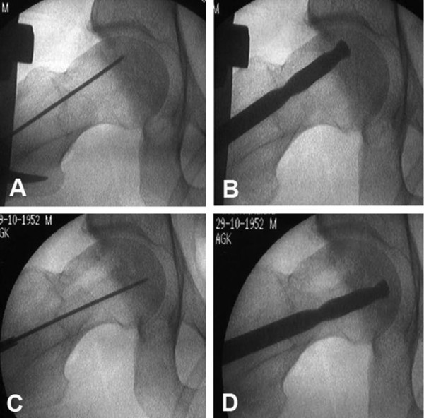
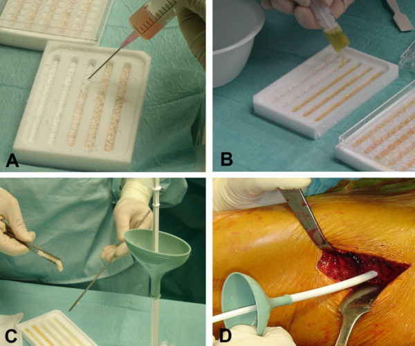
Briefly, bone marrow aspirates (100 mL) were harvested bilaterally from the posterior iliac crest. The aspirates were expanded for 12 days in autologous serum-supplemented medium by means of a good manufacturing practice (GMP) system (Aastrom Biosciences, Inc.). Fluorescence-activated cell sorting analysis of the expanded cells showed a significantly higher number of CFU-fibroblasts and cells expressing surface markers characteristic of MSCs (that is, CD90) and hematopoietic (that is, CD133) and endothelial (that is, CD90) progenitor cells when compared with the mononuclear fraction of native bone marrow aspirates, indicating an enrichment of these cell types [20]. Early AVN (ARCO stage II) was diagnosed in all treated patients. After surgical exposure of the proximal femur, a bone plug was removed by using a hollow diamond drill. For the treatment of small AVN lesions (ARCO stage IIA), a K-wire was placed centrally into the necrotic area and was used as a guide for a cannulated 10-mm drill bit. For the treatment of more extensive AVN (ARCO stage IIB and IIC), two drill holes were used (Figure 1A-D) [20]. Subsequently, β-TCP granula (Vitoss; Orthovita Inc., Malvern, PA, USA) were placed in a custom-made plastic tray and the TRC suspension was homogeneously seeded onto the granula (Figure 2A). For ease of handling, autologous plasma obtained during surgery was used to achieve physical bonding between the MSC-seeded β-TCP granula (Figure 2B). The tissue-engineered construct consisting of cells, β-TCP granula, and autologous plasma was administered to the necrotic area through a plastic funnel (Orthovita Inc.) (Figure 2C,D), and the drill hole was sealed with the preserved bony cylinder.
Figure 1.

Intraoperative fluoroscopy demonstrating the drilling prior to stem cell tricalcium phosphate matrix application. (A) A 3-mm K-wire is placed in the anterior portion of the necrosis and (B) overdrilled with a 10-mm cannulated drill. (C,D) The same procedure is repeated for the posterior part of the necrosis. Reprinted with permission from Schattauer [54].
Figure 2.

Preparation and application of the stem cell tricalcium phosphate (TCP) matrix. (A) The stem cell suspension is applied to the β-TCP granula. (B) This is followed by the addition of intraoperatively obtained autologous serum. (C,D) The matrix is transplanted without compression by using a push rod and a custom-made funnel-shaped applicator. Reprinted with permission from Schattauer [54].
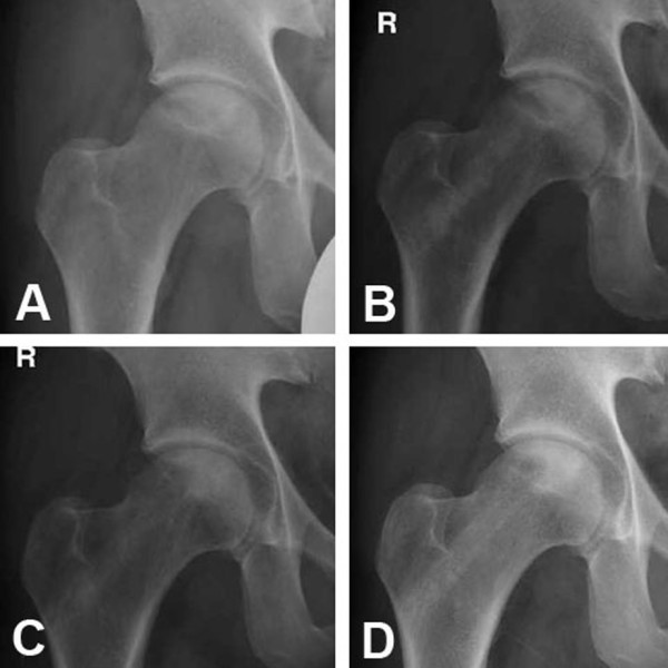
At present, a total of four patients have been treated by using the described technique without disease progression (Figure 3A-D) at the 24-month follow-up. A European Union-funded (FP-7) clinical phase I and II study is in preparation to evaluate the beneficial effects of bone marrow-derived progenitor cell application compared with standard core decompression in the treatment of early-stage AVN [21].
Figure 3.

Postoperative radiographs of a 38-year-old patient with avascular necrosis (ARCO stage II) of the right hip. (A) Postoperative x-ray. X-rays of the right hip joint at 6 months (B), 12 months (C), and 24 months (D) after treatment. Over time, the sclerotic and necrotic zones have decreased in size, especially on the lateral side. Despite the extensive defect, the femoral head has neither collapsed nor progressed to an ARCO stage III. After 24 months, the beta-tricalcium phosphate (β-TCP) granula have not undergone complete resorption. Reprinted with permission from Schattauer [54]. ARCO, Association Research Circulation Osseous.
Application of allogenic bone marrow-derived stem cells
The use of allogenic instead of autologous MSCs for the treatment of AVN appears attractive because of logistic and economic advantages given that these cells might be available as an 'off the shelf' product. However, allogenic MSCs harbor the danger of disease transmission and immunological rejection [22], as observed in organ transplantation. Therefore, a peculiar risk-benefit analysis of allogenic MSC-based strategies in large populations of patients has to be addressed, especially when aiming for non-life-threatening diseases such as AVN. Because alternative treatment options are lacking, allogenic MSC transplantation appears to be more suitable in severe disorders such as osteogenesis imperfecta. Cell engraftment and accelerated growth velocity have been shown in children with severe osteogenesis imperfecta after systemic administration of isolated allogenic bone marrow-derived MSCs [23,24].
Growth factor-based strategies
A variety of growth factors produced by osteogenic cells, platelets, and inflammatory cells - including bone morphogenetic proteins (BMPs) [25-27], insulin-like growth factor-1 and -2, transforming growth factor-β1 (TGF-β1), platelet-derived growth factor, and fibroblast growth factor-2 - are functionally involved in bone healing. The bone matrix serves as a reservoir for these growth factors, which are activated during matrix resorption by matrix metalloproteases. Additionally, the acidic environment that develops during the inflammatory process leads to activation of latent growth factors, which assist in chemo-attraction, migration, proliferation, and differentiation of MSCs into osteoblasts or chondroblasts [25-27]. All of these functions are driven by complex interactions among growth factors and other cytokines and are influenced by multiple regulatory factors. Thus, the use of osteogenic growth or differentiation factors for joint-preserving treatment of AVN is also a potentially promising approach [28].
Osteogenic growth factors
Bone morphogenetic proteins
Healing of osseous tissue is regulated mainly by members of the TGF-β superfamily. Specifically, BMPs act to induce the formation of both bone and cartilage by stimulating mesenchymal progenitor cells. However, only a subset of BMPs, notably BMP-2, -4, -7, and -9, have been shown to exhibit osteoinductive activity in de novo bone formation [25-28]. Lieberman and colleagues [29] treated 15 patients (17 hip joints at Ficat stage II or III) after core decompression with allogenic, antigen-extracted, autolyzed fibula grafts, combined with 50 mg of recombinant human BMP-2 and non-collagenous protein. During the mean follow-up of 53 months, three patients (one Ficat stage II and two Ficat stage III) required total hip replacement because of an aggravation of clinical symptoms corresponding with radiographic progression [29].
Mont and colleagues [30] augmented bony defects resulting from AVN with a triphasic bone substitute consisting of DBM, processed allograft bone chips, and a thermoplastic carrier plus the addition of BMP-7 in 19 patients (21 hip joints). Eighty-six percent of the treated hips were assessed as positive outcomes - Harris Hip Score (HHS) of at least 80 - at the final follow-up (mean follow-up of 48 months) [30]. The same group conducted a retrospective cohort study in 33 patients (39 hip joints: 22 at Ficat stage II and 17 at Ficat stage III) implanted with autologous, non-vascularized bone grafts loaded with BMP-7. At the final follow-up (mean of 36 months), only four out of 22 Ficat stage II hips and 11 out of 17 Ficat stage III hips had undergone hip arthroplasty. At present, whether the administration of BMPs results in better clinical outcomes remains unclear as the literature lacks controlled randomized studies that include appropriate numbers of patients and control groups [31].
In another study, Mont and colleagues [32] created an experimental bony defect at the antero- lateral aspect of the femoral head in canines. The defect was augmented with an autologous bone graft from the iliac crest in combination with 250 mg of BMP-7 per gram of autograft. In the control group, the defect remained untreated. Four months after surgery, all control animals showed a collapse of the femoral head with degeneration of the hyaline cartilage. Autograft augmentation with or without BMP-7 preserved the subchondral bone and the production of a surface layer of hyaline cartilage. Independently of BMP supplementation, radiographs revealed good to excellent osseous remodeling when autograft augmentation was performed [32].
The healing potential of BMP-2- and BMP-14-loaded collagen scaffolds was investigated by Simank and colleagues [33] in a sheep model. Necrosis of the femoral head was created by direct ethanol injection. After core decompression, BMP-laden scaffolds were transplanted and compared with non-loaded scaffolds. Three months after surgery, both growth factor-treated groups revealed better bone regeneration histomorphologically [33]. When the same animal model was used, BMP-14-loaded, hydroxyapatite-coated collagen scaffolds (Healos; DePuy, Warsaw, IN, USA) significantly improved bony remodeling of the necrotic area. In comparison, when the scaffold was applied without BMP-14, only partial defect filling with mainly fibrous tissue and persistence of the necrosis was seen after 12 weeks [34].
To achieve long-term availability of osteoinductive or angiogenic growth factors in a defect, it is also possible to deliver gene-modified cells synthesizing and secreting the desired protein. Genetic engineering strategies focusing on osteoinductive factors have emerged as efficient approaches to enhance bone formation. Two modalities are generally used: (a) direct in vivo delivery of gene constructs and (b) ex vivo transduction and subsequent transplantation of cells expressing the osteoinductive factor [4]. The choice of the gene delivery method depends on several factors, including the particular gene of interest, indication targeted, desired duration of gene expression, and nature of the delivery vector.
Tang and colleagues [35] induced bilateral AVN in goats by ligation of the lateral and medial circumflexing arteries and additional delivery of liquid nitrogen to the femoral head. The authors investigated the influence of MSCs, transduced with either BMP-2 or β-galactosidase and seeded onto β-TCP scaffolds on AVN consolidation. Three weeks after core decompression, the tissue-engineered constructs were transplanted. Whereas control animals (core decompression only) showed AVN progression with structural disintegration and collapse of the subchondral bone 4 months after treatment, no signs of such progression were observed in the cell-treated groups [35]. In the case of BMP-2 transduction, superior bone regeneration in the defects was observed when compared with the β-galactosidase group, as were significantly higher amounts of new bone and higher maximum compressive strength and bone density [35].
Angiogenic growth factors
Vascular endothelial growth factor
Vascular endothelial growth factor (VEGF) regulates numerous cellular events associated with angiogenesis and vasculogenesis, such as tissue remodeling, during embryonic development and in adults [36]. Yang and colleagues [37] delivered plasmids encoding VEGF immobilized on a collagen carrier into the necrotic area of the femoral head in rabbits. Significantly higher bone formation was reported 8 weeks after treatment in comparison with control animals that received a collagen carrier without VEGF plasmids. The successful in vivo transfection of local cells by the VEGF plasmids and expression of VEGF after surgical delivery, however, were shown up to 2 weeks only [37].
Other growth factors
Granulocyte colony-stimulating factor and stem cell factor
Granulocyte colony-stimulating factor (G-CSF) is a glycoprotein, growth factor, and cytokine with two isoforms, both of which stimulate the proliferation of bone marrow stromal cells and enhance their availability in the circulating blood volume [38]. Stem cell factor (SCF) is a cytokine that plays a critical role in regulating the differentiation of hematopoietic stem cells. After intramuscular application of G-CSF and SCF, Wu and colleagues [39] observed increased blood vessel formation (3.3-fold), a higher blood vessel density (2.6-fold), and increased bone formation in the necrotic area of steroid-induced AVN of rabbits in comparison with a non-treated control group. However, whether direct angiogenic/osteogenic differentiation of the mobilized progenitor cells accounted for the increased vascularization and bone formation or whether an increased trophic activity of these cells activated endogenous progenitor cells at the defect site was unclear [39].
Hepatocyte growth factor
Hepatocyte growth factor (HGF) is a multifunctional cytokine that regulates cell growth, cell motility, and morphogenesis and exhibits synergistic effects with VEGF [40]. Wen and colleagues [41] used a hormone-induced AVN rabbit model to compare the therapeutic effect of core decompression alone versus the additional augmentation of the necrotic area with HGF-transduced MSCs administered in fibrin glue. De novo bone formation with regular trabecular structure and enhanced vascularization of the necrotic bone was observed only in animals treated with HGF-transduced MSCs as shown by magnetic resonance imaging and computed tomography imaging and immunohistochemistry [41].
Anterograde osteochondral reconstruction in advanced stages of avascular necrosis
With the progression of untreated AVN (ARCO stage III and IV), structural disintegration of the subchondral bone occurs and leads to the collapse of the femoral head. A substantial degeneration of the hyaline cartilage has to be assumed at this stage, and retrograde strategies, such as core decompression and autologous bone grafting, offer only very limited prospects [42]. In most of these cases, total joint replacement remains the only treatment option [43]. The potential of regenerative, joint-preserving techniques, such as osteochondral transplantation (autografts or allografts), bone grafting followed by autologous chondrocyte implantation (ACI), or acellular matrix implantation, has not been exploited sufficiently so far. These techniques include a surgical dislocation of the hip to anterogradely access the osteochondral defect, which is more invasive and surgically demanding [42,44].
Osteochondral transplantation (mosaicplasty)
Meyers and colleagues [45,46] transplanted osteochondral autografts or allografts in 21 patients with progressed stages of AVN (Ficat stage II to IV) after hip dislocation and debridement of the osteochondral defect. The treatment was reported to be successful in 71% to 80% of all cases (follow-up for 1.5 to 5 years) but in only 50% of the patients with steroid-induced AVN [45,46]. Sotereanos and colleagues [47] treated a 36-year-old patient after ineffective core decompression and vascularized fibula graft augmentation. Three osteochondral cylinders were transferred from a non-weight-bearing area at the inferior-lateral femoral head into the defect following bone grafting. After 5 years, the HHS was 96, the patient was pain-free, and the range of motion was not limited [47].
Rittmeister and colleagues [48] performed an autologous osteochondral transfer (one to three grafts) in five patients with advanced AVN. After a mean follow-up of 57 months, the therapy failed in four patients and total hip arthroplasty was required. Only one patient was treated successfully, and hip function showed no limitations 31 months after surgery [48].
Autologous chondrocyte implantation
ACI has been proven to be a suitable technology for the treatment of full-thickness cartilage defects in the knee and ankle joint. Its therapeutic potential in the hip has not been addressed sufficiently so far. Only one case report on ACI at the femoral head has been published so far. Akimau and colleagues [49] treated a 31-year-old man with a diagnosed post-traumatic necrosis of the femoral head by using a modified first-generation ACI approach. Healthy hyaline cartilage (240 mg) was harvested arthroscopically from the ipsilateral knee joint, and chondrocytes were isolated and expanded in monolayer culture over a period of 3 weeks. In a second surgery, the femoral head was dislocated and exposed. After removal of the degenerated cartilage and necrotic bone, the defect was covered with a collagen type I membrane (Chondro-Gide; Geistlich, Wolhusen, Switzerland), and 6 × 10⁶ chondrocytes were injected underneath the membrane. The HHS increased from 45 preoperatively to 76 at 12 and 18 months after surgery. Arthroscopic biopsy revealed a 2 mm-thick repair tissue consisting of fibrous and, to a lesser extent, hyaline-like cartilage [49].
Acellular matrix implantation
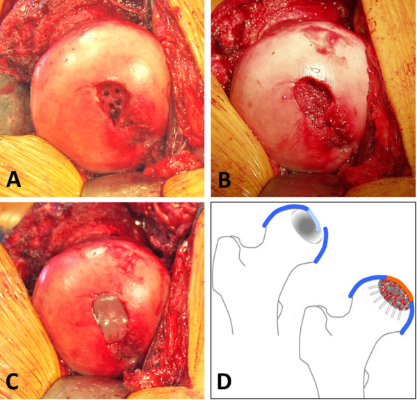
Matrix-based ACI has been shown to be a clinically successful method for the treatment of full-thickness cartilage defects by using a variety of scaffold materials [4,50,51]. In ACI, a previous cartilage biopsy harvest followed by ex vivo cell processing, which is timeconsuming and costly, is required. To circumvent these barriers, an innovative one-step procedure combining microfracturing [52] of the subchondral bone and coverage of the cartilage defect by using an acellular scaffold (Autologous Matrix-Induced Chondrogenesis) was developed [53]. We recently adopted this basic principle for the treatment of an advanced stage of AVN in the hip (ARCO stage III) as shown in Figure 4. The hip joint was dislocated after a trochanteric flip osteotomy was performed [44] and the femoral head was exposed (Figure 4). The osteochondral defect was debrided, the sclerotic bony ground was repeatedly penetrated by using a small drill bit, and the bony defect was augmented with autologous cancellous bone. Subsequently, the cartilage defect was replenished by using an acellular collagen type I hydrogel (CaReS-OneStep; Arthro Kinetics, Krems, Austria) [54]. So far, three patients have been treated with this technique with a follow-up of 9 months.
Figure 4.

Acellular matrix implantation in a patient with an osteochondral defect of the femoral head and an unstable cartilage defect (ARCO stage III). A trochanteric flip osteotomy [43] was performed to expose the femoral head. (A) Intraoperative view after surgical dislocation, debridement, and anterograde drilling into the sclerotic bone. (B) Aspect of the defect after bone grafting by using cancellous bone from the osteotomy. (C) The cartilaginous portion of the lesion was augmented with an acellular collagen type I hydrogel (CaReS®-OneStep; Arthro Kinetics, Krems, Austria). (D) Schematic illustration of the procedure. ARCO, Association Research Circulation Osseous.
Conclusions
Core decompression is the gold standard technique for the treatment of early-stage AVN of the femoral head [55-57] and reveals superior clinical outcome in comparison with non-operative treatment regimens [58,59]. The variable and unpredictable clinical outcome led to the development of complex surgical techniques, such as the transplantation of non-vascularized/vascularized autologous bone grafts, aiming [31,60] for a more reliable bone regeneration and preservation of a physiological joint function. Extensive research activities over the last decade explored the potential of mesenchymal progenitor cells [4-7] and growth factors [4,5,25,27,28,35,37,54], aiming for an autologous ex vivo or in vivo regeneration of a variety of musculoskeletal tissues.
Early promising clinical data on the application of concentrated bone marrow cells, isolated bone marrow-derived progenitor cells, and growth factors for the treatment of AVN have been reported [3,11-15,31,32]. However, so far, both cell- and growth factor-based strategies undertaken lacked randomized clinical trials to sufficiently validate their efficiency in comparison with conventional therapies. Whether and to what extent the observed bone regeneration resulted from the applied cells or growth factors remain uncertain.
In addition, ex vivo processed/expanded cells are classified as advanced therapy medicinal products (ATMPs) by the competent international and national regulatory agencies (for example, the FDA and the EMA). ATMPs have to fulfill specific quality and safety criteria that rely on standardized GMP conditions for cell processing, the performance of adequate preclinical animal models for the targeted disease, and controlled clinical phase I/II trials [16]. This increases time- and cost-consuming barriers on the way to FDA/EMA approval on the one hand but provides the opportunity to investigate the potential of cell-based strategies in musculoskeletal diseases guided by a controlled regulatory framework on the other hand. Also, owing to unresolved safety concerns, the clinical translation of gene therapy approaches for the treatment of AVN is currently not feasible at all.
The treatment of osteochondral defects in advanced stages of AVN, characterized by a collapse of the subchondral bone, remains an unresolved burden in orthopedic surgery [1-3]. As a result, the predominantly young patients usually require total hip arthroplasty [2,3,61]. At present, the applicability of regenerative joint-preserving techniques, such as matrix-based ACI or osteochondral transplantation, has not been adequately addressed. One reason is likely the technically demanding surgical hip dislocation required for anterograde access to the femoral head [44].
Applying tissue engineering-based techniques for joint preservation approaches could have great therapeutic potential in the restoration of femoral head integrity [4,28,54]. It is noteworthy that the few case reports recently published have shown heterogeneous results, ranging from complete therapeutic failure to full recovery with restored hip function [45-49]. Full evaluation of the promising therapeutic potential of these novel regenerative strategies for AVN will require rigorous randomized controlled trials that address stage-dependent treatment of the disease.
Abbreviations
β-TCP: beta-tricalcium phosphate; ACI: autologous chondrocyte implantation; ARCO: Association Research Circulation Osseous; ATMP: advanced therapy medicinal product; AVN: avascular necrosis; BMP: bone morphogenetic protein; CFU: colony-forming unit; DBM: demineralized bone matrix; EMA: European Medicines Agency; EPC: endothelial progenitor cell; FDA: US Food and Drug Administration; G-CSF: granulocyte colony-stimulating factor; GMP: good manufacturing practice; HGF: hepatocyte growth factor; HHS: Harris Hip Score; MSC: mesenchymal stem cell; SCF: stem cell factor; TGF-β1: transforming growth factor-beta 1; TRC: Tissue Repair Cells; VEGF: vascular endothelial growth factor.
Competing interests
The authors declare that they have no competing interests.
Authors' contributions
LR helped to prepare the outline, write the initial draft of the manuscript, and develop the acellular matrix implantation surgical technique presented here. LE, SR, and JCR helped to prepare the outline and write the initial draft of the manuscript. UN and MR helped to edit and revise both the outline and the draft of the manuscript, provided additional references and insights, and helped to develop the acellular matrix implantation surgical technique presented here. FJ, HW, OP, and RST helped to edit and revise both the outline and the draft of the manuscript and provided additional references and insights.
Additional information
The ARCO, Ficat, and Steinberg classifications rely on radiographic and magnetic resonance imaging to determine the severity of AVN of the femoral head. In general, stage/grade I and II reflect early AVN with an intact subchondral plate, whereas stage/grade III and IV describe more advanced AVN with a collapse of the subchondral plate [55]. The HHS is based on a standardized assessment form to evaluate the clinical function of the hip joint. The HHS is reported as 100 for an excellent hip function and 0 for a very bad hip function.
Contributor Information
Lars Rackwitz, Email: l-rackwitz.klh@uni-wuerzburg.de.
Lars Eden, Email: l-eden.klh@uni-wuerzburg.de.
Stephan Reppenhagen, Email: s-reppenhagen.klh@uni-wuerzburg.de.
Johannes C Reichert, Email: johannes.c.reichert@googlemail.com.
Franz Jakob, Email: f-jakob.klh@uni-wuerzburg.de.
Heike Walles, Email: heike.walles@uni-wuerzburg.de.
Oliver Pullig, Email: oliver.pullig@uni.wuerzburg.de.
Rocky S Tuan, Email: rst13@pitt.edu.
Maximilian Rudert, Email: m-rudert.klh@uni-wuerzburg.de.
Ulrich Nöth, Email: u-noeth.klh@uni-wuerzburg.de.
Acknowledgements
This project is funded, in part, by a grant from the European Union, 7th Framework Program HEALTH (FP-7), and VascuBone (LR, HW, FJ, MR, OP, and UN) and, in part, by a grant from the Commonwealth of Pennsylvania Department of Health (RST). The Department specifically disclaims responsibility for any analyses, interpretations, or conclusions. The surgical technique of stem cell application was developed in cooperation with Aastrom Biosciences, Inc.
References
- Mont MA, Hungerford DS. Non-traumatic avascular necrosis of the femoral head. J Bone Joint Surg Am. 1995;77:459–474. doi: 10.2106/00004623-199503000-00018. [DOI] [PubMed] [Google Scholar]
- Vail TP, Covington DB. In: Osteonecrosis: Etiology, Diagnosis, Treatment. Urbaniak JR, Jones JR, editor. Rosemont, IL: American Academy of Orthopedic Surgeons; 1997. The incidence of osteonecrosis; pp. 43–49. [Google Scholar]
- Lieberman JR, Berry DJ, Mont MA, Aaron RK, Callaghan JJ, Rajadhyaksha AD, Urbaniak JR. Osteonecrosis of the hip: management in the 21st century. Instr Course Lect. 2003;52:337–355. [PubMed] [Google Scholar]
- Nöth U, Rackwitz L, Steinert AF, Tuan RS. Cell delivery therapeutics for musculoskeletal regeneration. Adv Drug Deliv Rev. 2010;62:765–783. doi: 10.1016/j.addr.2010.04.004. [DOI] [PubMed] [Google Scholar]
- Baksh D, Song L, Tuan RS. Adult mesenchymal stem cells: characterization, differentiation, and application in cell and gene therapy. J Cell Mol Med. 2004;8:301–316. doi: 10.1111/j.1582-4934.2004.tb00320.x. [DOI] [PMC free article] [PubMed] [Google Scholar]
- Yan Z, Hang D, Guo C, Chen Z. Fate of mesenchymal stem cells transplanted to osteonecrosis of femoral head. J Orthop Res. 2009;27:442–446. doi: 10.1002/jor.20759. [DOI] [PubMed] [Google Scholar]
- Caplan AI, Dennis JE. Mesenchymal stem cells as trophic mediators. J Cell Biochem. 2006;98:1076–1084. doi: 10.1002/jcb.20886. [DOI] [PubMed] [Google Scholar]
- Brunt KR, Hall SR, Ward CA, Melo LG. Endothelial progenitor cell and mesenchymal stem cell isolation, characterization, viral transduction. Methods Mol Med. 2007;139:197–210. doi: 10.1007/978-1-59745-571-8_12. [DOI] [PubMed] [Google Scholar]
- Feng Y, Yang SH, Xiao BJ, Xu WH, Ye SN, Xia T, Zheng D, Liu XZ, Liao YF. Decreased in the number and function of circulation endothelial progenitor cells in patients with avascular necrosis of the femoral head. Bone. 2010;46:32–40. doi: 10.1016/j.bone.2009.09.001. [DOI] [PubMed] [Google Scholar]
- Schächinger V, Assmus B, Britten MB, Honold J, Lehmann R, Teupe C, Abolmaali ND, Vogl TJ, Hofmann WK, Martin H, Dimmeler S, Zeiher AM. Transplantation of progenitor cells and regeneration enhancement in acute myocardial infarction: final one-year results of the TOPCARE-AMI trial. J Am Coll Cardiol. 2004;44:1690–1699. doi: 10.1016/j.jacc.2004.08.014. [DOI] [PubMed] [Google Scholar]
- Hernigou P, Beaujean F. Treatment of osteonecrosis with autologous bone marrow grafting. Clin Orthop Relat Res. 2002;405:14–23. doi: 10.1097/00003086-200212000-00003. [DOI] [PubMed] [Google Scholar]
- Hernigou P, Manicom O, Poignard A, Nogier A, Filippini P, De Abreu L. Core decompression with marrow stem cells. Operative Tech Orthop. 2004;14:68–74. doi: 10.1053/j.oto.2004.03.001. [DOI] [Google Scholar]
- Hernigou P, Poignard A, Manicom O, Mathieu G, Rouard H. The use of percutaneous autologous bone marrow transplantation in nonunion and avascular necrosis of bone. J Bone Joint Surg Br. 2005;87:896–902. doi: 10.1302/0301-620X.87B7.16289. [DOI] [PubMed] [Google Scholar]
- Gangji V, Hauzeur JP. Treatment of osteonecrosis of the femoral head with implantation of autologous bone-marrow cells. Surgical technique. J Bone Joint Surg Am. 2005;87:106–112. doi: 10.2106/JBJS.D.02662. [DOI] [PubMed] [Google Scholar]
- Gangji V, De Maertelaer V, Hauzeur JP. Autologous bone marrow cell implantation in the treatment of non-traumatic osteonecrosis of the femoral head: Five year follow-up of a prospective controlled study. Bone. 2011;49:1005–1009. doi: 10.1016/j.bone.2011.07.032. [DOI] [PubMed] [Google Scholar]
- Committee for Advanced Therapies (CAT); CAT Scientific Secretariat. Schneider CK, Salmikangas P, Jilma B, Flamion B, Todorova LR, Paphitou A, Haunerova I, Maimets T, Trouvin JH, Flory E, Tsiftsoglou A, Sarkadi B, Gudmundsson K, O'Donovan M, Migliaccio G, Ancāns J, Maciulaitis R, Robert JL, Samuel A, Ovelgönne JH, Hystad M, Fal AM, Lima BS, Moraru AS, Turcáni P, Zorec R, Ruiz S, Akerblom L, Narayanan G, Kent A. et al. Challenges with advanced therapy medicinal products and how to meet them. Nat Rev Drug Discov. 2010;9:195–201. doi: 10.1038/nrd3052. [DOI] [PubMed] [Google Scholar]
- Müller I, Vaegler M, Holzwarth C, Tzaribatchev N, Pfister SM, Schütt B, Reize P, Greil J, Handgretinger R, Rudert M. Secretion of angiogenic proteins by human multipotent mesenchymal stromal cells and their clinical potential in the treatment of avascular osteonecrosis. Leukemia. 2008;22:2054–2061. doi: 10.1038/leu.2008.217. [DOI] [PubMed] [Google Scholar]
- Kawate K, Yajima H, Ohgushi H, Kotobuki N, Sugimoto K, Ohmura T, Kobata Y, Shigematsu K, Kawamura K, Tamai K, Takakura Y. Tissue-engineered approach for the treatment of steroid-induced osteonecrosis of the femoral head: transplantation of autologous mesenchymal stem cells cultured with beta-tricalcium phosphate ceramics and free vascularized fibula. Artif Organs. 2006;30:960–962. doi: 10.1111/j.1525-1594.2006.00333.x. [DOI] [PubMed] [Google Scholar]
- Steinberg ME, Hayken GD, Steinberg DR. A quantitative system for staging avascular necrosis. J Bone Joint Surg Br. 1995;77:34–41. [PubMed] [Google Scholar]
- Nöth U, Reichert J, Reppenhagen S, Steinert A, Rackwitz L, Eulert J, Beckmann J, Tingart M. Cell based therapy for the treatment of femoral head necrosis. Orthopade. 2007;36:466–471. doi: 10.1007/s00132-007-1087-2. [DOI] [PubMed] [Google Scholar]
- VascuBone homepage. http://www.vascubone.fraunhofer.eu
- Griffin MD, Ritter T, Mahon BP. Immunological aspects of allogeneic mesenchymal stem cell therapies. Hum Gene Ther. 2010;21:1641–1655. doi: 10.1089/hum.2010.156. [DOI] [PubMed] [Google Scholar]
- Horwitz EM, Prockop DJ, Fitzpatrick LA, Koo WW, Gordon PL, Neel M, Sussman M, Orchard P, Marx JC, Pyeritz RE, Brenner MK. Transplantability and therapeutic effects of bone marrow-derived mesenchymal cells in children with osteogenesis imperfecta. Nat Med. 1999;5:309–313. doi: 10.1038/6529. [DOI] [PubMed] [Google Scholar]
- Horwitz EM, Gordon PL, Koo WK, Marx JC, Neel MD, McNall RY, Muul L, Hofmann T. Isolated allogeneic bone marrow-derived mesenchymal cells engraft and stimulate growth in children with osteogenesis imperfecta: implications for cell therapy of bone. Proc Natl Acad Sci USA. 2002;99:8932–8937. doi: 10.1073/pnas.132252399. [DOI] [PMC free article] [PubMed] [Google Scholar]
- Reddi AH. Cartilage morphogenetic proteins: role in joint development, homoeostasis, and regeneration. Ann Rheum Dis. 2003;62:ii73–ii78. doi: 10.1136/ard.62.suppl_2.ii73. [DOI] [PMC free article] [PubMed] [Google Scholar]
- Tsumaki N, Tanaka K, Arikawa-Hirasawa E, Nakase T, Kimura T, Thomas JT, Ochi T, Luyten FP, Yamada Y. Role of CDMP-1 in skeletal morphogenesis: promotion of mesenchymal cell recruitment and chondrocyte differentiation. J Cell Biol. 1999;144:161–173. doi: 10.1083/jcb.144.1.161. [DOI] [PMC free article] [PubMed] [Google Scholar]
- Gruber R, Mayer C, Schulz W, Graninger W, Peterlik M, Watzek G, Luyten FP, Erlacher L. Stimulatory effects of cartilage-derived morphogenetic proteins 1 and 2 on osteogenic differentiation of bone marrow stromal cells. Cytokine. 2000;12:1630–1638. doi: 10.1006/cyto.2000.0760. [DOI] [PubMed] [Google Scholar]
- Mont MA, Jones LC, Einhorn TA, Hungerford DS, Reddi AH. Osteonecrosis of the femoral head. Potential treatment with growth and differentiation factors. Clin Orthop Relat Res. 1998;355:S314–S335. [PubMed] [Google Scholar]
- Lieberman JR, Conduah A, Urist MR. Treatment of osteonecrosis of the femoral head with core decompression and human bone morphogenetic protein. Clin Orthop Relat Res. 2004;429:139–145. doi: 10.1097/01.blo.0000150312.53937.6f. [DOI] [PubMed] [Google Scholar]
- Mont MA, Etienne G, Ragland PS. Outcome of nonvascularized bone grafting for osteonecrosis of the femoral head. Clin Orthop Relat Res. 2003;417:84–92. doi: 10.1097/01.blo.0000096826.67494.38. [DOI] [PubMed] [Google Scholar]
- Seyler TM, Marker DR, Ulrich SD, Fatscher T, Mont MA. Nonvascularized bone grafting defers joint arthroplasty in hip osteonecrosis. Clin Orthop Relat Res. 2008;466:1125–1132. doi: 10.1007/s11999-008-0211-x. [DOI] [PMC free article] [PubMed] [Google Scholar]
- Mont MA, Jones LC, Elias JJ, Inoue N, Yoon TR, Chao EY, Hungerford DS. Strutautografting with and without osteogenic protein-1: a preliminary study of a canine femoral head defect model. J Bone Joint Surg Am. 2001;83:1013–1022. [PubMed] [Google Scholar]
- Simank HG, Manggold J, Sebald W, Ries R, Richter W, Ewerbeck V, Sergi C. Bone morphogenetic protein-2 and growth and differentiation factor-5 enhance the healing of necrotic bone in a sheep model. Growth Factors. 2001;19:247–257. doi: 10.3109/08977190109001090. [DOI] [PubMed] [Google Scholar]
- Simank HG, Herold F, Schneider M, Maedler U, Ries R, Sergi C. Growth and differentiation factor 5 (GDF-5) composite improves the healing of necrosis of the femoral head in a sheep model. Analysis of an animal model. Orthopade. 2004;33:68–75. doi: 10.1007/s00132-003-0541-z. [DOI] [PubMed] [Google Scholar]
- Tang TT, Lu B, Yue B, Xie XH, Xie YZ, Dai KR, Lu JX, Lou JR. Treatment of osteonecrosis of the femoral head with hBMP-2-gene-modified tissue-engineered bone in goats. J Bone Joint Surg Br. 2007;89:127–129. doi: 10.1302/0301-620X.89B1.18350. [DOI] [PubMed] [Google Scholar]
- Ferrara N. Vascular endothelial growth factor: basic science and clinical progress. Endocr Rev. 2004;25:581–611. doi: 10.1210/er.2003-0027. [DOI] [PubMed] [Google Scholar]
- Yang C, Yang S, Du J, Li J, Xu W, Xiong Y. Vascular endothelial growth factor gene transfection to enhance the repair of avascular necrosis of the femoral head of rabbit. Chin Med J. 2003;116:1544–1548. [PubMed] [Google Scholar]
- Toth ZE, Leker RR, Shahar T, Pastorino S, Szalayova I, Asemenew B, Key S, Parmelee A, Mayer B, Nemeth K, Bratincsák A, Mezey E. The combination of granulocyte colony-stimulating factor and stem cell factor significantly increases the number of bone marrow-derived endothelial cells in brains of mice following cerebral ischemia. Blood. 2008;111:5544–5552. doi: 10.1182/blood-2007-10-119073. [DOI] [PMC free article] [PubMed] [Google Scholar]
- Wu X, Yang S, Duan D, Liu X, Zhang Y, Wang J, Yang C, Jiang S. A combination of granulocyte colony-stimulating factor and stem cell factor ameliorates steroid-associated osteonecrosis in rabbits. J Rheumatol. 2008;35:2241–2248. doi: 10.3899/jrheum.071209. [DOI] [PubMed] [Google Scholar]
- Gherardi E, Stoker M. Hepatocytes and scatter factor. Nature. 1990;346:228. doi: 10.1038/346228b0. [DOI] [PubMed] [Google Scholar]
- Wen Q, Ma L, Chen YP, Yang L, Luo W, Wang XN. Treatment of avascular necrosis of the femoral head by hepatocyte growth factor-transgenic bone marrow stromal stem cells. Gene Ther. 2008;15:1523–1535. doi: 10.1038/gt.2008.110. [DOI] [PubMed] [Google Scholar]
- Mont MA, Einhorn TA, Sponseller PD, Hungerford DS. The trapdoor procedure using autogenous cortical and cancellous bone grafts for osteonecrosis of the femoral head. J Bone Joint Surg Br. 1998;80:56–62. doi: 10.1302/0301-620X.80B1.7989. [DOI] [PubMed] [Google Scholar]
- Seyler TM, Cui Q, Mihalko WM, Mont MA, Saleh KJ. Advances in hip arthroplasty in the treatment of osteonecrosis. Instr Course Lect. 2007;56:221–233. [PubMed] [Google Scholar]
- Ganz R, Gill TJ, Gautier E, Ganz K, Krügel N, Berlemann U. Surgical dislocation of the adult hip a technique with full access to the femoral head and acetabulum without the risk of avascular necrosis. J Bone Joint Surg Br. 2001;83:1119–1124. doi: 10.1302/0301-620X.83B8.11964. [DOI] [PubMed] [Google Scholar]
- Meyers MH, Jones RE, Bucholz RW, Wenger DR. Fresh autogenous grafts and osteochondral allografts for the treatment of segmental collapse in osteonecrosis of the hip. Clin Orthop Relat Res. 1983;174:107–112. [PubMed] [Google Scholar]
- Meyers MH. Resurfacing of the femoral head with fresh osteochondral allografts. Long-term results. Clin Orthop Relat Res. 1985;197:111–114. [PubMed] [Google Scholar]
- Sotereanos NG, DeMeo PJ, Hughes TB, Bargiotas K, Wohlrab D. Autogenous osteochondral transfer in the femoral head after osteonecrosis. Orthopedics. 2008;31:177. doi: 10.3928/01477447-20080201-33. [DOI] [PubMed] [Google Scholar]
- Rittmeister M, Hochmuth K, Kriener S, Richolt J. Five-year results following autogenous osteochondral transplantation to the femoral head. Orthopade. 2005;34:320–326. doi: 10.1007/s00132-005-0776-y. [DOI] [PubMed] [Google Scholar]
- Akimau P, Bhosale A, Harrison PE, Roberts S, McCall IW, Richardson JB, Ashton BA. Autologous chondrocyte implantation with bone grafting for osteochondral defect due to posttraumatic osteonecrosis of the hip - a case report. Acta Orthop. 2006;77:333–336. doi: 10.1080/17453670610046208. [DOI] [PubMed] [Google Scholar]
- Brittberg M. Cell carriers as the next generation of cell therapy for cartilage repair: a review of the matrix-induced autologous chondrocyte implantation procedure. Am J Sports Med. 2010;38:1259–1271. doi: 10.1177/0363546509346395. [DOI] [PubMed] [Google Scholar]
- Schneider U, Rackwitz L, Andereya S, Siebenlist S, Fensky F, Reichert J, Löer I, Barthel T, Rudert M, Nöth U. A prospective multicenter study on the outcome of type I collagen hydrogel-based ACI (CaReS) for the repair of articular cartilage defects in the knee. Am J Sports Med. 2011;39:2558–2565. doi: 10.1177/0363546511423369. [DOI] [PubMed] [Google Scholar]
- Steadman JR, Rodkey WG, Rodrigo JJ. Microfracture: surgical technique and rehabilitation to treat chondral defects. Clin Orthop Relat Res. 2001;391:S362–S369. doi: 10.1097/00003086-200110001-00033. [DOI] [PubMed] [Google Scholar]
- Gille J, Schuseil E, Wimmer J, Gellissen J, Schulz AP, Behrens P. Mid-term results of Autologous Matrix Induced Chondrogenesis for treatment of focal cartilage defects in the knee. Knee Surg Sports Traumatol Arthrosc. 2010;18:1456–1464. doi: 10.1007/s00167-010-1042-3. [DOI] [PubMed] [Google Scholar]
- Nöth U, Reppenhagen S, Steinert A, Jakob F, Reichert J, Rudert M, Rackwitz L. Future strategies in the treatment of avascular necrosis of the femoral head [in German] Osteologie. 2010;19:53–59. [Google Scholar]
- Steinberg ME, Steinberg DR. Classification systems for osteonecrosis: an overview. Orthop Clin North Am. 2004;35:273–283. doi: 10.1016/j.ocl.2004.02.005. [DOI] [PubMed] [Google Scholar]
- Ficat RP. Idiopathic bone necrosis of the femoral head. Early diagnosis and treatment. J Bone Joint Surg Br. 1985;67:3–9. doi: 10.1302/0301-620X.67B1.3155745. [DOI] [PubMed] [Google Scholar]
- Fairbank AC, Bhatia D, Jinnah RH, Hungerford DS. Long-term results of core decompression for ischaemic necrosis of the femoral head. J Bone Joint Surg Br. 1995;77:42–49. [PubMed] [Google Scholar]
- Mont MA, Carbone JJ, Fairbank AC. Core decompression versus nonoperative management for osteonecrosis of the hip. Clin Orthop. 1996;324:169–178. doi: 10.1097/00003086-199603000-00020. [DOI] [PubMed] [Google Scholar]
- Koo KH, Kim R, Ko GH, Song HR, Jeong ST, Cho SH. Preventing collapse in early osteonecrosis of the femoral head. A randomised clinical trial of core decompression. J Bone Joint Surg Br. 1995;77:870–874. [PubMed] [Google Scholar]
- Chen CC, Lin CL, Chen WC, Shih HN, Ueng SW, Lee MS. Vascularized iliac bone-grafting for osteonecrosis with segmental collapse of the femoral head. J Bone Joint Surg Am. 2009;91:2390–2394. doi: 10.2106/JBJS.H.01814. [DOI] [PubMed] [Google Scholar]
- McGrory BJ, York SC, Iorio R, Macaulay W, Pelker RR, Parsely BS, Teeny SM. Current practices of AAHKS members in the treatment of adult osteonecrosis of the femoral head. J Bone Joint Surg Am. 2007;89:1194–1204. doi: 10.2106/JBJS.F.00302. [DOI] [PubMed] [Google Scholar]


