Abstract
AIM
To observe the effect on multifocal electroretinogram (mfERG) in persistently elevated intraocular pressure (IOP) by erigeron breviscapus extract (also named Dengzhanhua in Chinese) in rat models.
METHODS
The rat models with persistently elevated IOP were established by the method of Akira. Then, erigeron breviscapus extract was given for one month to observe the effect on mfERG in persistently elevated IOP in rats.
RESULTS
As elevated IOP went on, the mfERG changes were mainly in weaken of reaction density with progressive development. After intervention of erigeron breviscapus extract, the total peak latency of P1 wave had recovered to some extent and the difference was significant when compared with control group (P<0.05); the total response density and P1 wave response density in second circle had risen noticeably, which had significant differences than those of control group (P<0.05).
CONCLUSION
Erigeron breviscapus extract can improve the impaired visual function of persistently elevated IOP in rats, suggesting that this extract is the effective part of erigeron breviscapus for optic neuroprotection.
Keywords: erigeron breviscapus extract, glaucoma, multifocal electroretinogram, optic neuroprotection
INTRODUCTION
Glaucoma, as one of the major causes of blindness and characterized by variety of pathogenic factors and progressive optic nerve damage, has attracted much attention due to its irreversible blindness. Up till now, the exact mechanism of glaucomatous optic nerve damage is still not clarified. Researches on mechanism of glaucoma have revealed that multiple mechanisms could be involved in glaucomatous optic neuropathy. In 1996, Flammer[1] advanced that the combination of elevated IOP and deceleration of blood flow might be the main pathogenesis of glaucoma.
Erigeron breviscapus series is a kind of herbal medicine produced in Yunnan Province which belongs to genus erigeron and genera compositae. Modern pharmacological studies have shown that this herb can expand blood vessels, reduce vascular resistance, increase the blood flow, reduce whole blood viscosity, and improve the experimental microcirculations. Previous studies[2] have shown that persistently elevated IOP had distinct impact on P1 wave of the first order kernel of mfERG in rats. Through observing the effect of erigeron breviscapus extract on mfERG in persistently elevated IOP in rats, this experiment aimed to investigate its function for optic neuroprotection against elevated intraocular pressure.
MATERIALS AND METHODS
Materials
Animals
Twenty-four, female, 8-12 week old, Sprague Dawley rats (SD rats), weighed about 150-200g, conform the standards of the first class experimental animals, were fed with whole value grain feedstuff. Rats and the feedstuff were provided by Laboratory Animal Center of Chengdu University of Traditional Chinese Medicine. The raising room temperature was 10-15°C, relative humidity was 55-75%, with 12 consecutive hour light exposure, eat and drink freely. Inclusion criteria: 1) without external ocular diseases; 2) normal binocular direct light reflex and indirect light reflex.
Experimental equipments
Multifocal electroretinogram system: RETI scan (Version 3.11) produced by German Roland Co. TONO-PEN (pen tonometer); XL, produced by U.S. Medtronic Solan Co.; erigeron breviscapus extract purchased from Kunming, Yunnan chrysanthemum village medicine market. Erigeron breviscapus extract was paste, dissolved in distilled water placed in the refrigerator paste, provided by the College of Pharmacy, Chengdu University of Traditional Chinese Medicine. And then, it was heated in constant temperature water bath at 40°C for 30 minutes every time (1g approximately equals 43g crude material).
Methods
Animal grouping
Eight rats were randomly assigned to control group, and the rest 16 rats were left for model-making[3]. After the rat model of persistently elevated IOP was established, they were randomly divided into model group and erigeron breviscapus extract group (medicine group for short) with 8 rats in each group. The control group and the model group were free for water and food intake. In the medicine group, each rat was given 5 times of the adult daily dose (30g crude material /50kg body weight), that was 0.007g erigeron breviscapus extract dissolved in distilled water per 100g body weight, by gastric lavage from the 2nd day once a day for one consecutive month.
IOP
In this experiment, all IOP measurements were taken at the 3rd day before modeling, the moment when model was established, the 7th day after modeling respectively at the same time of each day (9 a.m.-2 p.m.) for a month.
mfERG
SD rats were given lower left intraperitoneal injection of 3% sodium pentobarbital at the dose of 1.5mL/kg for general anesthesia. Tropicamide was instilled to the surgical eyes to dilate pupil to 2-3mm, and 1% otetracaine was given for topical conjunctiva anesthesia. Stimulation: according to the methods of Ball and Nusinowitz[4],[5], the stimulation picture was displayed in a 21-inch screen looked like 61 concentric circled hexagons. The 61 hexagons had alternated light frames and dark frames according to the pseudo-random sequence generated by the computer binary m-sequence, and the local ERG responses of each grid was recorded and processed automatically by the computer. The stimulation use of bright and dark alternation with the ratio of 1:4, on other words, after one light frame, there were 4 dark frames and after dark frames each hexagon had bright and dark alternation randomly according to m-sequence. The above alternation repeated within 47 seconds as one time interval. Each test consisted of 4 time intervals. The pass frequency was 5Hz-300Hz and rats were placed on the plate 20cm opposing to the screen. Recording: the electrodes were all connected with the acupuncture needles. R+ polar of the acupuncture needle was pierced into the inferior edge of the cornea, while R− polar of the acupuncture needle was pierced into the midpoint of upper edge of the two eyes with tip affixed to the periosteum. The earthelectrode was placed subcutaneously in tail end. The result was amplified (×10,000) and filtered (1Hz/1 kHz). Analysis: the first large positive and negative waves, that were the P1 and N1 waves, were be served as analysis objects of all mfERG response curves. Conventional-analysis parameters and self-determined analysis parameters were applied for analysis. Conventional analysis parameters included the total waves and the concentric circles response waves; and self-determined analysis parameters were the response waves of different regions. The values were recorded by the wave response densities (i.e.: the amplitude of each unit area nV/deg2) and the wave latency (ms).
Statistical Analysis
Statistical software of SPSS 11.0 for windows was employed for analysis. Paired t test was used for before and after comparison, while one-way analysis of variance (LSD method) was conducted for comparison between groups.
RESULTS
IOP
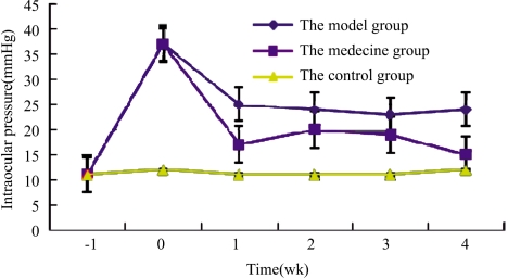
The intraocular pressures of the model group and the medicine group were notably elevated post-modeling and pre-treatment, which was around 37mmHg. But soon afterwards the pressures were significantly decreased. One week after modeling the intraocular pressures of the model group were stabilized to 24-25mmHg followed by some extent volatility. During the experiment, the overall intraocular pressures in the model group were significantly higher than that of the other groups. After one week's modeling the intraocular pressures of the medicine group were around 16-18mmHg followed by small amplitude of increase, then gradually declined with highest IOP around 20mmHg. The IOP of the control group was significantly lower than that of the other groups, which had a slight fluctuation between 11-12mmHg (Figure 1).
Figure 1. Intraocular pressure curves in rats before and after treatment.
mfERG changes
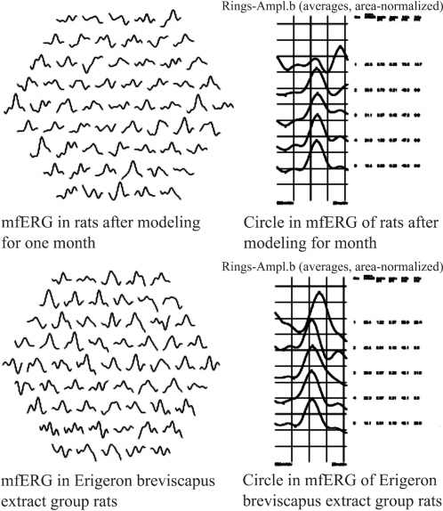
Based on preliminary results of previous experiments, the total wave peak latency, the total response density, P1 wave densities of the 2nd and 4th circles were selected for the intervention indicators of erigeron breviscapus extract. The results were shown as follows in Figure 2 and Table 1.
Figure 2. mfERG before and after treatment.
Table 1. mfERG changes affected by erigeron breviscapus extract in persistently elevated intraocular pressure in rats.
| Groups | Eye (n) | P1 wave |
P1 wave of the 2nd circle |
P1 wave of the 4th circle |
|
| Total peak latency(ms) | Total response density(nV/deg2) | Response density (ms) | Response density(nV/deg2) | ||
| Model | 16 | 58.01±9.8670b | 10.22±9.1417b | 15.95±12.0989b | 11.63±9.3884bb |
| Medicine | 16 | 50.85±5.3105a | 22.38±15.2867a,b | 29.22±14.3127a,b | 16.57±9.209b |
| Control | 16 | 49.09±2.3957 | 41.78±14.4258 | 60.41±26.0595 | 39.93±11.3192 |
aP<0.05 vs model group, bP<0.01 vs control group
Mean±SD
The results shown in Table 1 suggested that: 1) the total peak latency of P1 wave in mfERG of the model group was significantly longer than that of the control group with significantly difference (P<0.01). The total peak latency of P1 wave in mfERG of the medicine group had recovered to some extent, which had no significant difference compared with the control group (P>0.05) and no significant difference compared with the model group, either (P<0.05); 2) the total response density of P1 wave in mfERG of the model group and the medicine group had decreased compared with that of the control group and the difference was significant (P<0.01). The total response density of the medicine group had rebounded and the difference was significant compared with the model group (P<0.05); 3) the response density of P1 wave in the second concentric circles of mfERG in the model group and the medicine group had decreased compared with that of the control group and the difference was significant (P<0.01). While in the medicine group, the response density had rebounded compared with that of the model group, and the rebounding difference was significant (P<0.05); 4) in the model group and the medicine group, there was significant difference (P<0.01) in response density of P1 wave of the 4th concentric circle in mfERG compared with the control group. In the medicine group the response density had recovered to certain extent, but there was not significant difference compared with the model group (P>0.05).
DISCUSSION
Glaucoma is a degenerative disease of optic nerve. It is very important to clarify the pathogenesis and evaluate the effect of drug therapy and other interventions through observation of in vivo visual function damages. Generally speaking, this kind of optic nerve degenerative disease always had topography changes. Thereby, it is significant to recognize the local retinal responses for diagnosis and treatment.
Clinical researchers[6],[7] found that response density of P1 wave in first order kernel of fERG in glaucomatous and IOP elevated people was lower than that in normal people, however the peak latency showed no extending. The first order kernel and second order kernel of mfERG were sensitive methods to detect glaucoma, especially the quadrant reaction which could be sensitive enough to detect early abnormalities before glaucomatous visual field damage was observed. Some studies[8] also revealed when comparing glaucoma patients with normal people, the mean value of latency in first order kernel in the first positive wave was significantly longer (P<0.005) than that of the control group. But among these 18 patients, most of them had severe visual field defect although they were in normal range. Experimental studies showed that persistently high IOP had obvious effect on response density of P1 wave of the first order kernel in mfERG, and the changes were selective at the early time. As time went by, the affected area would gradually be expanded[9]. Experimental high IOP could lead to damages of optic disc and functions of periretinal area. Plus, examining in the position opposing optic disc center together with 5 circle wave analysis was valuable for the diagnosis of high IOP-induced optic nerve damage.
Erigeron breviscapus series is, herbal medicine produced in Yunnan Province which belongs to genus erigeron and genera compositae. Modern pharmacological studies have shown that this herb can expand blood vessels, reduce vascular resistance, increase the blood flow, reduce whole blood viscosity, and improve the experimental microcirculations. Since the 1990s, some scholars had given their endeavors to put it into use of glaucoma. The results revealed: erigeron breviscapus injection could improve the transport function of optic nerve axon under high intraocular pressure; Yimaikang(qingguangkang), with main components erigeron breviscapus, could protect retinal ganglion cells from apoptosis by increasing the activity of cytochrome oxidase of retinal ganglion cells and removing the free radicals produced by high intraocular pressure; clinical application had proved that erigeron breviscapus could improve the visual functions of IOP-controlled late stage glaucomatous patients, etc.[10]-[15].
In this experiment, the first order kernel of mfERG in persistently elevated IOP in rats was significantly abnormal, and erigeron breviscapus extract could partially restore its mfERG. These findings suggested that erigeron breviscapus extract had certain effect on improving visual function from impairment induced by persistently elevated intraocular pressure, and it was the effective part of erigeron breviscapus for optic neuroprotection.
Footnotes
Foundation item: National Major Science-Technology Project of Science and Technology Ministry-Major New Medicine Innovation (No. 2009ZX09103-369); National Natural Science Foundation of China (No. 30171172); Key Project of Education Department of Sichuan Province, China (No. 08ZA118)
REFERENCES
- 1.Flammer J. Vol. 1996. Basel: Karger; Ocular bloodflow; pp. 12–39. [Google Scholar]
- 2.Lu XJ, Duan JG, Liao PZ, Liu AQ, Zhang FW. Study on effect of multifocal electroretinogram in persistent elevated intraocular pressure rat model. The 3rd Global Chinese Ophthalmic Conference in Conjunction with 6th National Congress of Chinese Ophthalmological Society; p. 2006. [Google Scholar]
- 3.Lu XJ, Duan JG, Liao PZ, Zhang FW, Liu AQ. The effect of episcleral veins cauterization on intraocular pressure and retina in rat. Chinese Ophthalmic Research. 2005;23(4):386–388. [Google Scholar]
- 4.Ball S L, Petry HM. Noninvasive assessment of retinal function in rats using multifocal electroretinography. Invest Ophthalmol Vis Sci. 2000;41(2):610–617. [PubMed] [Google Scholar]
- 5.Nusinowitz S, Heckenlively JR. Rod multifocal electroretinograms in mice. Invest Ophthalmol Vis Sci. 1999;40(12):2848–2858. [PubMed] [Google Scholar]
- 6.Liu RJ, Yan L, Zhang MY. The performance of multifocal ERG of elevated intraocular pressure and glaucoma. Yanke Xin Jingzhan. 2001;21(4):292–293. [Google Scholar]
- 7.Yang L, Lu H, Yan L. Observation of primary open-angle glaucoma diagonosed by m-ERG. Yanke Xin Jingzhan. 2003;23(3):187–190. [Google Scholar]
- 8.Graham SL, Klistorner A. Electrophysiology: a review of signalorigins and applications to investigating glaucoma. Aust NZJ Ophthalmol. 1998;26(1):71–85. doi: 10.1046/j.1440-1606.1998.00082.x. [DOI] [PubMed] [Google Scholar]
- 9.Yang XG, Yu JG, Guo B. Changes of multifocal ERG in chronic high intraocular pressure rats. Yanke Xin Jingzhan. 2008;28(2):106–109. [Google Scholar]
- 10.Zhu YH, Jiang YQ, Liu ZH. The effect of Dengzhanxixin Injection on optic nerve axonal transport in experimental rats with elevated intraocular pressure. Zhonghua Yanke Zazhi. 2000;36(4):253–254. [Google Scholar]
- 11.Jiang YQ, Wu ZZ, Mo XJ. Discussion on the treatment of last stage glaucoma whose intraocular pressure is well controlled. Yanke Yanjiu. 1991;9:229. [Google Scholar]
- 12.Jia LJ, Jiang YQ, Wu ZZ. Observation of clinical effect of Qing Guang Kang Pian on the late stage primary glaucoma whose intraocular pressure (IOP) is well controlled. Zhonguo Shiyong Yanke Zazhi. 1994;12:269. [Google Scholar]
- 13.Jia LJ, Liu ZH, Luo XGc. The action of Qingguangkang Injection on retinal ganglion cells' metabolism function of acute experimental EIP rats. Zhonghua Yanke Zazhi. 1995;31:129. [Google Scholar]
- 14.Ye CH, Jiang YQ. Erigeron neuroprotective effects of glaucoma in clinical research. Yanke Yanjiu. 2003;21(3):307–311. [Google Scholar]
- 15.Wang NL, Sun XH, Li JZ. Multi-center clinical study in treatment of erigeron on glaucoma (in English) Guoji Yanke Zazhi (Int J Ophthalmol) 2004;5(4):587–592. [Google Scholar]


