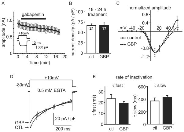
Figure 4. Gabapentin does not alter the density or kinetics of voltage-gated Ca2+ channel currents.
(A) Whole cell patch-clamp electrophysiology was used to record ICa elicited by 20 ms step depolarizations (see inset), and the peak amplitude (mean ± SEM) plotted against time. Acute application of 1mM gabapentin (indicated by black bar) had no effect on ICa amplitude. (B) Cells were treated with vehicle (ctl) or 1mM gabapentin (GBP) for 18–24 h prior to recording. Peak ICa was normalized to cell size (membrane capacitance) to yield current density. There was no change in current density (mean ± SEM) in gabapentin treated cells (n = 17) compared to control cells (n = 21) (p = 0.92). (C) The current-voltage relationship for ICa was not shifted by treatment with 1mM gabapentin for 18 – 24 h. ICa amplitude (mean ± SEM) was normalized to the peak inward current in each cell to facilitate comparison of the voltage-dependence. (D) Inactivation of ICa during a 500 ms step depolarization in control cells and cells treated for 18– 24 h with 1mM gabapentin. Traces represent the data from 8 control cells and 10 gabapentin treated cells (mean ± SEM). For clarity, error bars are shown for only a few data points. (E) Inactivation of ICa during a 500 ms step was fit with a double exponential decay and the time constants from each cell were pooled. There was no difference in either the fast (τ fast) (p = 0.28) or slow (τ slow) (p = 0.31) inactivation time constants between control (n = 8) and gabapentin treated cells (n = 10) (mean ± SEM).

