Abstract
Sterol regulatory element-binding protein-1c (SREBP-1c) enhances transcription of genes encoding enzymes of unsaturated fatty acid biosynthesis in liver. SREBP-1c mRNA is known to increase when cells are treated with agonists of liver X receptor (LXR), a nuclear hormone receptor, and to decrease when cells are treated with unsaturated fatty acids, the end products of SREBP-1c action. Here we show that unsaturated fatty acids lower SREBP-1c mRNA levels in part by antagonizing the actions of LXR. In cultured rat hepatoma cells, arachidonic acid and other fatty acids competitively inhibited activation of the endogenous SREBP-1c gene by an LXR ligand. Arachidonate also blocked the activation of a synthetic LXR-dependent promoter in transfected human embryonic kidney-293 cells. In vitro, arachidonate and other unsaturated fatty acids competitively blocked activation of LXR, as reflected by a fluorescence polarization assay that measures ligand-dependent binding of LXR to a peptide derived from a coactivator. These data offer a potential mechanism that partially explains the long-known ability of dietary unsaturated fatty acids to decrease the synthesis and secretion of fatty acids and triglycerides in livers of humans and other animals.
Keywords: fatty acid synthesis, arachidonic acid, nuclear receptors
Sterol regulatory element binding protein-1c (SREBP-1c) (1), also known as ADD-1 (2), is a membrane-bound transcription factor that functions at the interface between sterol and fatty acid metabolism. SREBP-1c enhances fatty acid synthesis by activating the genes encoding acetyl CoA carboxylase, fatty acid synthase (FAS), stearoyl CoA desaturase-1 (SCD-1), and other enzymes (3). The SREBP-1c gene is activated by insulin, which helps to explain the classic ability of insulin to enhance the conversion of glucose to fatty acids (4–6). In contrast, mRNA levels for SREBP-1c are decreased when cells are exposed to unsaturated fatty acids, the end products of SREBP-1c action (7–9).
Recent experiments have revealed that SREBP-1c mRNA levels are also controlled by oxysterols. In rodent liver and hepatoma cells, transcription of the SREBP-1c gene is stimulated by oxysterols that bind to the α and β forms of the liver X receptor (LXR α and β), two closely related members of the nuclear hormone receptor superfamily (10, 11). LXR activators include two naturally occurring sterols, 24(S),25-epoxycholesterol and 22(R)-hydroxycholesterol (12). LXR is also activated by T0901317, a synthetic nonsteroidal compound (10, 13). The levels of SREBP-1c mRNA declined dramatically when cultured rat hepatoma cells were treated with inhibitors of 3-hydroxy-3-methylglutaryl coenzyme reductase, which block the synthesis of endogenous LXR ligands (11). This inhibition was reversed when the cells were incubated with either T0901317 or mevalonate, the product of the reductase reaction. These data indicate that basal transcription of the SREBP-1c gene requires an endogenous sterol that activates LXR. The LXRs enhance transcription of the SREBP-1c gene by binding to a consensus recognition sequence in the enhancer region (10).
SREBP-1c belongs to a three-member family that includes SREBP-1a and SREBP-2. SREBP-1a and -1c are produced from a common gene that uses alternative promoters to produce different first exons that are spliced into a common second exon (14). The encoded proteins differ at their extreme NH2 termini, and this sequence difference affects their abilities to stimulate transcription of target genes. SREBP-1a is a strong activator of cholesterol and fatty acid biosynthesis, whereas SREBP-1c is selective for the fatty acid pathway (3, 15, 16). SREBP-2 is encoded by a separate gene, and it is relatively selective for cholesterol biosynthesis. Nonhepatic cells in tissue culture produce much more mRNA for SREBP-1a than for -1c (17). In contrast, the liver, which synthesizes lipids for export as well as for membrane maintenance, produces up to 10-fold more SREBP-1c than -1a (17).
All three SREBPs are synthesized as membrane-bound precursors of ≈1,150 aa in length (14). The NH2-terminal domain of ≈480 aa is a basic helix–loop–helix-leucine-zipper transcription factor. This domain is followed by a membrane attachment domain of ≈80 aa and a COOH-terminal regulatory domain of ≈590 aa. The SREBP precursors are initially bound to membranes of the endoplasmic reticulum. In sterol-depleted cells, they travel to the Golgi apparatus, where the NH2-terminal domains are released by two-step proteolysis, allowing them to enter the nucleus and activate transcription (14).
In view of the opposing effects of LXR agonists and unsaturated fatty acids on SREBP-1c mRNA levels, the current experiments were designed to test the hypothesis that unsaturated fatty acids act as direct antagonists of LXR. Consistent with this hypothesis, we show that the inhibitory effects of unsaturated fatty acids are reversed by LXR agonists in cultured rat hepatoma cells. We also present data to show that unsaturated fatty acids prevent agonists from enhancing the binding of LXR to a peptide derived from a coactivator that mediates transcriptional activation. Together, these findings provide evidence that unsaturated fatty acids reduce SREBP-1c mRNA by antagonizing the activation of LXR by endogenous ligands.
Materials and Methods
Materials.
We obtained fatty acids from Sigma-Aldrich and Nu Chek Prep, Elysian, MN; defatted BSA, 9-cis retinoic acid, chenodeoxycholic acid, and 17β-estradiol from Sigma-Aldrich; N-acetyl-leucinal-leucinal-norleucinal from Calbiochem; and [α-32P]CTP (800 Ci/mmol), redivue [α-32P]dCTP (3,000 Ci/mmol), and [α-32P]UTP (800 Ci/mmol) from Amersham Pharmacia. Delipidated FCS, sodium mevalonate, and sodium compactin were prepared as described (7, 18, 19). 24(S),25-Epoxycholesterol was synthesized by Julio Medina at Tularik.
Preparation of Albumin-Bound Fatty Acids.
A 5 or 10 mM stock solution of each fatty acid was prepared by diluting the free fatty acid in ethanol and precipitating it with the addition of NaOH (final concentration of 0.25 M) (7). Excess ethanol was evaporated under nitrogen gas, and the precipitated sodium salt was reconstituted with 0.9% (wt/vol) NaCl and stirred at room temperature for 10 min with defatted BSA [final concentration at 10% (wt/vol) in 0.15 M NaCl]. Each solution was adjusted to pH 7.4 with NaOH and stored in multiple aliquots at −20°C protected from light in tubes evacuated under nitrogen gas.
Culture and Fractionation of FTO-2B Cells.
The FTO-2B rat hepatoma cell line (obtained from R. E. Fournier, Fred Hutchinson Cancer Research Center, Seattle, WA) (20) was maintained in monolayer culture at 37°C in an 8% CO2 incubator. On day 0, cells were set up at a density of 7 × 10⁵ cells per 100-mm dish in medium A (1:1 mixture of Ham's F-12 medium and DMEM containing 100 units/ml penicillin and 100 μg/ml streptomycin sulfate) supplemented with 5% (vol/vol) FCS. On day 3, cells were washed with PBS and refed with medium B (medium A supplemented with 5% delipidated FCS) containing the appropriate additions. After incubation for 5 h at 37°C, cells received 25 μg/ml N-acetyl-leucinal-leucinal-norleucinal and were further incubated for 1 h. Cells were harvested and fractionated into nuclear extract and membrane fractions as described (21).
SDS/PAGE and Immunoblot Analysis.
Immunoblot analysis of endogenous rat SREBP-1 and -2 was carried out with 5 μg/ml of rabbit polyclonal antibodies against the NH2-terminal domains of either SREBP-1 (IgG-295) or SREBP-2 (IgG-302) (5). Bound antibodies were visualized with peroxidase-conjugated, affinity-purified donkey anti-rabbit IgG (Jackson ImmunoResearch), with the use of the SuperSignal CL-HRP substrate according to the manufacturer's instructions. SDS/PAGE gels (8%) were calibrated with prestained molecular weight markers (Bio-Rad). Blots were exposed to Kodak X-Omat film at room temperature.
Northern Blot Hybridization of mRNA.
Total RNA was isolated from two pooled dishes of FTO-2B cells with the use of an RNA Stat-60 kit (Tel-Test, Friendswood, TX). Cells were cultured as described above, except that N-acetyl-leucinal-leucinal-norleucinal was not added to the medium. Aliquots of total RNA (20 μg) from each sample were denatured with RNA sample loading buffer containing 50 μg/ml ethidium bromide, subjected to electrophoresis in a 1% agarose formaldehyde gel, and transferred to Hybond N+ membranes. The cDNA probes (22) were labeled with [α-32P]dCTP with the use of a Rediprimer II Random Labeling Kit (Amersham Pharmacia). The membranes were hybridized with the indicated 32P-labeled probe (2 × 10⁶ cpm/ml) for 2 h at 65°C with the use of Rapid-Hyb buffer (Amersham Pharmacia), washed twice with 0.1% (wt/vol) SDS/2× SSC at room temperature for 10 min, followed by 0.1% (wt/vol) SDS/0.1× SSC at 65°C for 15 min, and exposed at −80°C to Kodak X-Omat film with BioMax intensifying screens for the indicated time (1× SSC = 0.15 M sodium chloride/0.015 M sodium citrate, pH 7).
RNase Protection Assay.
Aliquots of total RNA (20 μg) were hybridized with [α-32P]CTP-labeled rat SREBP-1, SREBP-2, and β-actin cRNA probes at 68°C for 10 min with the use of a HybSpeed RPA Kit (Ambion, Austin, TX) as described (5, 11). The mixture was digested with RNase A/T1, and protected fragments corresponding to rat SREBP-1a (257 bp), -1c (160 bp), and -2 (110 bp) were separated on 8 M urea/5% polyacrylamide gels. Gels were dried and subjected to autoradiography with reflection film and BioMax intensifying screens at −80°C.
Nuclear Run-On Assay.
FTO-2B cells were set up at a density of 2 × 10⁶ cells per 150-mm dish in medium A supplemented with 5% FCS. After 60 h, the cells were washed with PBS and refed with medium B with or without 100 μM sodium arachidonate and 1 μM T0901317. After incubation for 6 h, the cells from four dishes were pooled and lysed in 0.5% (vol/vol) Nonidet P-40 lysis buffer (23). Nuclei were stored until use at −80°C in buffer containing 75 mM Hepes (pH 7.5), 40% (vol/vol) glycerol, 60 mM KCl, 5 mM MgCl2, 0.1 mM sodium EDTA, 0.5 mM spermine, 1 mM spermidine, and 0.5 mM DTT. Nuclear run-on assays were performed as described by labeling nuclei with [α-32P]UTP (24). Extracted RNA (≈8 × 10⁶ cpm) was hybridized at 42°C for 72 h to nitrocellulose membranes containing linearized cDNAs for rat SREBP-1 (3.4 kb), rat FAS (7.2 kb), and rat phosphoenolpyruvate carboxykinase (2.26 kb). Membranes were washed overnight in 0.1% SDS/0.1× SSC at 65°C.
Fluorescence Polarization Assay.
We used a previously described fluorescence polarization assay to measure agonist binding to LXRα and other nuclear receptors (13). The assay measures the reduction in the tumbling rate that occurs when a specific rhodamine-labeled peptide (8 aa) containing the coactivator signature motif LXXLL (25) interacts with a ligand-bound nuclear receptor. The ligand-binding domains of nuclear receptors LXRα, FXR, retinoid X receptorα (RXRα), and estrogen receptor β (ERβ) were produced in Escherichia coli as glutathione S-transferase (GST) fusion proteins and purified as described (26). A rhodamine-labeled LXXLL-containing peptide (10 nM) with the amino acid sequence ILRKLLQE was incubated with 500 nM GST-LXRα, GST-FXR, GST-RXRα, or GST-ERβ in 50 μl of buffer (10 mM Hepes/150 mM NaCl/2 mM MgCl2/5 mM DTT at pH 7.9) in 384-well black polypropylene plates as described (13). BSA-conjugated fatty acids were added to the reaction mix just before one of the following agonists was added: 300 nM 24(S),25-epoxycholesterol, 20 μM chenodeoxycholic acid, 100 nM 9-cis retinoic acid, or 100 nM 17-β estradiol. After overnight incubation at room temperature, fluorescence polarization (mP) was measured with a CRI Symmetry Fluorescent Plate Reader (CRI, Woburn, MA).
Gal4-Ligand Binding Domain Transient Transfection Assay.
Human embryonic kidney-293 (HEK-293) cells were transiently transfected in 24-well plates with the use of Effectene Transfection Reagent (Qiagen, Chatsworth, CA). Each well received 20–50 ng of a luciferase reporter construct containing four copies of the yeast transcription factor Gal4 response element and a TATA element derived from the thymidine kinase promoter together with 40 ng of an expression vector containing the Gal4 DNA binding domain fused to the ligand-binding domain of LXRα (amino acids 142–447), FXR (amino acids 105–472), or RXRα (amino acids 1–462) (13). An expression plasmid containing the β-galactosidase gene driven by the cytomegalovirus promoter (4 ng/well) was cotransfected for normalization.
Results
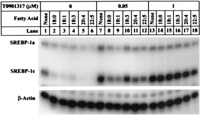
To measure the effects of fatty acids and LXR agonists on SREBP-1c mRNA levels, we studied FTO-2B cells, a line of cultured rat hepatoma cells that resembles the liver in producing relatively high levels of the SREBP-1c transcript (SREBP-1c:1a ratio of 3:1 when grown in 5% delipidated FCS). The cells were incubated for 6 h in medium containing delipidated FCS, which is nearly devoid of fatty acids (<0.5 μM) and cholesterol (<0.5 μg/ml). We supplemented the medium with varying concentrations of fatty acids and then measured the amounts of the three SREBP transcripts with the use of an RNase protection assay (11). In the absence of fatty acids, the amount of SREBP-1c mRNA was high (Fig. 1, lane l), and it was reduced in the presence of a variety of fatty acids added at 100 μM concentrations (Fig. 1, lanes 2–6). Active fatty acids included stearate (18 carbons, 0 double bonds) as well as monounsaturated and polyunsaturated fatty acids of 18, 20, or 22 carbons. The effectiveness of stearate was unexpected on the basis of prior studies with cultured kidney-derived cells from humans (HEK-293 cells) (7) and monkeys (CV-1 cells) (27). The effect of stearate in the FTO-2B cells was reproducible in multiple experiments, and it may be attributable to the high levels of SCD-1 activity in these cells, which allows them to convert stearate to oleate.
Figure 1.
SREBP mRNA expression in FTO-2B cells treated with different fatty acids in the absence or presence of LXR agonist T0901317, as measured by RNase protection. On day 0, FTO-2B cells were set up in medium A containing 5% FCS as described in Materials and Methods. On day 3, cells were switched to medium B containing 100 μM of the indicated fatty acid (added in a final concentration of 0.1% BSA) in the presence or absence of the indicated concentration of LXR agonist T0901317 (added in a final concentration of 0.1% DMSO). All dishes of cells received 0.1% BSA and 0.1% DMSO. After incubation for 6 h at 37°C, the cells from three dishes were harvested and pooled for RNA isolation. Aliquots of pooled total RNA (20 μg) were hybridized for 10 min at 68°C to the indicated 32P-labeled cRNA probe. RNase-protected fragments were separated by gel electrophoresis and exposed to film at −80°C for 6 h.
When the fatty acids were added to the FTO-2B cells together with the LXR ligand T0901317, their ability to suppress SREBP-1c mRNA was blocked (Fig. 1). This block was evident at 0.05 μM T0901317 (Fig. 1, lanes 8–12) and complete at 1 μM (Fig. 1, lanes 14–18). All of these effects were relatively specific for SREBP-1c. The SREBP-1a transcript was only slightly affected by fatty acids and T0901317 (Fig. 1), and the SREBP-2 transcript was not affected at all (see Fig. 3B).
Figure 3.
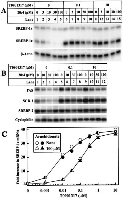
Suppression of mRNAs encoding SREBP-1c and two target genes by varying concentrations of arachidonate in the absence and presence of T0901317. On day 0, FTO-2B cells were set up in medium A containing 5% FCS. (A and B) On day 3, cells were switched to medium B containing the indicated concentration of sodium arachidonate and T0901317. All dishes of cells received 0.1% BSA and 0.1% DMSO. After incubation for 6 h, the total RNA from two dishes was pooled and isolated. (A) Aliquots of total RNA (20 μg) were hybridized for 10 min at 68°C to the indicated 32P-labeled cRNA probe. RNase-protected fragments were separated by gel electrophoresis and exposed to film at −80°C for 8 h (SREBP-1a and -1c) or 6 h (β-actin). (B) Aliquots of total RNA (20 μg) were subjected to electrophoresis and Northern blot hybridization with the indicated 32P-labeled probe, and filters were exposed to film at −80°C for 3 h (FAS), 4 h (SCD-1), or 8 h (SREBP-2 and cyclophilin). (C) On day 2, cells were switched to medium B containing 50 μM compactin and 50 μM sodium mevalonate. On day 3, cells were refed with the same medium containing the indicated concentrations of T0901317 and arachidonate. All dishes of cells received 0.1% BSA and 0.1% DMSO. After incubation for 6 h, total RNA from duplicate dishes was separately isolated, aliquots (20 μg) were analyzed as described in A, and the bands for SREBP-1c were quantified with a Fuji Bio-Imaging analyzer and normalized to the β-actin control. Values for each of the duplicate incubations are shown.
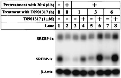
Fig. 2 shows that the LXR ligand reversed the fatty acid suppression in rapid fashion. In this experiment, cells were preincubated with arachidonate (20:4) for 6 h, during which time the SREBP-1c mRNA declined (lane 2). The mRNA rose markedly within 1 h after the addition of T0901317, even though the arachidonate was maintained in the culture medium (lane 4). The increase persisted at 3 and 6 h (lanes 6 and 8).
Figure 2.
T0901317 rapidly reverses the suppressive effect of arachidonate on SREBP-1c mRNA. On day 0, FTO-2B cells were set up in medium A containing 5% FCS. On day 3, the cells were switched to medium B and preincubated for 6 h in the absence or presence of 100 μM sodium arachidonate in a final concentration of 0.1% BSA. Two groups of cells were then harvested (lanes 1 and 2), and the remaining cells (lanes 3–8) were treated with either 1 μM T0901317 in 0.1% DMSO or 0.1% DMSO alone for the indicated time. Total RNA from three dishes was pooled and isolated. Aliquots of total RNA (20 μg) were hybridized for 10 min at 68°C to the indicated 32P-labeled cRNA probe. RNase-protected fragments were separated by gel electrophoresis and exposed to film at −80°C for 6 h.
Fig. 3 A and B show the effects of various concentrations of arachidonate on SREBP-1c mRNA levels as measured by RNase protection (A) and on the levels of mRNA for two target genes, FAS and SCD-1, as measured by Northern blotting (B). In the absence of an LXR ligand, arachidonate suppressed the SREBP-1c mRNA maximally at 10 μM, and a slightly higher concentration (30 μM) gave maximal suppression of the FAS and SCD-1 mRNAs. T0901317 at concentrations of 0.1 and 10 μM overcame the inhibitory effect of arachidonate on the SREBP-1c mRNA and the mRNAs for the two target genes. Arachidonate had minor effects on the SREBP-1a mRNA (A), and it had no effect on the SREBP-2 mRNA (B). As controls, we studied the mRNA for β-actin (A) and cyclophilin (B), neither of which was affected by arachidonate or T0901317.
Fig. 3C shows that arachidonate behaved as a competitive inhibitor of T0901317 in inducing SREBP-1c mRNAs. We preincubated the cells with compactin, an inhibitor of 3-hydroxy-3-methylglutaryl CoA reductase, and a low concentration of mevalonate, which permits synthesis of nonsterol isoprenes but not sterols. This treatment deprives cells of an endogenous sterol ligand for LXR, and therefore it lowers SREBP-1c mRNA levels (11). In the absence of arachidonate, T0901317 increased SREBP-1c mRNA maximally at 1 μM. In the presence of 100 μM arachidonate, the T0901317 dose–response curve was shifted ≈10-fold to the right, but the maximal level of stimulation was unchanged.
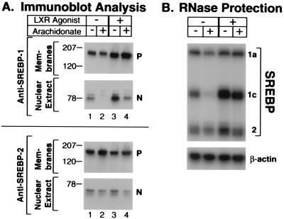
Previous studies in HEK-293 cells (which produce SREBP-1a predominantly) indicated that unsaturated fatty acids decrease nuclear SREBP-1 protein levels by two mechanisms: mRNA suppression and partial inhibition of proteolytic cleavage (7). To determine the effects of T0901317 on these responses, we measured the amounts of the membrane-bound precursor and the nuclear forms of SREBP-1 in FTO-2B cells by immunoblotting with the best available antibody, which does not distinguish between the SREBP-1a and -1c isoforms (Fig. 4A). When added for 6 h in the absence of T0901317, arachidonate markedly decreased the amount of nuclear SREBP-1 (Fig. 4A Upper, lane 2). The amount of the membrane-bound precursor form of SREBP-1 did not change. T0901317 increased both the precursor and nuclear forms of SREBP-1. In the presence of T0901317, arachidonate decreased the nuclear form of SREBP-1c only partially (Fig. 4A, lane 4). As expected, in the same experiment arachidonate decreased the SREBP-1c mRNA in the absence, but not the presence, of T0901317 (Fig. 4B). The partial reduction of nuclear SREBP-1 protein by arachidonate in the presence of T0901317 may represent the arachidonate-mediated inhibition of SREBP processing. Arachidonate had minor effects on the amount of nuclear SREBP-2 in the absence and presence of T0901317 (Fig. 4A Lower).
Figure 4.
Immunoblot analysis (A) and RNase protection assay (B) of SREBPs in FTO-2B cells incubated with arachidonate and LXR agonist T0901317. On day 0, cells were set up in medium A supplemented with 5% FCS. On day 3, cells were switched to medium B supplemented with 100 μM sodium arachidonate and 1 μM T0901317 as indicated. The medium in all cultures contained 0.1% BSA and 0.1% DMSO. (A) After a 5-h incubation at 37°C, cells received a direct addition of N-acetyl-leucinal-leucinal-norleucinal at 25 μg/ml and were harvested 1 h later. Membrane and nuclear extract fractions were prepared and subjected to SDS/PAGE (68 μg protein per lane) and immunoblot analysis as described in Materials and Methods. Filters were exposed to film at room temperature for 5 s. P and N denote precursor and nuclear cleaved forms of SREBPs, respectively. (B) After incubation at 37°C for 6 h, cells were harvested, and aliquots of total RNA from pooled dishes (20 μg) were hybridized for 10 min at 68°C to 32P-labeled cRNA probes for rat SREBP-1, SREBP-2, and β-actin as described in Materials and Methods. RNase protected fragments were separated by gel electrophoresis and exposed to film for 6 h at −80°C.
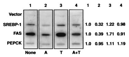
To demonstrate directly that arachidonate and T0901317 affect SREBP-1c gene transcription, we incubated FTO-2B cells with these reagents for 6 h, isolated nuclei, and pulse-labeled them with [α-32P]UTP. The data showed that arachidonate reduced SREBP-1 transcription by 68%, and this reduction was reversed by T0901317 (Fig. 5). Arachidonate had a similar inhibitory effect on transcription of the FAS gene, which is a target of SREBP-1. There was no effect on the phosphoenolpyruvate carboxykinase gene, which served as a control (Fig. 5).
Figure 5.
Nuclear run-on assays of nascent 32P-labeled RNA transcripts isolated from the nuclei of FTO-2B cells treated with arachidonate in the absence or presence of T0901317. Cells were set up for experiments as described in Materials and Methods. After incubation for 6 h in medium B with or without 100 μM sodium arachidonate (A) in the absence or presence of 1 μM T0901317 (T) as indicated, the cells from four 150-mm dishes were pooled, lysed, and subjected to nuclear run-on assay as described in Materials and Methods. After hybridization with the indicated cDNA probe, the washed filters were exposed to Kodak X-Omat film with a Bio-Max (Kodak) intensifying screen at −80°C for 24 h. Relative levels of the transcripts for each gene (shown at right) were quantified with a Fuji Bio-Imaging analyzer. A blank value corresponding to the vector band was subtracted from all values.
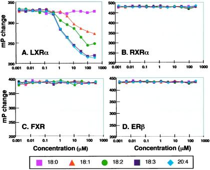
To test directly the hypothesis that arachidonate blocks ligand-mediated activation of LXR, we performed a series of in vitro assays. Previous studies showed that activation of nuclear hormone receptors increases the ability of the receptors to bind to coactivators that contain the consensus sequence leucine-X-X-leucine-leucine (LXXLL), where X is any amino acid (25). This binding can be studied in vitro with the use of a fluorescence polarization assay that measures the binding of a fluorescently labeled LXXLL peptide to the nuclear receptor (13). Binding decreases the tumbling rate of the fluorophore, and thus it leads to enhanced fluorescence polarization. Fig. 6A shows an assay in which the binding of the LXXLL peptide to LXRα was stimulated by the natural ligand 24(S),25-epoxycholesterol. The addition of three polyunsaturated fatty acids (18:2, 18:3, and 20:4) decreased binding of the peptide, as manifested by a decrease in fluorescence polarization (mP change). A noticeable effect was observed at fatty acid concentrations of 1–10 μM. Oleate (18:1) was less effective, and stearate (18:0) had no effect. The specificity of this response was tested by showing that the fatty acids did not affect binding of the LXXLL peptide to three other nuclear hormone receptors: RXRα (stimulated by 9-cis retinoic acid) (Fig. 6B), FXR (stimulated by chenodeoxycholic acid) (Fig. 6C), and the ERβ (stimulated by 17-β estradiol) (Fig. 6D).
Figure 6.
Fatty acids block activation of LXRα, but not other nuclear receptors, as measured in a fluorescence polarization assay. The indicated BSA-conjugated fatty acid was evaluated for its ability to inhibit the binding of an LXXLL-containing peptide to LXRα, RXRα, FXR, or ERβ in the presence of the corresponding agonist: (A) 3 μM 24(S),25-epoxycholesterol for LXRα; (B) 100 nM 9-cis retinoic acid for RXRα; (C) 20 μM chenodeoxycholic acid for FXR; or (D) 100 nM 17-β estradiol for ERβ. Data are presented as the increase in fluorescence polarization (mP) normalized to the BSA control.
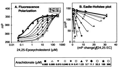
To determine whether arachidonate acts as a competitive antagonist of the activating ligand, we measured fluorescence polarization as a function of the concentration of 24(S),25-epoxycholesterol at varying concentrations of arachidonate (Fig. 7A). The data showed that arachidonate had its greatest inhibiting effect at low concentrations of 24(S),25-epoxycholesterol, but high concentrations of the activator were able to overcome the arachidonate effect. An Eadie–Hofstee plot of these data (Fig. 7B) showed that all lines converged at a common intercept on the vertical axis, indicating that arachidonate was indeed acting as a competitive antagonist of 24(S),25-epoxycholesterol. We calculated that the effect of arachidonate was half-maximal at 1.5 μM.
Figure 7.
Arachidonate competitively inhibits LXR activation by 24(S),25-epoxycholesterol as measured by fluorescence polarization. (A) The indicated concentration of sodium arachidonate (20:4) was evaluated for its ability to inhibit the binding of an LXXLL-containing peptide to LXRα in the presence of the indicated concentration of 24(S),25-epoxycholesterol. Data are presented as the increase in fluorescence polarization (mP) normalized to the BSA control. (B) Eadie–Hofstee plot of the data in A. A blank value for fluorescence polarization in the absence of 24,25-epoxycholesterol was subtracted from all measured values.
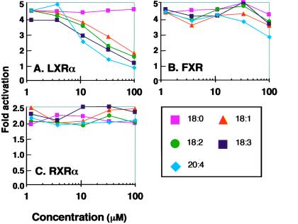
A way to test the effects of regulators on nuclear hormone receptors in vivo is through the use of nuclear receptor-Gal4 fusion proteins (13). Accordingly, we transfected HEK-293 cells with a cDNA encoding a chimeric protein that contains the DNA binding domain of Gal4 and the ligand-binding and transcription-activating domains of LXRα. We also transfected a cDNA encoding luciferase driven by a promoter containing four copies of a Gal4 binding sequence. When the transfected cells were incubated with the LXR ligand 24(S),25-epoxycholesterol, luciferase activity rose by 4- to 5-fold (Fig. 8A). Addition of each of the unsaturated fatty acids lowered luciferase activity to the basal level. Stearate (18:0) had no effect, most likely because HEK-293 cells have low levels of desaturase activity. As controls, we studied Gal4 fusion proteins containing the ligand-binding domain of FXR (Fig. 8B) and RXRα (Fig. 8C). When incubated with their respective ligands, both of these proteins stimulated the luciferase reporter gene. The fatty acids had no significant effect on these activations.
Figure 8.
Fatty acids selectively antagonize transcription-stimulating activity of LXR in HEK-293 cells. Cells were transiently cotransfected with a Gal4-driven luciferase reporter construct; an expression vector containing the Gal4 DNA binding domain fused to the ligand-binding domain of LXRα (A), FXR (B), or RXRα (C) as indicated; and a control plasmid containing the β-galactosidase gene driven by the cytomegalovirus promoter. Six to eight hours after transfection, the natural ligands (5 μM 24(S),25-epoxycholesterol for LXRα, 25 μM chenodeoxycholic acid for FXR, and 50 nM 9-cis retinoic acid for RXRα) and the indicated fatty acids were added to the medium in complex with BSA as described in the legend to Fig. 1. After incubation for 20 h, the cells were harvested for assay. Data are presented as relative luciferase activity normalized to β-galactosidase activity.
Discussion
The current results establish that unsaturated fatty acids can act as competitive antagonists of LXR in cultured rat hepatoma and human HEK-293 cells and in cell-free assays that reflect LXR activation. This antagonism appears to explain, at least partially, the ability of unsaturated fatty acids to lower the levels of mRNA for SREBP-1c (7–9), the transcription of which has been shown to depend on an endogenous LXR ligand (11). The lowered SREBP-1c, in turn, leads to a fall in mRNAs for enzymes responsible for synthesizing unsaturated fatty acids, thus completing a feedback loop.
A noteworthy difference between the in vivo and in vitro assays of LXR antagonism relates to the effect of stearate (18:0), which acted as an LXR antagonist in intact FTO-2B cells (Fig. 1), but not in the in vitro assays (Fig. 6) or in the HEK-293 cells (Fig. 8). This difference may reflect a higher level of desaturase activity in the FTO-2B cells, which convert stearate to oleate. This hypothesis has yet to be tested.
The in vitro assays were performed with fatty acids bound to albumin. Under these conditions, arachidonate had a half-maximal effect at 1.5 μM, which is lower than the 10 μM concentration that was required when added to intact cells (Fig. 3). The levels of free fatty acids within cells are generally thought to be low, and they are largely bound to intracellular binding proteins. It is possible that fatty acids are more effective when bound to these physiological carriers. It is also possible that the true antagonists are not free fatty acids, but rather are their CoA or carnitine thioesters. We attempted to inhibit LXR activation in vitro with the use of fatty acyl CoAs, obtaining negative results in the fluorescence polarization assay.
In addition to transcriptional inhibition mediated by LXR antagonism, unsaturated fatty acids also appear to lower SREBP-1 mRNA by accelerating its degradation. This effect of fatty acids has been demonstrated directly by studies of mRNA decay rates in the presence of α-amanitin in freshly isolated rat hepatocytes (9). It is likely that such an effect also occurred in the current studies, inasmuch as arachidonate inhibited SREBP-1 transcription by about 3-fold, as determined by the nuclear run-on assay (Fig. 5) at a time when the mRNA level declined by 6-fold, as determined by Northern blotting (data not shown). Arachidonate also inhibits the proteolytic processing of SREBP-1a and -1c in HEK-293 cells (7). Thus, arachidonate appears to lower nuclear SREBP-1c levels by at least three mechanisms in animal cells.
When fed to experimental animals, polyunsaturated acids of the n-3 and n-6 classes have long been known to inhibit fatty acid and triglyceride synthesis and to lower plasma triglyceride levels (28). Accumulating evidence, some of which is presented here, strongly suggests that these effects are mediated by a reduction in the levels of nuclear SREBP-1c in liver. In contrast, LXR agonists raise plasma triglycerides in experimental animals, apparently through induction of hepatic SREBP-1c (13). Agents that lower nuclear SREBP-1c levels, either by antagonizing LXR action in the liver or by some other means, should be effective in lowering plasma triglyceride levels in humans.
Acknowledgments
We thank Jay Horton and David Mangelsdorf for their critical review of the manuscript and Voe Hannah for helpful suggestions in the initial phases of the study. Lisa Beatty and Christine Alvares provided invaluable help with cultured cells. This research was made possible by grants from the National Institutes of Health (HL20948) and the Perot Family Foundation. R.A.D.-B. is the recipient of a postdoctoral fellowship from the Jane Coffin Childs Memorial Fund for Medical Research.
Abbreviations
- ER
estrogen receptor
- FAS
fatty acid synthase
- GST
glutathione S-transferase
- HEK-293 cells
human embryonic kidney-293 cells
- LXR
liver X receptor
- RXR
retinoid X receptor
- SCD-1
stearoyl-CoA desaturase-1
- SREBP
sterol regulatory element-binding protein
References
- 1.Yokoyama C, Wang X, Briggs M R, Admon A, Wu J, Hua X, Goldstein J L, Brown M S. Cell. 1993;75:187–197. [PubMed] [Google Scholar]
- 2.Kim J B, Spiegelman B M. Genes Dev. 1996;10:1096–1107. doi: 10.1101/gad.10.9.1096. [DOI] [PubMed] [Google Scholar]
- 3.Shimano H, Horton J D, Shimomura I, Hammer R E, Brown M S, Goldstein J L. J Clin Invest. 1997;99:846–854. doi: 10.1172/JCI119248. [DOI] [PMC free article] [PubMed] [Google Scholar]
- 4.Foretz M, Guichard C, Ferre P, Foufelle F. Proc Natl Acad Sci USA. 1999;96:12737–12742. doi: 10.1073/pnas.96.22.12737. [DOI] [PMC free article] [PubMed] [Google Scholar]
- 5.Shimomura I, Bashmakov Y, Ikemoto S, Horton J D, Brown M S, Goldstein J L. Proc Natl Acad Sci USA. 1999;96:13656–13661. doi: 10.1073/pnas.96.24.13656. [DOI] [PMC free article] [PubMed] [Google Scholar]
- 6.Shimomura I, Hammer R E, Ikemoto S, Brown M S, Goldstein J L. Nature (London) 1999;401:73–76. doi: 10.1038/43448. [DOI] [PubMed] [Google Scholar]
- 7.Hannah V C, Ou J, Luong A, Goldstein J L, Brown M S. J Biol Chem. 2001;276:4365–4372. doi: 10.1074/jbc.M007273200. [DOI] [PubMed] [Google Scholar]
- 8.Mater M K, Thelen A P, Pan D A, Jump D B. J Biol Chem. 1999;274:32725–32732. doi: 10.1074/jbc.274.46.32725. [DOI] [PubMed] [Google Scholar]
- 9.Xu J, Teran-Garcia M, Park J H Y, Nakamura M T, Clarke S D. J Biol Chem. 2001;276:9800–9807. doi: 10.1074/jbc.M008973200. [DOI] [PubMed] [Google Scholar]
- 10.Repa J J, Liang G, Ou J, Bashmakov Y, Lobaccaro J-M A, Shimomura I, Shan B, Brown M S, Goldstein J L, Mangelsdorf D J. Genes Dev. 2000;14:2819–2830. doi: 10.1101/gad.844900. [DOI] [PMC free article] [PubMed] [Google Scholar]
- 11.DeBose-Boyd R A, Ou J, Goldstein J L, Brown M S. Proc Natl Acad Sci USA. 2001;98:1477–1482. doi: 10.1073/pnas.98.4.1477. [DOI] [PMC free article] [PubMed] [Google Scholar]
- 12.Janowski B A, Grogan M J, Jones S A, Wisely G B, Kliewer S A, Corey E J, Mangelsdorf D J. Proc Natl Acad Sci USA. 1999;96:266–271. doi: 10.1073/pnas.96.1.266. [DOI] [PMC free article] [PubMed] [Google Scholar]
- 13.Schultz J R, Tu H, Luk A, Repa J J, Medina J C, Li L, Schwendner S, Wang S, Thoolen M, Mangelsdorf D J, et al. Genes Dev. 2000;14:2831–2838. doi: 10.1101/gad.850400. [DOI] [PMC free article] [PubMed] [Google Scholar]
- 14.Brown M S, Goldstein J L. Cell. 1997;89:331–340. doi: 10.1016/s0092-8674(00)80213-5. [DOI] [PubMed] [Google Scholar]
- 15.Pai J, Guryev O, Brown M S, Goldstein J L. J Biol Chem. 1998;273:26138–26148. doi: 10.1074/jbc.273.40.26138. [DOI] [PubMed] [Google Scholar]
- 16.Horton J D, Shimomura I, Brown M S, Hammer R E, Goldstein J L, Shimano H. J Clin Invest. 1998;101:2331–2339. doi: 10.1172/JCI2961. [DOI] [PMC free article] [PubMed] [Google Scholar]
- 17.Shimomura I, Shimano H, Horton J D, Goldstein J L, Brown M S. J Clin Invest. 1997;99:838–845. doi: 10.1172/JCI119247. [DOI] [PMC free article] [PubMed] [Google Scholar]
- 18.Brown M S, Faust J R, Goldstein J L, Kaneko I, Endo A. J Biol Chem. 1978;253:1121–1128. [PubMed] [Google Scholar]
- 19.Goldstein J L, Basu S K, Brown M S. Methods Enzymol. 1983;98:241–260. doi: 10.1016/0076-6879(83)98152-1. [DOI] [PubMed] [Google Scholar]
- 20.Wynshaw-Boris A, Lugo T G, Short J M, Fournier R E K, Hanson R W. J Biol Chem. 1984;259:12161–12169. [PubMed] [Google Scholar]
- 21.DeBose-Boyd R A, Brown M S, Li W-P, Nohturfft A, Goldstein J L, Espenshade P J. Cell. 1999;99:703–712. doi: 10.1016/s0092-8674(00)81668-2. [DOI] [PubMed] [Google Scholar]
- 22.Shimano H, Horton J D, Hammer R E, Shimomura I, Brown M S, Goldstein J L. J Clin Invest. 1996;98:1575–1584. doi: 10.1172/JCI118951. [DOI] [PMC free article] [PubMed] [Google Scholar]
- 23.Sambrook J, Russell D W. Molecular Cloning: A Laboratory Manual. Plainview, NY: Cold Spring Harbor Lab. Press; 2001. [Google Scholar]
- 24.Zhang J, Ou J, Bashmakov Y, Horton J D, Brown M S, Goldstein J L. Proc Natl Acad Sci USA. 2001;98:3756–3761. doi: 10.1073/pnas.071054598. . (First Published March 20, 2001, 10.1073/pnas.071054598) [DOI] [PMC free article] [PubMed] [Google Scholar]
- 25.Heery D M, Kalkhoven E, Hoare S, Parker M G. Nature (London) 1997;387:733–736. doi: 10.1038/42750. [DOI] [PubMed] [Google Scholar]
- 26.Smith D B, Johnson K S. Gene. 1988;67:31–40. doi: 10.1016/0378-1119(88)90005-4. [DOI] [PubMed] [Google Scholar]
- 27.Worgall T S, Sturley S L, Seo T, Osborne T F, Deckelbaum R J. J Biol Chem. 1998;273:25537–25540. doi: 10.1074/jbc.273.40.25537. [DOI] [PubMed] [Google Scholar]
- 28.Jump D B, Clarke S D. Annu Rev Nutr. 1999;19:63–90. doi: 10.1146/annurev.nutr.19.1.63. [DOI] [PubMed] [Google Scholar]


