Abstract
Tuberculosis (TB) remains a major global disease, and diabetes which is documented to increase susceptibility to TB threefold, is also becoming pandemic. This susceptibility has been attracting extensive research interest. The increased risk of TB in diabetes may serve as a unique model to understand host susceptibility to specific pathogens in humans. To examine this rationale, we investigated expression of reported TB candidate genes in a longitudinal diabetes study. Two genes HK2 and CD28 emerged as potential culprits in diabetes-increased TB susceptibility.
Keywords: CD28, HK2, host susceptibility, type 2 diabetes, transcriptome, tuberculosis
Tuberculosis (TB) control is a major health issue in Low and Middle Income Countries (LMIC). Diabetes triples the risk of TB by predisposing to both primary and reactivation TB (1–2). The sheer number of people with diabetes is vastly larger than those infected with HIV. Diabetes is set to become the major global driving force for spread of TB and threatens TB control(3). Understanding the molecular mechanisms of host susceptibility to TB is an important knowledge gaining process, for the ultimate aim of improving control of this stubborn disease. Genetic association studies (GAS), especially the rise of genome-wide association studies (GWAS) in recent years, have been a great success in acquiring new understanding of human common diseases. However, in the case of tuberculosis, results from GAS tend to lack replicability. A recent, well-designed GWAS study using specimens from two large cohorts identified a genetic association signal from a gene-poor region(4). However, genetic associations previously reported from candidate gene studies were not replicated with statistical significance. Host susceptibility to TB in these studies was largely defined by the pathogenesis and virulence of the bacilli, and the intensity of exposure. Other significant confounding factors include aging, diabetes status, HIV infection, nutritional status, and administration of immunosuppressive drugs. Therefore, much more research effort and support is justified for understanding the contribution of host genes to the susceptibility to TB. The fact that TB is a major health issue mainly in Low and Middle Income Countries (LMIC) increases the difficulty in obtaining substantial support for TB GAS research.
To understand the molecular mechanisms underlying the risk of TB in diabetes and to develop a unique model for these studies we investigated expression of reported TB candidate genes in a longitudinal diabetes study within our Cameron County Hispanic Cohort (CCHC: n=2500) consisting of community-recruited, randomly-selected Mexican Americans from a population on the US/Mexico border where we find high rates of both diabetes and tuberculosis. In this same population we have shown that about 30% of TB cases can be attributed to diabetes(5). We followed up 9 CCHC individuals (3 males and 6 females, age range from 32 to 74 years of age with the median of 57) from impaired fasting glucose (pre-diabetes: fasting blood glucose 100–126 mg/dl) to frank clinical diabetes (fasting blood glucose >126 mg/dl). We performed quarterly visits over three years at which clinical status was reviewed and blood specimens taken. The study was approved by the Committee for the Protection of Human Subjects of the University of Texas Health Science Center at Houston (UTHealth), and written informed consent was obtained from each participant.
Gene expression in white blood cells taken at each quarterly visit was profiled using the Illumina HumanHT-12 Beachip (Illumina, San Diego). We observed expression profiles of RNA samples that were mainly clustered by individual, not by disease status, as shown by the principal component analysis. Because of the individual variance, we performed the statistical analysis by pairwise Z test(6) to compare expression levels in prediabetes with diabetes for each individual.
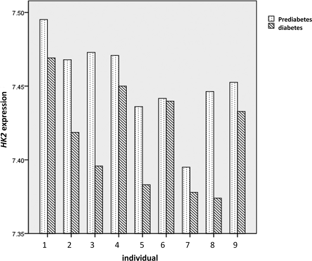
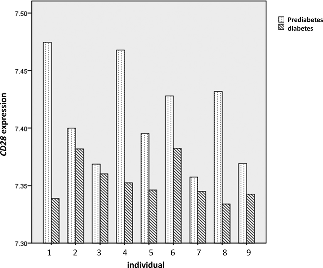
Two sets of TB candidate genes were examined respectively. The first set was 22 TB candidate genes highlighted by previous genetic studies(4). Eight genes were undetectable in our assay and there was no statistical significance in gene expression observed among the remaining 14 genes (Supplementary Table 1). The second set was 312 genes recently identified as the blood transcriptional signature of human tuberculosis by Berry et al.(7) Forty-five of these genes were undetectable in our assay. Among the remaining 267 genes, 13 were identified as having nominal statistical significance (P<0.05). After correction for the multiple comparisons with the Benjamini and Hochberg False Discovery Rate (FDR) using QVALUE software(8), two genes HK2 and CD28, were identified as having statistically significant reduction in expression in all nine participants as they transitioned to diabetes (Supplementary Table 2). As shown in Fig.1, the dramatic and consistent individual variance of both genes highlighted the robust design of this longitudinal study. Compared with pre-diabetes status, the expression of both genes in diabetes decreased in each individual without exception (HK2: P=9.91×10⁻⁶, q value=3.19×10⁻³; CD28: P=1.89 ×10⁻⁴, q value=3.05×10⁻²).
Fig. 1.
The expression of HK2 (a) and CD28 (b) in each participant. These are the semi-quantification results after data normalization. Obvious variations were observed among the different individuals, which was concordant with our PCA finding of the whole transcriptome profiles. However, compared with pre-diabetes status, the expression of both genes in diabetes decreased in each individual without exception.
The gene HK2 encodes hexokinase 2 which is a critical mediator of aerobic glycolysis. Aerobic glycolysis is the unique energy source for macrophages. Roles for HK2 in the development of insulin resistance and diabetes have been demonstrated in experimental studies(9). Decreased expression of HK2 may impair macrophage function, thus increasing the risk of tuberculosis. The gene CD28 encodes T-cell antigen CD28. Th1 response plays a key role in activating macrophages in immunity against TB. After phagocytizing Mtb, proinflammatory macrophages present mycobacterial antigens to the T cell receptors (TCR) on the surfaces of CD4+ T lymphocytes. With the co-stimulation by IL-12, mycobacterial antigens activate naïve CD4+ T cells and induce the generation of Th1 cells. In this process, the activation of T lymphocytes by the TCR complex after antigen recognition requires the co-stimulation by CD28(10). Decreased CD28 expression in diabetes may thus impair CD4+ T-cell activation and the Th1 response, thus increase TB susceptibility.
The pandemic of diabetes has become a major global driver of TB. Understanding the diabetes-caused susceptibility of TB will enable more precise and therefore more efficient prevention and treatment of diabetes-associated TB. We highlight two molecular mechanismsrepresented by HK2 and CD28 in our longitudinal diabetes study. We are pursuing additional molecular mechanisms in our continuing studies of transition to diabetes. We acknowledge the small sample size of this report. Our continuing studies will not only increase our sample size but repeat our findings using more sensitive technology for mRNA quantification to achieve a degree of accurate quantification and assess reproducibility across a larger sample. In addition to the risk of TB from diabetes, we suggest that the study of the risk of TB from diabetes at the level of gene expression may be able to serve as a robust and accessible model for understanding the mechanisms of host susceptibility to TB. These host gene interactions are otherwise extremely difficult to study in humans.
Supplementary Material
Acknowledgments
We apologize to colleagues whose work could not be cited owing to space limitations. We thank our cohort recruitment team, particularly Rocio Uribe, Elizabeth Braunstein and Julie Ramirez. We also thank Marcela Montemayor and other laboratory staff for their contribution, and Christina Villarreal for administrative support. We thank Valley Baptist Medical Center, Brownsville for providing us space for our Center for Clinical and Translational Science Clinical Research Unit. We also thank the community of Brownsville and the participants who so willingly participated in this study in their city.
This work was supported by MD000170 P20 funded from the National Center on Minority Health and Health Disparities (NCMHD), and the Centers for Translational Science Award 1U54RR023417-01 from the National Center for Research Resources (NCRR). H.Q.Q is funded by the Center for Clinical and Translational Sciences at the University of Texas Health Science Center at Houston. The funders had no role in study design, data collection and analysis, decision to publish, or preparation of the manuscript.
Footnotes
Conflict of Interest statement: None declared.
References
- 1.Dooley KE, Chaisson RE. Tuberculosis and diabetes mellitus: convergence of two epidemics. Lancet Infect Dis. 2009;9:737–746. doi: 10.1016/S1473-3099(09)70282-8. [DOI] [PMC free article] [PubMed] [Google Scholar]
- 2.Jeon CY, Murray MB. Diabetes Mellitus Increases the Risk of Active Tuberculosis: A Systematic Review of 13 Observational Studies. PLoS Med. 2008;5:e152. doi: 10.1371/journal.pmed.0050152. [DOI] [PMC free article] [PubMed] [Google Scholar]
- 3.Bailey SL, Grant P. 'The tubercular diabetic': the impact of diabetes mellitus on tuberculosis and its threat to global tuberculosis control. Clin Med. 2011;11:344–347. doi: 10.7861/clinmedicine.11-4-344. [DOI] [PMC free article] [PubMed] [Google Scholar]
- 4.Thye T, Vannberg FO, Wong SH, Owusu-Dabo E, Osei I, Gyapong J, Sirugo G, Sisay-Joof F, Enimil A, Chinbuah MA, Floyd S, Warndorff DK, Sichali L, Malema S, Crampin AC, Ngwira B, Teo YY, Small K, Rockett K, Kwiatkowski D, Fine PE, Hill PC, Newport M, Lienhardt C, Adegbola RA, Corrah T, Ziegler A, Morris AP, Meyer CG, Horstmann RD, Hill AVS. Genome-wide association analyses identifies a susceptibility locus for tuberculosis on chromosome 18q11.2. Nat Genet. 2010;42:739–741. doi: 10.1038/ng.639. [DOI] [PMC free article] [PubMed] [Google Scholar]
- 5.Restrepo BI, Camerlin AJ, Rahbar MH, Wang W, Restrepo MA, Zarate I, Mora-Guzman F, Crespo-Solis JG, Briggs J, McCormick JB, Fisher-Hoch SP. Cross-sectional assessment reveals high diabetes prevalence among newly-diagnosed tuberculosis cases. Bull World Health Organ. 2011;89:352–359. doi: 10.2471/BLT.10.085738. [DOI] [PMC free article] [PubMed] [Google Scholar]
- 6.Cheadle C, Vawter MP, Freed WJ, Becker KG. Analysis of microarray data using Z score transformation. J Mol Diagn. 2003;5:73–81. doi: 10.1016/S1525-1578(10)60455-2. [DOI] [PMC free article] [PubMed] [Google Scholar]
- 7.Berry MPR, Graham CM, McNab FW, Xu Z, Bloch SAA, Oni T, Wilkinson KA, Banchereau R, Skinner J, Wilkinson RJ, Quinn C, Blankenship D, Dhawan R, Cush JJ, Mejias A, Ramilo O, Kon OM, Pascual V, Banchereau J, Chaussabel D, O'Garra A. An interferon-inducible neutrophil-driven blood transcriptional signature in human tuberculosis. Nature. 2010;466:973–977. doi: 10.1038/nature09247. [DOI] [PMC free article] [PubMed] [Google Scholar]
- 8.Storey JD. A direct approach to false discovery rates. Journal of the Royal Statistical Society. Series B. 2002;64:479–498. [Google Scholar]
- 9.Printz RL, Koch S, Potter LR, O'Doherty RM, Tiesinga JJ, Moritz S, Granner DK. Hexokinase II mRNA and gene structure, regulation by insulin, and evolution. Journal of Biological Chemistry. 1993;268:5209–5219. [PubMed] [Google Scholar]
- 10.Alegre ML, Frauwirth KA, Thompson CB. T-cell regulation by CD28 and CTLA- Nat Rev Immunol. 2001;1:220–228. doi: 10.1038/35105024. [DOI] [PubMed] [Google Scholar]
Associated Data
This section collects any data citations, data availability statements, or supplementary materials included in this article.


