
Figure 3. IGCR1/CBE Mutations lead to VHDJH and VHD rearrangements in thymocytes.
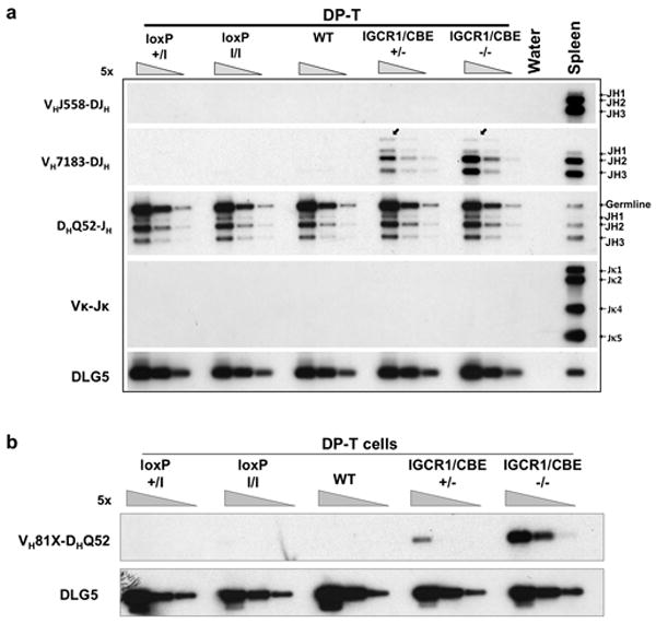
(a) PCR analyses of VH family rearrangements in sorted DP thymocytes and total splenic B cells from indicated mice with DLG5 as a loading control. Bands corresponding to rearrangements to various JHs are indicated on right. Igκ rearrangement (VκJκ) served as a control for B cell contamination. (b) Semi-quantitative PCR analyses of direct VH-D rearrangements in sorted DP-T cells from indicated mice. Assays are diagrammed in Supp. Figs. 6a and 8a.