



Introduction
Two cases of paraplegia due to primary spinal cord epidural non-Hodgkin's lymphoma (NHL) are presented. This is a rare cause of paraplegia. The clinical presentation, investigations and management of this rare condition are outlined. A review of the literature is presented.
Primary spinal epidural NHL should be considered in patients who give a history of back pain; followed by rapid development of features of spinal cord compression; have normal plain x-rays but whose CAT / myelogram reveal an extradural mass. Urgent surgical decompression followed by chemotherapy and radiotherapy improves the outcome of such patients.
Primary epidural Non Hodgkin's Lymphoma (NHL) with no evidence of parenchymal central nervous system (CNS) or systemic involvement is rare. Although it may present as a localised disease it is in fact a systemic disease. This disease entity must be differentiated from the more common clinical situation of primary CNS parenchymal lymphoma with meningeal involvement or systemic lymphoma, such as adult cell T lymphoma, complicated by lymphomatous meningitis. A 20-year experience with NHL at the Memorial Sloan Kettering Cancer Centre showed that primary epidural NHL accounted for 2 out of 256 (1.9%) of NHL cases1. A 10-year experience (1979–1989) at the Mayo Clinic report primary spinal epidural NHL accounting for 6.6% of all cases of intraspinal NHL2. Levitt et al report that among 592 cases of NHL seen between 1967 and 1977, 52 patients (11%) had CNS lymphomatous involvement. However they did not specify how many were primary spinal epidural NHL3.
With the current AIDS/HIV pan epidemic it is expected that the incidence of primary spinal NHL will increase. Distinguishing primary spinal NHL from an infectious spinal mass may be difficult as both would present with similar features of meningitis, fever, night sweats and weight loss.
Cases
LT
39-year-old self-employed businessman admitted to Queen Elizabeth Central Hospital on 9th January 2002 with loss of function in the lower limbs and inability to walk since mid December 2001. He started having mid thoracic back pain in October 2001. By end November 2001 he developed weakness in the lower limbs but was able to walk and was admitted at a rural mission hospital. Within two weeks, by mid December 2001, he developed almost complete loss of function in the lower limbs and was unable to walk. He had control of his bowels and the bladder and sensation was preserved.
He denied any trauma and systemically he was well with no significant constitutional features. He had no significant past medical history.
He looked well nourished and was afebrile. He had tenderness in the dorsal spine but there was no deformity. Neurological assessment of the lower limbs revealed power of 2/5; increased tone; hyperreflexia with sustained clonus; up going plantar reflexes; normal sensation. He was classified as Fraenkel grade C of paraplegia.
Plain x-rays of the spine were normal. Myelography revealed a right-sided posterolateral extradural compressive mass at T9 – T10 (Fig 1a, 1b). A CT scan, done some days after the myelogram, confirmed the compressive mass (Fig 1c).The rest of his investigations are as outlined in table 1.
Figure 1a.

Figure 1b.

Myelogram for LT showing a right posterolateral compressive mass at T9 – T10 level. No evidence of bone destruction noted.
Figure 1c.

CT scan at level of mass. A right sided lesion is seen displacing the spinal cord to the left. There is an extension through the intervertebral foramen
Table.
Investigations and results
| INVESTIGATIONS | CASE: LT | CASE: MC |
| Full Blood Count / Picture |
WBC 5.0; Hb 9.7; Plat 345 Nomochronic, normocytic anaemia; neutrophils 68%; lymph 28%; mono 4% |
WBC 3.2; Hb 4.9; Plat 2 |
| ESR | Not done | 60 mm/Hr |
| Blood culture | Not done | Bacillussp.? Contaminant |
| Lumbar puncture | Not Done | Clear CSF; protein++++; no organism seen; culture no growth |
| VDRL | Non Reactive | Non Reactive |
| Urinalysis / Stool | Nomal | Normal |
| HIV | Positive | Negative |
| Plain Spine x-ray | Normal | Normal |
| CAI / Myelogram | Extradural compressive mass at T9, T10 on the posterolateral aspect on the right. |
Extradural compressive mass T2 to T7 posterity |
| Chest x-ray | Nomal | Normal |
| Abdominal Ultrasound | Normal | Normal |
| Histology | Non Hodgkin's Lymphoma / lymphocytic lymphoma |
Non Hodgkin's lymphoma |
He was taken to theatre on 28th January 2002. He underwent hemilaminectomy at T9 and T10 on the right side. There was an extradural fibrinous soft tissue mass causing spinal cord compression. The tumour peeled off easily from the dura. Histology confirmed this to be non-Hodgkin's lymphoma. Chemotherapy using a CHOP (cyclophosphamide, adriamycin, vincristine, prednisolone) regime; six cycles over six months was recommended. However the patient has not been able to purchase the drugs and chemotherapy has not been commenced.
ILLUSTRATIONS
He made good neurological recovery post operatively and was discharged to a rehabilitation unit on 11th march 2002 mobilising with a walking frame. He was graded as Fraenkel D on discharge to the rehabilitation unit and he continues to improve.
He is also considering commencing HIV/AIDS anti retroviral medications.
MC
17-year-old secondary school student transferred from a rural mission hospital to Queen Elizabeth Central Hospital on 25th February 2002 with a two-day history of sudden and complete loss of function in the lower limbs; urinary retention and faecal incontinence.
He had complained of upper thoracic back pain of a non-traumatic origin since November 2001. This was associated with general malaise. On 4th February 2001 he was admitted on the medical ward at QECH with increased back pain and fever. He had an elevated ESR; was mildly anaemic: a negative blood culture; and a negative HIV test. After a course of intravenous antibiotics his fever subsided and he was discharged mobilising 7 days later. On 22nd February 2002 he was admitted at a mission hospital with sudden and complete loss of function of the lower limbs and was transferred to QECH on 25th February 2002.
Systemically he was unwell with weight loss; persistent fever; neck pain and stiffness suggestive of meningitis. He had flaccid paralysis of the lower limbs with loss of sensation from T4 distally. He had no bowel or bladder control. He was assessed as Fraenkel A. He had developed small pressure sores on the sacrum and the greater trochanter areas. Blood cultures grew bacillus species, which was thought to be a contaminant. He was anaemic with haemoglobin of 4.9. A lumbar puncture revealed clear CSF with increased protein; no organism seen; and a negative culture. Plain x-rays of the spine were normal. Other investigations are as shown in table 1.
He was commenced on intravenous antibiotics including chloramphenicol, penicillin and flucloxacillin. A dose of praziquantel was given. His fever persisted and on 7th March, two weeks after admission, TB treatment was commenced empirically. A privately sponsored CT Myelogram was done on 7th March 2002 and revealed a posterior extradural compressive mass extending from T2 to T7 (Fig 2a, 2b, 2c, 2d). The diagnosis was now considered to be spinal abscess and intravenous antibiotics as well as the TB treatment, were continued.
Figure 2a.
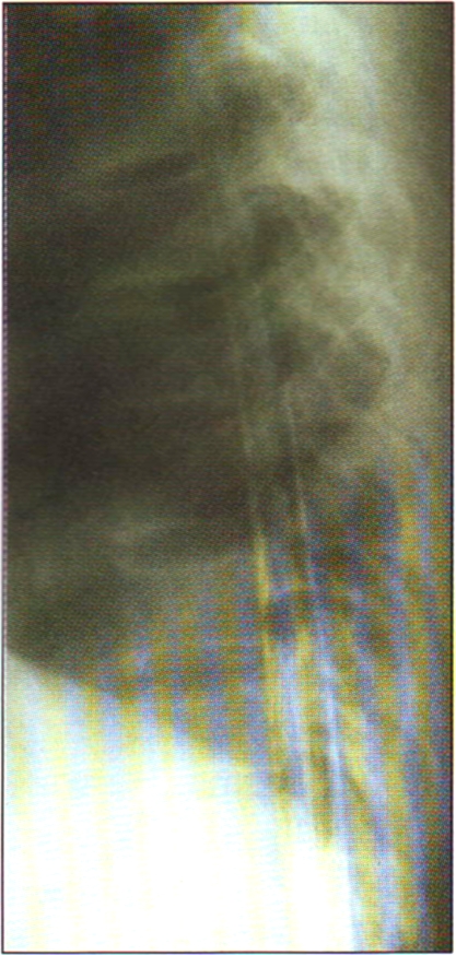
Lateral view myelogram of MC showing a posterior compressive mass extending over several vertebral segments.
Figure 2b.

Figure 2c.

Figure 2d.

CT scans at various levels. Fig 2b shows a posterior mass displacing the cord anteriorly. Fig 2c and 2d are scans at levels above and below the mass respectively showing that the spinal cord is centrally located in the spinal canal
An orthopaedic consultation was first made on 7th March 2002. On reviewing the CT Myelogram the opinion was that the diagnosis was epidural lymphoma. He was now noted to also have weakness of the upper limbs and was developing weakness of chest wall muscles. After discussing with the parents it was felt that surgery would help to arrive at a tissue diagnosis but was unlikely to reverse the neurological deficit. The general poor prognosis was explained. They felt we should go ahead with surgery because if lymphoma were confirmed, they would consider buying the chemotherapy.
He received four units of blood prior to surgery and he was taken to theatre on 19th March 2002. Laminectomies were done brillianerally from T3 to T5. A soft, fibrinous, epidural partially removed ant the spinal cord decompressed. Total excision was not possible due to the extent of the lesion. Histology of the tissue confirmed a diagnosis of non-Hodgkin's lymphoma. Worsening pressure sores and breakdown of the surgical wound complicated the postoperative period. The neurological deficit in the lower limbs did not improve, however power in the upper limbs returned to normal. Post operatively he required transfusion of four units of blood. Chemotherapy using a CHOP regime was recommended but the parents could not afford the medications. His general condition deteriorated and he died on 26th April 2002 from a combination of anaemia and septicaemia from infected pressure sores.
Discussion
Clinical Presentation / Features
Primary spinal epidural NHL can present at any age although it is more common in elderly patients with a median age 70 years. A prodrome period of back pain with a median duration ot months is common. This is followed by an acute neurological deterioration with a median duration of 6 days2.
Neurological findings may include paraparesis or paraplegia; a discrete sensory level; hyperreflexia; clonus and loss of sphincter control. In most patients, diagnosis of primary spinal epidural NHL is made when neurologic deficit is already established and histological specimens have been obtained at surgery.
Investigations
Blood tests:
Routine blood investigations include FBC, ESR and peripheral blood film. These may reveal a normocytic, normochronic anaemia; pancytopaenia and atypical cells; elevated ESR. Bone marrow assessment is important to exclude systemic lymphoma. Blood culture is necessary to exclude septicaemia in patient presenting with features of meningitis and fever. VDRL and HIV tests should be done after appropriate counselling.
Lumbar puncture
Analysis of CSF is important. Young et al 4 report that CSF abnormalities are found in 97% of patients of NHL with CNS involvement. Routine microscopy, culture and sensitivity testing of CSF must be done. This may reveal raised protein and cell count with a predominance of lymphocytes. Cytology of the CSF may be done but has been shown to be positive in only 67% of cases of lymphoma with CNS involvement4. India ink staining should be done to exclude cryptococcal meningitis.
Urine and stool
Urine and stool analysis are important to investigate schistosomiasis as a cause of paraparesis / paraplegia.
Radiological
Plain x - rays of the spine are invariably normal with no evidence of bone destruction. Myelography and CAT scan will reveal an extradural mass compressing the spinal cord, usually extending over more than one vertebral segment. MRI scan should be done where available to further delineate the extent of the lesion. Abdominal ultrasound, chest x-ray, CAT scan of the abdomen and chest are necessary to exclude systemic disease and for staging the disease.
Histology
Histological specimens from primary epidural NHL are obtained at the time of surgical decompression of the spinal cord. CAT guided biopsy of lesion may be attempted prior to surgery if the lesion is accessible.
In patients with NHL and CNS involvement, between 80% and 98% have a diffuse histological subtype CNS involvement in nodular lymphoma is uncommon and is reported in only about 3% of patients with NHL 4.
For proper sub typing immunohistochemistry and immunocytochemistry are important tests.
Treatment
Most patients with primary epidural NHL present with rapidly progressive or advanced neurological deficit. Diagnosis is usually confirmed by histological specimens taken at time of emergency surgical decompression of the spinal cord. Surgery is therefore important for decompression and tissue diagnosis. Surgery involves laminectomy and resection of the tumour. Spinal irradiation and systemic chemotherapy are important adjuvant treatment, which have been shown to increase disease free survival. Intrathecal chemotherapy can be considered in relapse cases. The ten-year experience at the Mayo clinic report shows that 8 patients had primary spinal epidural NHL and all underwent decompressive laminectomies, subtotal tumour resection and local spinal radiotherapy. Four patients died and four remain alive and well and free from disease. The twenty-year experience at the Memorial Sloan - Kettering Cancer Centre showed that 5 patients had primary spinal epidural NHL. Two patients died and three remained alive and diseases free after a median follow up period of ten years. Response in AIDS / HIV patients is similar to that seen in non-AIDS patients 5.
Prognosis
Some poor prognostic factors especially with respect to resolution of paraplegia and disease relapse include:
Rapidly progressive paraplegia with lack of motor function at presentation (Fraenkel A, B) and long duration of complete paraplegia before presentation have a poor chance of neurological recovery.
Poorly differentiated, diffuse histological grade is associated with increased relapse rate.
These two cases raise several discussion points. Primary spinal epidural NHL can present at any age, although the median age in most series is 70 years. MC is a young man who is HIV negative whereas LT is middle aged man and is HIV positive. There is a belief that with the HIV / AIDS pan epidemic the number of cases of primary spinal epidural NHL will increase. The literature suggests that the response to chemotherapy and radiation in AIDS patients is similar to non-AIDS patients.
Both patients reveal the difficulty in diagnosing this condition. During the prodrome period of back pain both patients did seek medical attention. However by the time they were referred to the central hospital, neurologic deficit had been present for some time. A high index of clinical suspicion is needed to consider this rare condition as a differential diagnosis in patients presenting with paraplegia. The other major constraint in arriving at a diagnosis is the lack of radiological investigations especially Myelography. In both these patients CT Myelography was done after some delay because the patients had to pay for it themselves at a private institution because QECH did not have Myelography contrast media and the CT scan is out of commission.
MC had a more rapid progression and on presentation had no motor function and already had developed some pressure sores. Although surgery was done, it was known that this was mainly to aid in tissue diagnosis and would not reverse the neurological deficit. His neurological deficit has remained. LT had a slower progression and had residual motor function at the time of presentation. Surgical decompression has resulted in improvement of the neurological deficit.
Chemotherapy in both cases has to be purchased by the patients, as these drugs are not on the hospitals pharmacy list. They are expensive and both patients were not able to purchase the medications. This undoubtedly will affect outcome in patient LT. It is debatable whether chemotherapy would have prevented the death of patient MC. Radiotherapy has been shown to be a useful adjuvant therapy but unfortunately is not available in Malawi. Primary spinal epidural NHL should be considered in patients who give a history of back pain; followed by rapid development of features of spinal cord compression; have normal plain x-rays but whose CAT / myelogram reveal an extradural mass. Urgent surgical decompression followed by chemotherapy and radiotherapy improves the outcome of such patients.
References
- 1.Mora J, Wollner N. Primary epidural non-Hodgkin's lymphoma: spinal cord compression syndrome as the initial form of presentation in childhood non-Hodgkin's lymphoma. Med Pediatr Oncol. 1999 Feb;32(2):102–105. doi: 10.1002/(sici)1096-911x(199902)32:2<102::aid-mpo6>3.0.co;2-y. [DOI] [PubMed] [Google Scholar]
- 2.Lyons MK, O'Neill BP, Marsh WR, Kurtin PJ. Primary spinal epidural non - Hodgkin's lymphoma: report of eight patients and review of the literature. Neurosurgery. 1992 May;30(5):675–680. doi: 10.1097/00006123-199205000-00004. [DOI] [PubMed] [Google Scholar]
- 3.Levitt LJ, Dawson DM, Rosenthal DS, Moloney WC. CNS involvement in the non - Hodgkin's lymphomas. Cancer. 1980 Feb;45(3):545–552. doi: 10.1002/1097-0142(19800201)45:3<545::aid-cncr2820450322>3.0.co;2-6. [DOI] [PubMed] [Google Scholar]
- 4.Young RC, Howser DM, Anderson T, et al. Central nervous system complications of non-Hodgkin's lymphoma. The potential role for prophylactic therapy. Am J Med. 1979 Mar;66(3):435–443. doi: 10.1016/0002-9343(79)91065-9. [DOI] [PubMed] [Google Scholar]
- 5.Chamberlain MC, Kormanik PA. AIDS - related central nervous system lymphomas. J Neurooncol. 1999 Jul;43(3):269–276. doi: 10.1023/a:1006262720665. [DOI] [PubMed] [Google Scholar]