Summary
Most cases with chronic subdural hematoma (CSDH) are treated by simple irrigation and drainage, then more than eighty percent of them result in good recovery. But we sometimes encounter intractable cases with hematoma re-collection, which is considered of repeated bleeding from macrocapillary in the hematoma capsule. Embolization of the middle meningeal artery (MMA) is considered to be useful to eliminate the blood supply to this structure. The authors experienced seven cases of intractable CSDH treated by MMA embolization and no recurrence took place in all cases for up to 15 months. Endovascular treatment may be a good alternative modality for recurrent CSDH.
key words: chronic subdural hematoma, middle meningeal artery (MMA), transarterial embolization
Introduction
Burr hole surgery with irrigation and drainage is usually curative for CSDH1. However some of them have recurrent blood collection requiring repeated evacuation. Individual and clinical factors such as poor brain re-expansion, alcoholism, liver dysfunction and coagulopathies are associated with recurrence of CSDH2.We embolized the MMA which is presumed to be a main source of blood supply for the outer membrane of the hematoma. Then we observed clinical sequel after this intervention.
Patients and Methods
Of the 80 patients with CSDH who underwent burr hole surgery at our hospital between November 2004 and December 2005, seven patients underwent embolization of the MMA. This treatment was planned when 1) the third recurrence with progressed subdural hematoma collection, 2) the second recurrence with a risk factors such as bleeding tendency and hemodialysis.
Interventions
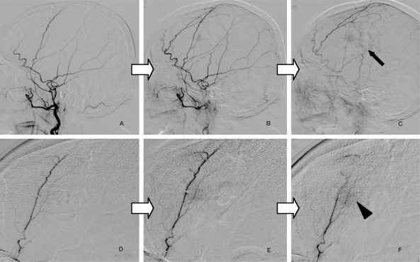
The 5F guiding catheter (Guider, Boston Scientific) was placed near the origin of the MMA. A flow guided type microcatheter (Magic or Baltacci, BALT, France) was advanced to the MMA for superselective angiography. It demonstrated diffuse abnormal vascular network at early arterial phase and foggy staining at late venous phase in all cases. This abnormal finding was easy to be missed when contrast media was injected slowly from main trunk of the external carotid artery. We confirmed the location of the origin of the recurrent meningeal artery on the MMA with the compression of the ipsilateral internal carotid artery. Then we advanced the microcatheter to distal portion of each branch of MMA and pulled the catheter slowly while injecting 20% NBCA. We paid attention to the dangerous anastomosis with a recurrent meningeal artery and petrosal branch when we injected NBCA. We tried to embolize all of the branches of MMA on the side of CSDH.
Case Presentation
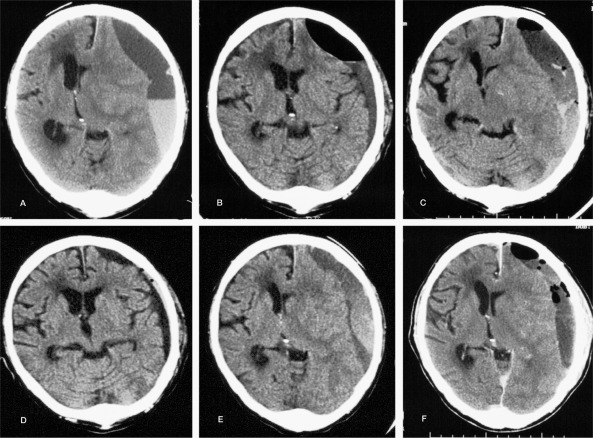
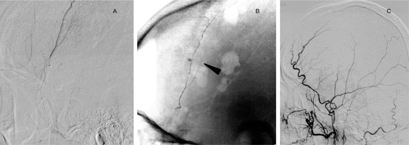
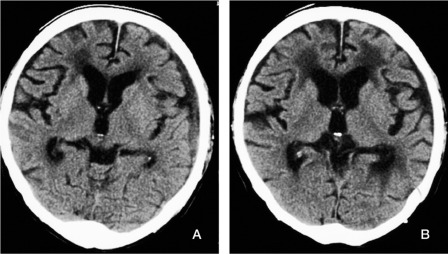
A 78-year-old man with traumatic CSDH had chronic renal failure which required regular hemodialysis. He underwent repeated burrhole drainage surgery but had the third recurrence in about a month (figure 1). The hematoma collected rapidly, so we decided to embolize the MMA. The superselective angiogram of the MMA demonstrated abnormal vascular networks (figure 2). Then an endovascular embolization of the left MMA using 20% NBCA was performed (figure 3). The abnormal vascular findings was no longer seen and recurrence was not seen postoperatively for sixteen months (figure 4).
Figure 1.
Preembolization CT scans of the presenting case. Preoperative (A) and postoperative (B) CT scans at first drainage. Hematoma is evacuated. Preoperative (C) and postoperative(D) CT scans at second drainage. Preoperative (E) and postoperative(F) CT scans at third drainage. Third recurrence in spite of repeated burr hole surgery.
Figure 2.
Left external carotid artery angiography (ECAG) (A,B,C). Left superselective MMA angiography (D,E,F). Abnormal vascular networks are demonstrated especially at the late artery phase (arrow, arrow head).
Figure 3.
A) Superselective angiogram of the left MMA, lateral projection. 20%NBCA injected into MMA: started from peripheral branch of MMA, sliding back to proximal trunk. B) Lateral view of skull X-ray shows NBCA cast (arrow head). C) Left ECAG after the embolization of the MMA. The MMA is completely embolized and the abnormal vascular network is no longer seen.
Figure 4.
A) CT scan three months after embolization showed noted decrease of hematoma. B) CT scan 16 months after embolization demonstrated almost complete resolusion of the hematoma.
Discussion
Recurrence rate of CSDH after treatment with burr hole irrigation and drainage is reported about 10% (1.7% to 16.2%) and the recurrence interval is from one to eight weeks (4 weeks on the average ). Table 1 summarizes the risk factors for CSDH recurrence 3. The mechanism of CSDH growth have been understood based on pathological investigations 4. Selective MMA angiography demonstrates diffuse dilatation of the MMA and visualization of scattered abnormal vascular networks, which reflect macrocapillary, sinusoidal channel layer and giant capillary in the outer membrane 5. The outer membrane is consist of two or three layers. A large number of macrocapillaries infiltrate into those layers on the side of the hematoma cavity 6,7. Direct arterial pressure of the MMA causes rupture of macrocapillaries and arterial bleeding into the hematoma cavity. For that reason the embolization of the MMA for the patients with refractory CSDH has recently been reported as a useful treatment8. Low concentration, 20% NBCA mixed with lipiodol is preferred. We found this concentration of NBCA very suitable to embolize peripheral vessels of the MMA with low risk of catheter adhesion. Endovascular treatment may be more effective for the multilocular hematoma that need treatment with multiple drainage tubes or endoscopic decapsulation. We are planning to investigate how much this endovascular treatment can contribute to serious clinical cases such as patients with large hematoma or with abnormal coagulopathies. Further study is needed to determine the efficacy of this modality of treatment for recurrent CSDH.
Table 1.
Risk factors of the CSDH recurrence.
| Clinical factors |
| • age |
| • alcoholism |
| • history of head injury |
| • interval from onset of initial symptoms to hospitalization |
| Laboratory findings |
| • thrombocytepenia |
| • deficiency of coagulation factors |
| • liver dysfunction |
| CT, MRI findings |
| • brain atrophy |
| • intracranial hypotension |
| • hematoma (multilocular, multilayers) |
| Operative findings |
| • additional procedure (drainage tube) |
| • postoperative residual air |
Conclusions
We have had good outcome of patients with intractable CSDH who underwent endovascular treatment. Embolization of the MMA which eliminate the blood supply to abnormal structure in hematoma cavity may be useful procedure of intractable CSDH.
References
- 1.Tsutsumi K, Maeda K, et al. The relationship and closed system drainage in the recurrence of chronic subdural hematoma. J Neurosurg. 1997;87:870–875. doi: 10.3171/jns.1997.87.6.0870. [DOI] [PubMed] [Google Scholar]
- 2.Matsumoto K, Akagi K, et al. Recurrence factors for chronic subdural hematoma after burr-hole craniostomy and closed system drainage. Neurol Res. 1999;21:277–280. doi: 10.1080/01616412.1999.11740931. [DOI] [PubMed] [Google Scholar]
- 3.Shiomi N, Sasajima H, et al. Relationship of Postoperative Residural Air and Recurrence in Chronic Subdural Hematoma. No Shinkeigeka, 2001;29(1):39–44. [PubMed] [Google Scholar]
- 4.Shimoji T, Sato K, et al. A Pathogenesis of Chronic Subdural Hematoma; It`s relationship to subdural membrane. No Shinkeigeka, 1992;20(2):131–137. [PubMed] [Google Scholar]
- 5.Nagahori T, Nishijima T, et al. Histological Study of the Outer Membrane of Chronic Subdural hematoma: Possible mechanism for expansion of hematoma cavity. No Shinkeigeka, 1993;21(8):697–701. [PubMed] [Google Scholar]
- 6.Tanaka T, Fujimoto S, et al. Superselective Angiographic Findings of Ipsilateral Middle Meningeal Artery of Chronic Subdural Hematoma in adults. No Shinkeigeka, 1998;26(4):339–347. [PubMed] [Google Scholar]
- 7.Tanaka T, Kaimori M, et al. Histological Study of Vascular Stracture between the Dura Mater and the Outer Membrane in Chronic Subdural Hematoma in an Adult. No Shinkeigeka, 1999;27(5):431–436. [PubMed] [Google Scholar]
- 8.Takahashi K, Muraoka K, et al. Middle Meningeal Artery Embolization for Refractory Chronic Subdural Hematoma: 3 case reports. No Shinkeigeka, 2002;30(5):535–539. [PubMed] [Google Scholar]


