Abstract
The ability of bifidobacteria to establish in the intestine of mammals is among the main factors considered to be important for achieving probiotic effects. The role of surface molecules from Bifidobacterium bifidum taxon in mucin adhesion capability and the aggregation phenotype of this bacterial species was analyzed. Adhesion to the human intestinal cell line HT29 was determined for a collection of 12 B. bifidum strains. In four of them—B. bifidum LMG13195, DSM20456, DSM20239, and A8—the involvement of surface-exposed macromolecules in the aggregation phenomenon was determined. The aggregation of B. bifidum A8 and DSM20456 was abolished after treatment with proteinase K, this effect being more pronounced for the strain A8. Furthermore, a mucin binding assay of B. bifidum A8 surface proteins showed a high adhesive capability for its transaldolase (Tal). The localization of this enzyme on the surface of B. bifidum A8 was unequivocally demonstrated by immunogold electron microscopy experiments. The gene encoding Tal from B. bifidum A8 was expressed in Lactococcus lactis, and the protein was purified to homogeneity. The pure protein was able to restore the autoaggregation phenotype of proteinase K-treated B. bifidum A8 cells. A recombinant L. lactis strain, engineered to secrete Tal, displayed a mucin- binding level more than three times higher than the strain not producing the transaldolase. These findings suggest that Tal, when exposed on the cell surface of B. bifidum, could act as an important colonization factor favoring its establishment in the gut.
INTRODUCTION
Bifidobacteria are members of the intestinal microbiota of humans, and some strains are able to exert health-promoting effects, thus being considered to have some probiotic features (2, 21). They are especially relevant in infants, constituting one of the predominant populations in their gut microbiota (48). Because of their beneficial effects, the functional characteristics of specific strains are being extensively studied with the aim of selecting those more suitable for different target populations, such as children, the elderly, or people suffering from immune disorders (3, 25, 31). In this regard, it is generally believed that a Bifidobacterium strain intended for use as a probiotic needs to survive gastrointestinal transit and colonize, at least transiently, the gut (45).
Intestinal colonization factors have extensively been studied in pathogenic bacteria, as well as in gut commensals and probiotics (42, 50). While in enteric pathogens they are undesirable traits and their presence constitutes an important safety issue, probiotic bacteria (i.e., Lactobacillus and Bifidobacterium strains) use them to persist in the intestine, enabling these microorganisms to accomplish their effects. In fact, some of these characteristics, such as adherence to mucus and/or human epithelial cells and cell lines, are recommended by the Food and Agriculture Organization of the United Nations–World Health Organization (FAO/WHO) as in vitro tests to screen potential probiotics (2).
Adhesion to the intestinal epithelium has been extensively studied in Lactobacillus and, to a lesser extent, in Bifidobacterium. Specialized intestinal epithelial cells secrete mucin, a complex glycoprotein mixture that is the main component of mucus and protects the mucosal surface limiting bacterial entry (32). However, some Lactobacillus display on their surface adhesins able to mediate the attachment to the mucus layer (4). This process is mainly mediated by proteins, although saccharide moieties and lipoteichoic acids have also been involved (50, 53). The proteins playing a role in the mucus adhesion phenotype of lactobacilli are mainly secreted and surface-associated proteins, either anchored to the membrane through a lipid moiety or embedded in the cell wall (14, 40, 52, 53). More recently, the potential contribution of the so-called moonlighting proteins, those able to switch between unrelated functions depending on the cell location, has also been described, i.e., elongation factor Tu and GAPDH (glyceraldehyde-3-phosphate dehydrogenase) (15, 19, 41). Regarding bifidobacteria, the involvement of surface proteins in the interaction with human plasminogen or enterocytes has been reported in Bifidobacterium animalis subsp. lactis and Bifidobacterium bifidum, respectively. Under certain circumstances, these proteins could play a role in facilitating the colonization of the human gut through degradation of the extracellular matrix of cells or by facilitating a close contact with the epithelium (5, 6, 7, 17, 42). Also, a pilus-type IV mediated host colonization and persistence mechanism has recently been demonstrated in Bifidobacterium breve (30).
In addition, autoaggregation was shown to play a role in favoring persistence and survival of intestinal bacteria in the human gastrointestinal tract (12). The aggregation phenotype of some probiotic bacteria can also promote coaggregation with pathogens, thus contributing to the removal of the pathogen in the oral or intestinal environment and decreasing its potential virulence (23, 44). Several aggregation factors have been described in Lactobacillus, and for some of them a relation between the aggregation phenotype and the persistence of the microorganisms in the gut has been reported (14, 51, 54). B. bifidum has a strong autoaggregation phenotype under in vitro conditions, although the molecular basis behind this phenomenon has not yet been elucidated (8). Remarkably, some authors have demonstrated a strong association between mucin adhesion and autoaggregation; cells with high adhesion capability possess a strong aggregation phenotype (14, 27).
In this context, the aim of the present study was to evaluate novel B. bifidum surface proteins able to contribute to its intestinal colonization capability.
MATERIALS AND METHODS
Bacterial strains and growth conditions.
The strains and plasmids used throughout the present study are listed in Table 1. B. bifidum strains were grown in MRS broth (Difco/BD Diagnostic Systems, Sparks, MD) supplemented with 0.25% (wt/vol) l-cysteine (Sigma Chemical Co., St. Louis, MO) (MRSc) and were incubated at 37°C in an anaerobic chamber (Whitley MG500 anaerobic workstation; DW Scientific, Shipley, United Kingdom) in an atmosphere of 10% CO2, 10% H2, and 80% N2. Lactococcus lactis strains were grown in M17 broth (Oxoid, Ltd., Hampshire, United Kingdom) containing 0.5% (wt/vol) glucose (GM17) at 32°C. Chloramphenicol (5 mg liter−1) was added to the medium for the selection of recombinant L. lactis strains containing pNZ plasmids. For the expression of the B. bifidum transaldolase gene (tal) in the strains L. lactis NZ9000-HTal and NZ9000-SecTal, 0.001% (vol/vol) of the supernatant from an overnight culture of the nisin-producing strain L. lactis NZ9700 (containing ∼10 ng of nisin A ml−1) was added to the cultures at an optical density at 600 nm (OD600) of 0.2, and the inductions were performed until the cultures reached an OD600 of ∼0.8.
Table 1.
Strains and plasmids used in this study
| Strain or plasmida | Relevant origin, phenotype, and/or genotypeb | Source or reference |
|---|---|---|
| Strains | ||
| B. bifidum | ||
| LMG13195 | Intestine of infant | |
| DSM20239 | Breast-fed infant | |
| DSM20456T | Breast-fed infant | 26 |
| DSM20082 | Intestine of adult | |
| DSM20215 | Intestine of adult | |
| A8 | Dairy product | 46 |
| D119 | Human feces | 46 |
| L22 | Human feces | 46 |
| JCM7002 | Human feces | |
| JCM7003 | Human feces | |
| KCTC3357 | Human feces | |
| KCTC5082 | Human feces | |
| L. lactis | ||
| NZ9000 | L. lactis MG1363 derivative carrying pepN::nisRK | 11 |
| NZ9700 | Nisin-producing strain | 22 |
| NZ9000-HTal | Cmr; NZ9000 derivative carrying pNZ8048-HTal | This study |
| NZ9000-SecTal | Cmr; NZ9000 derivative carrying pNZ8110-SecTal | This study |
| Plasmids | ||
| pNZ8048 | Cmr; inducible expression vector, nisA promoter | 11 |
| pNZ8110 | Cmr; pNZ8048 derivative, Usp45 signal peptide | 11, 49 |
| pNZ8048-HTal | Cmr; pNZ8048 derivative, carrying His tag tal | This study |
| pNZ8110-SecTal | Cmr; pNZ8110 derivative, carrying tal | This study |
LMG, Belgian Coordinated Collections of Microorganisms; DSMZ, Deutsche Sammlung von Mikroorganismen und Zellkulturen GmbH; JCM, Japan Collection of Microorganisms; KCTC, Korean Collection for Type Cultures.
Cmr, chloramphenicol resistance.
Characterization of B. bifidum strains. (i) Adhesion of B. bifidum strains to HT29 cells.
The cell line was maintained in McCoy broth (Sigma) supplemented with 3 mM l-glutamine, 10% (vol/vol) heat-inactivated fetal bovine serum, and a mixture of antibiotics (50 mg of penicillin, 50 mg of streptomycin, 50 mg of gentamicin, and 1.25 mg of amphotericin B liter−1). Medium and supplements were purchased from Sigma. Incubations took place at 37°C in 5% CO2 in an SL water jacketed CO2 incubator (Sheldon Mfg., Inc., Cornelius, OR). The culture medium was changed every 2 days, and the cell lines were trypsinized with 0.25% trypsin-EDTA solution (Sigma) according to standard procedures (10). For adhesion experiments, 105 cells ml−1 were seeded in 24-well plates, incubated to confluence, and used at a mean age of 13 ± 1 days old.
Twelve B. bifidum strains and the reference strain B. animalis subsp. lactis BB12, grown in 10 ml of MRSc for 24 h, were harvested by centrifugation, washed twice with Dulbecco phosphate-buffered saline (PBS) buffer (Sigma), and resuspended in McCoy medium without antibiotics at a concentration of ∼108 CFU ml−1. The bacterial suspensions were added in a ratio of epithelial cells to bacteria of 1:10. The plates were incubated for 1 h at 37°C in 5% CO2 in a Heracell 240 incubator (Thermo Electron LDD GmbH, Langenselbold, Germany). After the incubation period, the wells were gently washed three times with Dulbecco PBS buffer to remove the nonattached bacteria. Trypsin-EDTA solution was added to release the adhered cells, and bacterial counts were carried out in agar-MRSc to determine the number of adhered bacteria. The results were expressed as the percentage of bacteria adhered with respect to the amount of bacteria added to the monolayer (CFU of adhered bacteria × 100/CFU of added bacteria). Experiments were repeated at least twice, using two independent bacterial cultures and two different HT29 plates. In each plate, the samples were also tested in duplicate.
(ii) Aggregation phenotype of selected B. bifidum strains.
The B. bifidum strains A8, DSM20456, DSM20239, and LMG13195 were grown in 50 ml of MRSc for 18 h under standard conditions. Cultures were harvested by centrifugation, washed with PBS buffer (Oxoid), and resuspended in PBS buffer until an OD600 of 1 was reached. Aliquots of 10 ml were transferred to five tubes, harvested by centrifugation, and resuspended under the following conditions: condition 1, PBS buffer with 1 mg of proteinase K (EC 3.4.21.64; Molecular Biochemicals, Lewes, United Kingdom) ml−1; condition 2, PBS buffer with 1 mg of lipase (EC 3.1.1.3; Sigma) ml−1; condition 3, PBS buffer (control); condition 4, acetate buffer (pH 4.5) containing 5 mg of sodium metaperiodate (Merck, Darmstadt, Germany) ml−1; and condition 5, acetate buffer (pH 4.5) (control for the metaperiodate treatment). All bacterial suspensions were incubated for 30 min at 37°C. After incubation, the bacterial suspensions were washed with acetate buffer (pH 3.8) and resuspended in 5 ml of the same buffer. Then, each tube was incubated in a bath at 37°C, measuring the OD600 at different times (0, 60, 120, and 180 min). Aggregation percentage was calculated as follows: [(OD600 of the control bacterial suspension/OD600 of the bacterial suspension after treatment)] × 100. The influence of the different treatments on cell viability was checked by plate counting before and after the treatments. Experiments were carried out at least in triplicate.
For the determination of the influence of surface proteins or pure transaldolase (see below) on the aggregation phenotype, the strain B. bifidum A8 was grown in 100 ml of MRSc for 18 h and subjected to a proteinase K treatment under the conditions described above. Several aliquots of the proteinase-treated suspensions in 5 ml of acetate buffer (pH 3.8) were incubated in a bath at 37°C. After 1 h of incubation, surface proteins extracted after Na2CO3 treatment (10 μg), pure transaldolase from B. bifidum A8 (1 or 10 μg), bovine serum albumin (BSA; 1 or 10 μg), or pure histidine-tagged arabinofuranosidase (1 or 10 μg) (28) were added, and the OD600 was measured every hour for 4 h.
Functional characterization of surface proteins from B. bifidum A8. (i) Extraction of surface proteins by carbonate wash or sodium hydroxide treatment.
Cultures (50 ml) of B. bifidum A8, grown in MRSc for 18 h, were harvested by centrifugation and washed with acetate buffer (pH 3.8), and the pellets were subjected to two different treatments to extract proteins noncovalently bound to the cell surface. Cells from 50 ml of culture were resuspended in 5 ml of 100 mM Na2CO3 buffer, and cell suspensions were incubated for 1 h on ice and harvested by centrifugation (Na2CO3 fraction) (35). Another washed cell pellet from 50 ml of culture was resuspended in 5 ml of 0.01 M NaOH, and an incubation at 37°C for 30 min under gentle agitation was performed (NaOH fraction) (36). The protease inhibitors EDTA and phenylmethylsulfonyl fluoride were added to all of the solutions at final concentrations of 5 and 1 mM, respectively. After these treatments, the supernatants were collected and filtrated through 0.45-μm-pore-size filters. The protein concentrations were determined by using a BCA protein assay kit (Pierce, Rockford, IL). Protein (40 μg) was precipitated using a methanol-chloroform protocol (55), solubilized in Laemmli buffer, and separated by SDS-PAGE. The protein profiles were visualized by standard Coomassie staining, and the proteins were identified by tandem mass spectrometry in a matrix-assisted laser desorption ionization–time of flight apparatus (Servicio de Proteómica, CNIC, Madrid, Spain).
(ii) Mucin binding experiments.
Adhesion to mucin was performed as described previously (41). In brief mucin (2 mg, type III; Sigma) was used to coat the wells of F96 Maxisorp Immunoplates (Nunc, Roskilde, Denmark) for 1 h at 37°C and overnight at 4°C. The wells were then blocked with 1% (wt/vol) BSA (Sigma) in PBS for 1 h at 37°C and washed twice with PBS, and 100 μg of B. bifidum A8 surface proteins, from both NaOH and Na2CO3 fractions, was added to each well, followed by incubation at 37°C for 1 h. Unbound proteins were removed by several washes with PBS and, subsequently, 60 μl of 1% (wt/vol) SDS was added to each well, followed by incubation at 37°C for 1 h to extract the bound protein. Proteins that were able to adhere to the mucin layer were analyzed by SDS-PAGE, visualized by silver staining, and identified by a mass spectrometry analysis (Servicio de Proteómica, CNIC). At least two different surface protein extracts (biological replicates from two independent cultures) from each fraction (NaOH-extracted proteins and Na2CO3-extracted proteins) were used for mucus-binding assays, and several technical replicates with each extract were performed per plate.
(iii) Cloning of the transaldolase gene, protein purification, and Western blotting.
The transaldolase gene (tal) was amplified from the genomic DNA of B. bifidum A8 by PCR using high-fidelity Platinum Pfx DNA polymerase (Invitrogen, Barcelona, Spain) and the primers TalF (5′-TCCGATTCATGACTGAAGCAACTCAGCGCACG-3′; the NcoI site is underlined) and TalHisR (5′-TCGGCTTCTAGATCAATGGTGATGGTGATGGTGGGCGTTCACGCGGTCGATG-3′; the XbaI site is underlined, including the codons for the synthesis of a His6 tag). The PCR product was purified with a QIAquick PCR purification KIT (Qiagen, Ltd., Strasse, Germany), digested with RcaI and XbaI (RcaI yields compatible ends with NcoI), and ligated with the NcoI/XbaI-digested pNZ8048 vector, yielding pNZ8048-HTal. A similar protocol was used to clone tal into pNZ8110, which contains the coding sequence for the Usp45 signal peptide (49) and allows the secretion of the expressed protein outside the cell. In this case, the gene was amplified by using the primers SecTalF (5′-CCGAATGCCGGCATGACTGAAGCAACTCAGCG-3′) and SecTalR (5′-CCGAATGCCGGCTCAGGCGTTCACGCGGTCGA-3′), both containing an NaeI site (underlined). NaeI-digested pNZ8110 was ligated with the NaeI-digested PCR product, and the fragment orientation was checked by restriction analysis, yielding pNZ8110-SecTal. Plasmids were electroporated into L. lactis NZ9000 according to procedures described elsewhere (28), yielding the recombinant strains NZ9000-HTal and NZ9000-SecTal. The absence of PCR-introduced mutations was verified in the two plasmid constructs by sequencing both DNA strands on an ABI Prism sequencer (Applied Biosystems). Expression and purification of the histidine-tagged protein from NZ9000-HTal protein extracts was performed according to the method of Margolles and de los Reyes-Gavilán (28). After purification, the pure protein was dialyzed against PBS (Oxoid) and stored at −80°C.
For Western blot analysis, rabbit polyclonal IgG obtained against purified Tal was used for the primary immunoreaction. The polyclonal antibodies were obtained in the Laboratory of Biotechnological and Biomedical assays of the University of Oviedo (Oviedo, Asturias, Spain). Previous experiments showed a good specificity of the serum, and a single band with the proper molecular weight was detected by confronting the serum to total protein extracts of B. bifidum A8, using standard Western blot procedures. In brief, for Western blot analysis of the different L. lactis protein extracts (supernatant, cytoplasmic, and surface-associated proteins), ∼30 μg of protein was run on SDS-PAGE gels, the samples were then transferred to polyvinylidene difluoride membranes (Amersham Biosciences, Buckinghamshire, United Kingdom), and immunoreactions were performed as previously described (39). The blots were developed with 4-chloro-1-naphthol.
(iv) Mucin binding experiments of recombinant L. lactis.
L. lactis NZ9000 carrying the plasmid pNZ8110 (control; empty plasmid) or pNZ8110-SecTal were grown in 10 ml of GM17 at 32°C to an OD600 0.2. The expression of the gene was induced as indicated above, and the cells were harvested by centrifugation and resuspended in PBS to OD600 of 1. Next, 100-μl portions of lactococci suspensions were added to each well of mucin-coated F96 Maxisorp Immunoplates, followed by incubation at 37°C for 1 h. After the wells were washed twice to remove nonattached bacteria, the well content was resuspended in 100 μl of PBS, and lactococcus counts were determined in GM17 agar. The results are expressed as the percentage of bacteria adhered with respect to the amount of bacteria added (% of CFU bacteria adhered/CFU of bacteria added). Experiments were carried out using two biological replicates, each tested in duplicate in two mucin-coated wells.
(v) Electron microscopy and immunogold labeling.
To visualize the cell surface-associated Tal of B. bifidum A8, the conditions described previously were used (6). B. bifidum A8 was grown in 10 ml of MRSc for 18 h, harvested by centrifugation, washed twice with PBS, and rotated in PBS–1% BSA for 30 min. Cells were harvested by centrifugation, resuspended in 1 ml of rabbit polyclonal anti-Tal IgG (1:100 in PBS–1% BSA), and incubated for 1 h. After incubation the cells were washed three times with PBS, resuspended in PBS–1% BSA, and incubated for 30 min at room temperature. The bacteria were then resuspended in 1 ml of anti-rabbit IgG coupled to 12-nm gold particles (Jackson Immunoresearch Europe, Ltd., Suffolk, United Kingdom) (1:40 in PBS–1% BSA) and incubated for 45 min. The bacteria were then collected, washed twice in PBS–1% BSA, and fixed in PBS containing 4% glutaraldehyde and 2% paraformaldehyde for 3 h. Labeled samples were stored at 4°C until microscopy visualization, which was carried out at the Servicio de Microscopía CNB-CSIC (Cantoblanco, Madrid, Spain). As a negative control, the same treatment was performed in the absence of rabbit polyclonal anti-Tal IgG.
RESULTS AND DISCUSSION
Twelve strains of B. bifidum from different origins (infant feces, adult feces, and dairy products) were studied in order to determine phenotypic traits able to distinguish the different strains. First, their ability to adhere to HT29 cells (Table 2) was evaluated. This cell line has an enterocyte-like differentiation and is considered to be representative of surface or villus type cells also presenting mucus-producing Goblet cells. The adhesion of B. bifidum to HT29 ranged between 0.13 and 74.79% depending on the strain, while the reference strain B. animalis subsp. lactis BB12 displayed an adhesion of 1.03% ± 0.43%. In general, the majority of B. bifidum strains, compared to other bifidobacteria, showed good adhesion properties to HT29 cells (10, 33). To our surprise, some strains, such as L22 and LMG13195, displayed extremely high adhesion capabilities (more than half of the cell population remained attached to the enterocyte layer after the adhesion assay). This could be related to the ability of some B. bifidum strains to degrade and metabolize mucin (34, 46, 47). The breakdown of the glycoprotein linkages within the mucin by some B. bifidum strains could facilitate access to the outer mucus layer and likely contributes to a tighter contact with the colonocyte.
Table 2.
Adhesion of B. bifidum strains to HT29 cells
| Strain | Adhesion to HT29 (%) ± SDa |
|---|---|
| A8 | 6.90 ± 2.17 |
| D119 | 15.56 ± 2.03 |
| DSM20082 | 20.33 ± 14.11 |
| DSM20215 | 40.14 ± 10.74 |
| DSM20239 | 0.13 ± 0.03 |
| DSM20456 | 3.25 ± 1.14 |
| JCM7002 | 1.84 ± 0.51 |
| JCM7003 | 1.41 ± 0.49 |
| KCTC3357 | 26.56 ± 2.79 |
| KCTC5082 | 30.05 ± 11.73 |
| L22 | 60.68 ± 28.22 |
| LMG13195 | 74.79 ± 15.07 |
Adherence is represented as the percentage of bacteria adhered with respect to the amount of bacteria added. Strains selected for further characterization are indicated in boldface.
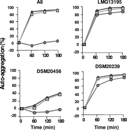
Taking into account the different adhesion capabilities of our strains, four of them were selected for further studies: B. bifidum LMG13195 (high adhesion), B. bifidum DSM20239 (low adhesion), and B. bifidum A8 (intermediate adhesion), including the type strain B. bifidum DSM20456 for comparison purposes. Since the adhesion capability has been found to be linked to cell autoaggregation, the involvement of surface-exposed macromolecules in the aggregation phenomena of the four selected strains was studied first. Initial experiments led us to conclude that the four selected strains displayed a strong autoaggregation phenotype (data not shown), which is in accordance with previous results that detected high levels of autoaggregation in B. bifidum species (8). In this regard, the type of surface-exposed molecules may have a critical role in the aggregation of the bacteria (16). To clarify this point, the nature of the molecules involved in the aggregation of our strains was preliminarily characterized. Different treatments were carried out in order to determine the involvement of carbohydrate moieties (metaperiodate), proteins (proteinase K), and lipids (lipase). All experiments were performed in acetate buffer (pH 3.8) since the aggregation phenotype of B. bifidum is strictly dependent on the pH (8, 16). The treatment with metaperiodate was lethal for B. bifidum, and viable cells were not recovered after the treatment; for this reason, the role of carbohydrate moieties in aggregation could not be tested under our experimental conditions. The rest of the treatments did not affect the viability of the B. bifidum strains (data not shown). None of the treatments affected the autoaggregation ability of B. bifidum LMG13195 and DSM20239 (Fig. 1). However, the aggregation of B. bifidum A8 and DSM20456 was abolished after a treatment with proteinase K, the effect being more pronounced for strain A8, suggesting the involvement of surface-exposed proteins in the aggregation phenotype. In this regard, it is worth mentioning that previous studies suggested that proteins have a key role in the autoaggregating ability of B. bifidum, since protease-treated cells lost this characteristic phenotype (8).
Fig 1.
Representative plots displaying autoaggregation percentages of selected B. bifidum strains after proteinase K or lipase treatments. Symbols: diamonds, control (PBS); squares, lipase; circles, proteinase K.
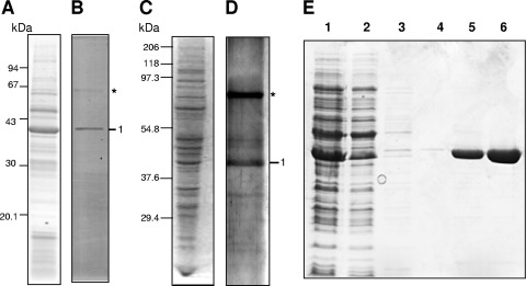
A correlation seems to exist between mucin adhesion and autoaggregation in some Lactobacillus strains (14, 27). Thus, our next attempt was to characterize in depth the relationship between these two features in B. bifidum. In order to pursue this, B. bifidum A8 was used as a model strain, since the abolishment of its aggregation phenotype was very pronounced after protease treatment. The mucin adhesion assays of surface-associated proteins from B. bifidum A8 extracted with two different methods, NaOH and Na2CO3 treatments, led to the isolation of a protein that was collected, almost exclusively, from the mucin layer, suggesting a high binding affinity (Fig. 2). The protein was identified by mass spectrometry analysis as transaldolase (Tal, BBPR_1029) (46). This enzyme is involved in the pentose phosphate pathway and is responsible for the conversion of fructose-6-P and erythrose-4-P into sedoheptulose-7-P and glyceraldehyde-3-P, the latter entering directly into the glycolytic pathway (43). Although its cytoplasmic location is generally agreed, some clues point to the fact that in several Bifidobacterium species it could be released in the extracellular milieu. In a previous work from our group 14 proteins were identified in the extracellular proteome of B. longum NCIMB8809 (37), all of them displayed a predicted noncytoplasmic location (membrane, cell wall, or extracellular) excluding Tal, which lacks any apparent signal peptide. Furthermore, Tal was also detected in the extracellular proteome of the probiotic bacteria B. animalis subsp. lactis BB12 (13). In addition, in the present study we demonstrated the presence of Tal on the surface of B. bifidum A8 by visualizing the enzyme with immunogold particles (Fig. 3). In this regard, recent studies have indicated that many enzymes of carbon catabolism, either from prokaryotic or eukaryotic cells, are often cell wall associated and are able to perform “moonlighting” functions. For instance, in tumor cells the surface exposed α-enolase acts as a plasminogen receptor, mediating plasmin activation, extracellular matrix degradation, and supporting anaerobic proliferation (9). The same role for α-enolase has been suggested in pathogenic bacteria, facilitating tissue invasion via extracellular matrix degradation (18). Also, surface-localized GAPDH acts as a mucin binding protein in several bacteria (1, 41). Interestingly, B. longum transaldolase is highly underproduced in acidic pH (38) and, theoretically, this should facilitate its passage through the stomach, preventing adhesion and directing it to the large gut, its natural habitat.
Fig 2.
SDS-PAGE of B. bifidum A8 surface proteins extracted with NaOH (A) or with Na2CO3 (C) and proteins recovered from the mucus layer after the mucus binding assay with the surface proteins extracted with NaOH (B) or with Na2CO3 (D). Markers: 1, transaldolase; *, BSA. (E) SDS-PAGE of samples taken during transaldolase purification. Lanes: 1, cytoplasmic protein extract of L. lactis NZ9000-HTal; 2, flowthrough; 3 and 4, protein eluted from the Ni-NTA resin after washing with 10 mM imidazole (lane 3) and 30 mM imidazole (lane 4); 5, pre-elution step; 6, purified histidine-tagged Tal eluted from the Ni-NTA column with 150 mM imidazole.
Fig 3.
Visualization of extracellular transaldolase of B. bifidum A8 by immunogold electron microscopy. (A) Control, B. bifidum A8 marked only with antibody against rabbit IgG carrying gold particles of 12 nm. (B and C) B. bifidum A8 labeled first with polyclonal rabbit IgG antibody against transaldolase and second with antibody against rabbit IgG carrying gold particles of 12 nm. Bar, 100 nm.
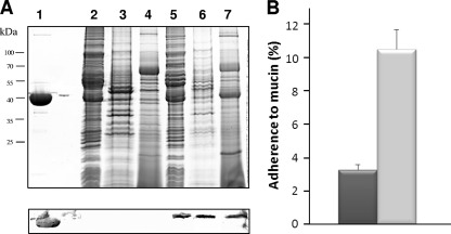
The data obtained in the present study point to a role of the surface-exposed transaldolase as a mucin binding protein in B. bifidum. To demonstrate this hypothesis, bifidobacterial tal was cloned, and the mucin adhesion ability of the recombinant bacterium was studied. The lack of proper molecular tools to construct isogenic mutants in B. bifidum led us to select L. lactis NZ9000 as a host, since this bacterium has been shown to be an adequate host to produce several Bifidobacterium proteins and, furthermore, it naturally lacks a transaldolase gene (24, 28, 29). A Western blot analysis confirmed that the protein was present in the three subcellular locations of the recombinant strain and absent in the control strain containing the empty plasmid pNZ8110 (Fig. 4A). Furthermore, the L. lactis strain expressing Tal displayed a mucin binding level more than three times higher than the nonproducing strain (Fig. 4B). Therefore, these experiments further support the role of Tal as an adhesin able to bind mucin. In relation to this, it has been suggested that not only proteinaceous factors play a role in the adhesion phenotype of B. bifidum, and the partial involvement of sugar moieties has also been indicated (16).
Fig 4.
(A) SDS-PAGE (upper panel) and Western blot (lower panel) analyses of different protein fractions from L. lactis NZ9000-SecTal. Lanes (upper panel): 1, 10 μg of purified transaldolase; 2, 3, and 4, cytoplasmic, surface-associated, and secreted protein fractions of L. lactis NZ9000 containing pNZ8110, respectively; lanes 5, 6, and 7, cytoplasmic, surface-associated, and secreted protein fractions of L. lactis NZ9000 containing pNZ8110-SecTal, respectively. The lower panel shows Western blotting using of the same samples run in a twin gel. (B) Percentage of adhesion to mucin of L. lactis NZ9000 containing pNZ8110 (dark gray) and L. lactis NZ9000 containing pNZ8110-SecTal (light gray). The plots represent the means and standard deviations of four measurements.
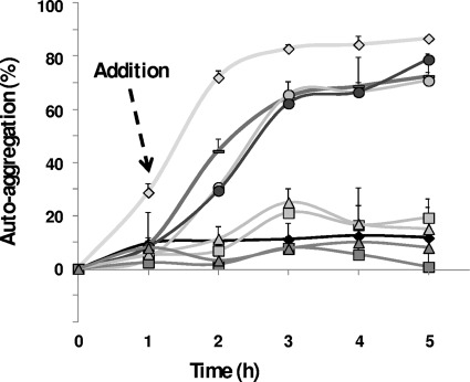
Finally, since mucin adhesion and autoaggregation are two characteristics often related in bacteria, the role of Tal in the autoaggregation phenotype of B. bifidum A8 was also checked. As seen in Fig. 5, proteinase K-treated cells, which have lost the intrinsic autoaggregation trait, restored this phenotype by adding a surface protein extract from the same bacterium or pure Tal, but not by adding histidine-tagged arabinofuranosidase (tested to rule out the possible effect of a histidine tag in the aggregation phenotype) or BSA. This indicates that Tal is a specific aggregation factor in B. bifidum A8 that can contribute, at least partially, to the strong autoaggregation phenotype found in some strains of this Bifidobacterium species.
Fig 5.
Autoaggregation ability of proteinase K-treated B. bifidum A8 in the presence of different proteins or surface protein extracts. Symbols: gray diamond, control (non-proteinase K-treated) strain; black diamond, proteinase K-treated strain; —, addition of 10 μg of A8 surface proteins; gray circle, addition of 1 μg of A8 transaldolase; black circle, addition of 10 μg of A8 transaldolase; light gray box, addition of 1 μg of BSA; dark gray box, addition of 10 μg of BSA; light gray triangle, addition of 1 μg of histidine-tagged arabinofuranosidase; dark gray triangle, addition of 10 μg of histidine-tagged arabinofuranosidase.
In conclusion, to the best of our knowledge the findings reported in this article are the first to suggest an extracellular role for transaldolase in Bifidobacterium. Until now there was no information about putative aggregation factors in bifidobacteria, and specific mucin binding proteins had not been described. We have demonstrated, in vitro, that the extracellular transaldolase from B. bifidum A8 can fulfill both functions. Bifidobacterial transaldolase is abundant in the infant gut, and its dominance in the feces of newborn infants might be related to the high abundance of bifidobacteria in this population group (20). Transaldolase, as has been demonstrated for other glycolytic enzymes, can be recruited on the cell surface, and it could be secreted via a nonclassical secretion mechanism or a thus-far-uncharacterized translocation pathway that remains to be elucidated. Our findings suggest that Tal, when exposed on the cell surface, could act as an important colonization factor favoring the establishment of B. bifidum in the intestinal tract.
ACKNOWLEDGMENTS
This study was financed by European Union FEDER funds and the Spanish Plan Nacional de I+D (grants AGL2007-61805, AGL2009-09445, and AGL2010-14952). I.G.-R. was the recipient of an FPI grant, and B.S. was the recipient of a Juan de la Cierva postdoctoral contract, both from the Spanish Ministerio de Ciencia e Innovación.
Strains NZ9000 and NZ9700 and plasmids pNZ8048 and pNZ8110 were kindly provided by Oscar Kuipers and Jan Kok (Department of Genetics, University of Groningen).
Footnotes
Published ahead of print 23 March 2012
REFERENCES
- 1. Alvarez RA, Blaylock MW, Baseman JB. 2003. Surface localized glyceraldehyde-3-phosphate dehydrogenase of Mycoplasma genitalium binds mucin. Mol. Microbiol. 48:1417–1425 [DOI] [PubMed] [Google Scholar]
- 2. Araya M, Morelli L, Reid G, Sanders ME, Stanton C. 2002. Guidelines for the evaluation of probiotics in foods. Food and Agriculture Organization of the United Nations-World Health Organization, Geneva, Switzerland: http://www.who.int/foodsafety/fs_management/en/probiotic_guidelines.pdf [Google Scholar]
- 3. Arboleya S, et al. 2011. Characterization and in vitro properties of potentially probiotic Bifidobacterium strains isolated from breast-milk. Int. J. Food Microbiol. 149:28–36 [DOI] [PubMed] [Google Scholar]
- 4. Buck BL, Altermann E, Svingerud T, Klaenhammer TR. 2005. Functional analysis of putative adhesion factors in Lactobacillus acidophilus NCFM. Appl. Environ. Microbiol. 71:8344–8351 [DOI] [PMC free article] [PubMed] [Google Scholar]
- 5. Candela M, et al. 2007. Binding of human plasminogen to Bifidobacterium. J. Bacteriol. 189:5929–5936 [DOI] [PMC free article] [PubMed] [Google Scholar]
- 6. Candela M, et al. 2009. Bifidobacterial enolase, a cell surface receptor for human plasminogen involved in the interaction with the host. Microbiology 155:3294–3303 [DOI] [PubMed] [Google Scholar]
- 7. Candela M, et al. 2010. DnaK from Bifidobacterium animalis subsp. lactis is a surface-exposed human plasminogen receptor upregulated in response to bile salts. Microbiology 156:1609–1618 [DOI] [PubMed] [Google Scholar]
- 8. Canzi E, Guglielmetti S, Mora D, Tamagnini I, Parini C. 2005. Conditions affecting cell surface properties of human intestinal bifidobacteria. Antonie Van Leeuwenhoek 88:207–219 [DOI] [PubMed] [Google Scholar]
- 9. Capello M, Ferri-Borgogno S, Cappello P, Novelli F. 2011. α-Enolase: a promising therapeutic and diagnostic tumor target. FEBS J. 278:1064–1074 [DOI] [PubMed] [Google Scholar]
- 10. de los Reyes-Gavilán CG, et al. 2011. Adhesion of bile-adapted Bifidobacterium strains to the HT29-MTX cell line is modified after sequential gastrointestinal challenge simulated in vitro using human gastric and duodenal juices. Res. Microbiol. 162:514–519 [DOI] [PubMed] [Google Scholar]
- 11. de Ruyter PG, Kuipers OP, de Vos WM. 1996. Controlled gene expression systems for Lactococcus lactis with the food-grade inducer nisin. Appl. Environ. Microbiol. 62:3662–3667 [DOI] [PMC free article] [PubMed] [Google Scholar]
- 12. Fisher K, Phillips C. 2009. The ecology, epidemiology, and virulence of Enterococcus. Microbiology 155:1749–1757 [DOI] [PubMed] [Google Scholar]
- 13. Gilad O, Svensson B, Viborg AH, Stuer-Lauridsen B, Jacobsen S. 2011. The extracellular proteome of Bifidobacterium animalis subsp. lactis BB-12 reveals proteins with putative roles in probiotic effects. Proteomics 11:2503–2514 [DOI] [PubMed] [Google Scholar]
- 14. Goh YJ, Klaenhammer TR. 2010. Functional roles of aggregation-promoting-like factor in stress tolerance and adherence of Lactobacillus acidophilus NCFM. Appl. Environ. Microbiol. 76:5005–5012 [DOI] [PMC free article] [PubMed] [Google Scholar]
- 15. Granato D, et al. 2004. Cell surface-associated elongation factor Tu mediates the attachment of Lactobacillus johnsonii NCC533 (La1) to human intestinal cells and mucins. Infect. Immun. 72:2160–2169 [DOI] [PMC free article] [PubMed] [Google Scholar]
- 16. Guglielmetti S, et al. 2009. Study of the adhesion of Bifidobacterium bifidum MIMBb75 to human intestinal cell lines. Curr. Microbiol. 59:167–172 [DOI] [PubMed] [Google Scholar]
- 17. Guglielmetti S, et al. 2008. Implication of an outer surface lipoprotein in adhesion of Bifidobacterium bifidum to Caco-2 cells. Appl. Environ. Microbiol. 74:4695–4702 [DOI] [PMC free article] [PubMed] [Google Scholar]
- 18. Itzek A, et al. 2010. Contribution of plasminogen activation toward the pathogenic potential of oral streptococci. PLoS One 5:e13826. [DOI] [PMC free article] [PubMed] [Google Scholar]
- 19. Jeffery CJ. 2003. Moonlighting proteins: old proteins learning new tricks. Trends Genet. 19:415–417 [DOI] [PubMed] [Google Scholar]
- 20. Klaassens ES, de Vos WM, Vaughan EE. 2007. Metaproteomics approach to study the functionality of the microbiota in the human infant gastrointestinal tract. Appl. Environ. Microbiol. 73:1388–1392 [DOI] [PMC free article] [PubMed] [Google Scholar]
- 21. Kleerebezem M, Vaughan EE. 2009. Probiotic and gut lactobacilli and bifidobacteria: molecular approaches to study diversity and activity. Annu. Rev. Microbiol. 63:269–290 [DOI] [PubMed] [Google Scholar]
- 22. Kuipers OP, Beerthuyzen M, Siezen RJ, de Vos WM. 1993. Characterization of the nisin gene cluster nisABTCIPR of Lactococcus lactis. Requirement of expression of the nisA and nisI genes for development of immunity. Eur. J. Biochem. 216:281–291 [DOI] [PubMed] [Google Scholar]
- 23. Lang C, et al. 2010. Specific Lactobacillus/mutans Streptococcus co-aggregation. J. Dent. Res. 89:175–179 [DOI] [PubMed] [Google Scholar]
- 24. Linares DM, Kok J, Poolman B. 2010. Genome sequences of Lactococcus lactis MG1363 (revised) and NZ9000 and comparative physiological studies. J. Bacteriol. 192:5806–5812 [DOI] [PMC free article] [PubMed] [Google Scholar]
- 25. López P, González-Rodríguez I, Gueimonde M, Margolles A, Suárez A. 2011. Immune response to Bifidobacterium bifidum strains support Treg/Th17 plasticity. PLoS One 6:e24776. [DOI] [PMC free article] [PubMed] [Google Scholar]
- 26. Ludwig W, Kirchhof G, Weizenegger M, Weiss N. 1992. Phylogenetic evidence for the transfer of Eubacterium suis to the genus Actinomyces as Actinomyces suis comb. nov. Int. J. Syst. Bacteriol. 42:161–165 [DOI] [PubMed] [Google Scholar]
- 27. Mackenzie DA, et al. 2010. Strain-specific diversity of mucus-binding proteins in the adhesion and aggregation properties of Lactobacillus reuteri. Microbiology 156:3368–3378 [DOI] [PubMed] [Google Scholar]
- 28. Margolles A, de los Reyes-Gavilán CG. 2003. Purification and functional characterization of a novel α-l-arabinofuranosidase from Bifidobacterium longum B667. Appl. Environ. Microbiol. 69:5096–5103 [DOI] [PMC free article] [PubMed] [Google Scholar]
- 29. Margolles A, Moreno JA, van Sinderen D, de Los Reyes-Gavilán CG. 2005. Macrolide resistance mediated by a Bifidobacterium breve membrane protein. Antimicrob. Agents Chemother. 49:4379–4381 [DOI] [PMC free article] [PubMed] [Google Scholar]
- 30. O'Connell Motherway M, et al. 2011. Functional genome analysis of Bifidobacterium breve UCC2003 reveals type IVb tight adherence (Tad) pili as an essential and conserved host-colonization factor. Proc. Natl. Acad. Sci. U. S. A. 108:11217–11222 [DOI] [PMC free article] [PubMed] [Google Scholar]
- 31. Ouwehand AC, et al. 2008. Bifidobacterium microbiota and parameters of immune function in elderly subjects. FEMS Immunol. Med. Microbiol. 53:18–25 [DOI] [PubMed] [Google Scholar]
- 32. Podolsky DK. 1985. Oligosaccharide structures of human colonic mucin. J. Biol. Chem. 260:8262–8271 [PubMed] [Google Scholar]
- 33. Preising J, et al. 2010. Selection of bifidobacteria based on adhesion and anti-inflammatory capacity in vitro for amelioration of murine colitis. Appl. Environ. Microbiol. 76:3048–3051 [DOI] [PMC free article] [PubMed] [Google Scholar]
- 34. Ruas-Madiedo P, Gueimonde M, Fernández-García M, de los Reyes-Gavilán CG, Margolles A. 2008. Mucin degradation by Bifidobacterium strains isolated from the human intestinal microbiota. Appl. Environ. Microbiol. 74:1936–1940 [DOI] [PMC free article] [PubMed] [Google Scholar]
- 35. Ruiz L, Couté Y, Sánchez B, de los Reyes-Gavilán CG, Sanchez JC, Margolles A. 2009. The cell-envelope proteome of Bifidobacterium longum in an in vitro bile environment. Microbiology 155:957–967 [DOI] [PubMed] [Google Scholar]
- 36. Sánchez B, et al. 2009. Identification of surface proteins involved in the adhesion of a probiotic Bacillus cereus strain to mucin and fibronectin. Microbiology 155:1708–1716 [DOI] [PubMed] [Google Scholar]
- 37. Sánchez B, et al. 2008. A preliminary analysis of Bifidobacterium longum exported proteins by two-dimensional electrophoresis. J. Mol. Microbiol. Biotechnol. 14:74–79 [DOI] [PubMed] [Google Scholar]
- 38. Sánchez B, et al. 2007. Low-pH adaptation and the acid tolerance response of Bifidobacterium longum biotype longum. Appl. Environ. Microbiol. 73:6450–6459 [DOI] [PMC free article] [PubMed] [Google Scholar]
- 39. Sánchez B, de los Reyes-Gavilán CG, Margolles A. 2006. The F1F0-ATPase of Bifidobacterium animalis is involved in bile tolerance. Environ. Microbiol. 8:1825–1833 [DOI] [PubMed] [Google Scholar]
- 40. Sánchez B, González-Tejedo C, Ruas-Madiedo P, Urdaci MC, Margolles A. 2011. Lactobacillus plantarum extracellular chitin-binding protein and its role in the interaction between chitin, Caco-2 cells, and mucin. Appl. Environ. Microbiol. 77:1123–1126 [DOI] [PMC free article] [PubMed] [Google Scholar]
- 41. Sánchez B, Schmitter JM, Urdaci MC. 2009. Identification of novel proteins secreted by Lactobacillus plantarum that bind to mucin and fibronectin. J. Mol. Microbiol. Biotechnol. 17:158–162 [DOI] [PubMed] [Google Scholar]
- 42. Sánchez B, Urdaci MC, Margolles A. 2010. Extracellular proteins secreted by probiotic bacteria as mediators of effects that promote mucosa-bacteria interactions. Microbiology 156:3232–3242 [DOI] [PubMed] [Google Scholar]
- 43. Sánchez B, Zúñiga M, González-Candelas F, de los Reyes-Gavilán CG, Margolles A. 2010. Bacterial and eukaryotic phosphoketolases: phylogeny, distribution, and evolution. J. Mol. Microbiol. Biotechnol. 18:37–51 [DOI] [PubMed] [Google Scholar]
- 44. Schachtsiek M, Hammes WP, Hertel C. 2004. Characterization of Lactobacillus coryniformis DSM 20001T surface protein Cpf mediating coaggregation with and aggregation among pathogens. Appl. Environ. Microbiol. 70:7078–7085 [DOI] [PMC free article] [PubMed] [Google Scholar]
- 45. Tuomola E, Crittenden R, Playne M, Isolauri E, Salminen S. 2001. Quality assurance criteria for probiotic bacteria. Am. J. Clin. Nutr. 73(Suppl 2):S393–S398 [DOI] [PubMed] [Google Scholar]
- 46. Turroni F, et al. 2010. Genome analysis of Bifidobacterium bifidum PRL2010 reveals metabolic pathways for host-derived glycan foraging. Proc. Natl. Acad. Sci. U. S. A. 107:19514–19519 [DOI] [PMC free article] [PubMed] [Google Scholar]
- 47. Turroni F, Milani C, van Sinderen D, Ventura M. 2011. Genetic strategies for mucin metabolism in Bifidobacterium bifidum PRL2010: an example of possible human-microbe co-evolution. Gut Microbes 2:183–189 [DOI] [PubMed] [Google Scholar]
- 48. Vaishampayan PA, et al. 2010. Comparative metagenomics and population dynamics of the gut microbiota in mother and infant. Genome Biol. Evol. 6:53–66 [DOI] [PMC free article] [PubMed] [Google Scholar]
- 49. van Asseldonk M, et al. 1990. Cloning of usp45, a gene encoding a secreted protein from Lactococcus lactis subsp. lactis MG1363. Gene 95:155–160 [DOI] [PubMed] [Google Scholar]
- 50. Vélez MP, De Keersmaecker SC, Vanderleyden J. 2007. Adherence factors of Lactobacillus in the human gastrointestinal tract. FEMS Microbiol. Lett. 276:140–148 [DOI] [PubMed] [Google Scholar]
- 51. Voltan S, et al. 2007. Aggregating phenotype in Lactobacillus crispatus determines intestinal colonization and TLR2 and TLR4 modulation in murine colonic mucosa. Clin. Vaccine Immunol. 14:1138–1148 [DOI] [PMC free article] [PubMed] [Google Scholar]
- 52. von Ossowski I, et al. 2010. Mucosal adhesion properties of the probiotic Lactobacillus rhamnosus GG SpaCBA and SpaFED pilin subunits. Appl. Environ. Microbiol. 76:2049–2057 [DOI] [PMC free article] [PubMed] [Google Scholar]
- 53. von Ossowski I, et al. 2011. Functional characterization of a mucus-specific LPXTG surface adhesin from probiotic Lactobacillus rhamnosus GG. Appl. Environ. Microbiol. 77:4465–4472 [DOI] [PMC free article] [PubMed] [Google Scholar]
- 54. Walter J, Schwab C, Loach DM, Gänzle MG, Tannock GW. 2008. Glucosyltransferase A (GtfA) and inulosucrase (Inu) of Lactobacillus reuteri TMW1.106 contribute to cell aggregation, in vitro biofilm formation, and colonization of the mouse gastrointestinal tract. Microbiology 154:72–80 [DOI] [PubMed] [Google Scholar]
- 55. Wessel D, Flügge UI. 1984. A method for the quantitative recovery of protein in dilute solution in the presence of detergents and lipids. Anal. Biochem. 138:141–143 [DOI] [PubMed] [Google Scholar]


