Abstract
Influenza A viruses containing the promoter mutations G3A/C8U in a given segment express increased levels of the corresponding viral protein during infection due to increased levels of mRNA or cRNA species. The replication of these recombinant viruses is attenuated, and they have an enhanced shedding of noninfectious particles and are incapable of antagonizing interferon (IFN) effectively. Our findings highlight the possibility of increasing influenza virus protein expression and the need for a delicate balance between influenza viral replication, protein expression, and assembly.
TEXT
Influenza A virus is an orthomyxovirus with a genome consisting of eight single-stranded RNA segments in negative orientation coding for at least 10 proteins (4, 33). All eight influenza A viral RNA (vRNA) segments possess a highly conserved sequence at their 3′ and 5′ noncoding regions (14, 15, 27). These ends are partially complementary, resulting in the formation of a secondary RNA “corkscrew/panhandle” structure that is critical for promoter activity (5, 6, 10, 13–15, 24, 30). Recognition of this structure by the polymerase complex, composed of the viral PB1, PB2, and PA proteins (18), results in either (i) an exact complementary copy of the vRNA, the cRNA, which is in turn copied by the viral polymerase into vRNA, or (ii) transcription of viral mRNAs (16, 19, 20). Our current understanding of the influenza A virus promoter has been obtained mostly by introducing mutations into this region in minigenome-based replication assays, consisting of expression of a reporter gene from a virus-like RNA by the influenza virus polymerase (9, 10, 17, 21, 22). However, these methods cannot evaluate how mutations in the conserved noncoding regions impact the virus life cycle and its pathogenesis. Furthermore, many mutations in the influenza viral promoter result in nonviable isolates, as evidenced by the difficulty of generating such mutants. Thus, extending the in vitro reporter-based findings to an in vivo setting is limited, and only a few recombinant viruses bearing promoter mutations have been generated (2, 3, 12, 28).
In this study, we describe for the first time the generation of two recombinant viruses that have a G-to-A and a C-to-U substitution in positions 3 and 8 of the 3′ end in one of the eight viral RNA segments. These mutations have been previously found to yield enhanced reporter gene expression in influenza virus CAT reporter assays (25) and increased vRNA and cRNA levels while considerably inhibiting mRNA levels in in vitro assays (31).
G3A/C8U double mutation results in increased reporter gene expression.
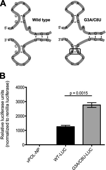
We first confirmed the previous published results on the impact of the G3A/C8U double mutation on influenza virus minigenome assays using a plasmid encoding a firefly luciferase reporter gene flanked by the wild-type or a G3A/C8U mutant noncoding region derived from the influenza virus NS gene (Fig. 1A). The minigenome expression plasmids were cotransfected into HEK293 cells together with influenza virus NP, PB1, PB2, and PA protein expression plasmids and with a transfection control renilla luciferase expression plasmid. Reporter gene activity was measured using a dual luciferase reporter assay (Promega, Madison, WI), as previously described (29). As a negative control, transfections were performed in the absence of the plasmid coding for NP. As expected, this influenza virus minireplicon assay reveals a significant increase (P = 0.0015) in reporter gene expression when the G3A/C8U double mutation is present (Fig. 1B) and is here referred to as super promoter (SP).
Fig 1.
The G3A/C8U mutation results in a significant increase in influenza virus minireplicon driven gene expression. (A) Depiction of the corkscrew structure adopted by wild-type influenza A viral segments (left panel) and the two mutations introduced in the promoter (right panel). These mutations result in a vRNA promoter that more closely resembles the cRNA promoter (note the base pairs that form the stem of the 5′ end of the vRNA molecule). (B) Minireplicon assays were performed in order to determine the effect of the G3A/C8U double mutation on reporter gene expression. WT-LUC, the reporter gene flanked by the wild-type noncoding sequences. G3A/C8U-LUC, the reporter gene flanked by the mutated noncoding sequences. As a negative control, the plasmid encoding the viral NP protein was omitted (vPOL-NP). The experiment was repeated four times. Error bars represent standard errors.
G3A/C8U double mutation enhances mRNA and cRNA levels in the context of recombinant influenza viruses.
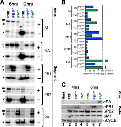
In order to study the impact of these mutations in infectious viruses, two recombinant influenza A viruses bearing the SP double mutation in their PB1 or PA segment (PB1SP or PASP, respectively) were rescued in the influenza A/Puerto Rico/8/1934 (PR8) virus background, using standard reverse genetics techniques (11). Note that while position 4 is polymorphic (U/C) and has been shown to affect promoter function (23), we did not modify the sequence found in the wild-type control PR8 in our study at this particular location. The effect on the multiple RNA species generated during the influenza virus life cycle was studied by primer extension using the AMV primer extension kit (Promega, Madison, WI). HEK293 cells were infected with the indicated viruses at a multiplicity of infection (MOI) of 2. At 6 and 12 h following infection, cells were harvested and their RNA was extracted with TRIzol (Invitrogen, Carlsbad, CA). Primers complementary to positive or negative RNA species were obtained, 32P labeled, and used in reverse transcriptase (RT) reactions as previously described (26). RT products were separated in a denaturing polyacrylamide gel containing 8% acrylamide (19:1, acrylamide:bis), 7 M urea, and Tris-borate-EDTA (TBE). Signal intensities were determined by exposing a phosphorimager cassette that was subsequently developed using a Typhoon scanner (GE Healthcare, Piscataway, NJ).
The presence of the SP mutations in a single gene (either PB1 or PA) results in a dramatic decrease in the vRNA level of all other segments (Fig. 2A) with the exception of those segments bearing the SP mutations, PB1 and PA, in PB1SP and PASP viruses, respectively (Fig. 2A). Densitometric analysis for the representative experiment for the negative-sense RNA species is provided in Fig. 2B.
Fig 2.
The G3A/C8U double mutation modulates the expression of viral RNA species and increases viral protein expression. (A) Primer extension analysis was performed by reverse transcription using two primers specific to the positive (cRNA and mRNA) and negative (vRNA) species in the same tube for each virus (PR8, PB1SP, or PASP) for the indicated segments (M, NA, PB1, PB2, and PA) at 6 and 12 h postinfection. A representative of three independent primer extension experiments is shown. The band corresponding to the mRNA (m) migrates more slowly (upper band of each doublet) due to the presence of the cap compared to the uncapped cRNA (c) species. The vRNA (v) is also indicated for each segment analyzed. (B) Densitometric analysis for the vRNA species of all segments of the representative experiment in panel A as a percentage of wild-type PR8 control is provided. (C) MDCK cells were infected at an MOI of 1 with PR8, PB1SP, or PASP viruses, and viral protein expression was analyzed 4 and 8 h postinfection. As a control, MDCKs were mock infected with phosphate-buffered saline (PBS) supplemented with bovine albumin.
The SP mutations resulted in a significant upregulation in the level of positive-sense RNA species for PB1 in PB1SP virus and for PA in the PASP virus compared to PR8 at both time points examined (Fig. 2A). The slight differences (approximately 2-fold higher or lower) seen in the level of mRNA and cRNA species for those segments bearing a wild-type promoter in PB1SP and PASP viruses are likely accounted for by the delayed replication of SP viruses.
Interestingly, our results differ slightly from those of Vreede and colleagues (31). In their report using an in vitro assay they revealed that the G3A/C8U mutations result in a decrease in mRNA and an increase in cRNA species. In our hands we see an enrichment for both positive-sense RNA species in PB1 in PB1SP and in PA in PASP compared to results for wild-type PR8. However, while PB1SP PB1 mRNA levels are higher than its cRNA levels, we observe the opposite trend for PA in PASP, where there is a higher level of cRNA than mRNA. These differences may be related to the identity of the segment being overexpressed or the different assays used for the evaluation of these mutations in our study and that of Vreede et al.
Our results suggest that polymerase complexes favor the replication of SP segments in detriment of wild-type promoter-bearing segments. This enhanced recognition results in a subsequent increase of both mRNA and cRNA species.
G3A/C8U double mutation results in an increased level of viral protein expression in the context of recombinant influenza viruses.
To determine if the increased level of positive-sense RNAs in PB1SP and PASP viruses correlates with an augmentation of PB1 and PA proteins, respectively, HEK293 cells infected with these viruses or with PR8 were harvested 4 and 8 h postinfection and analyzed by SDS-PAGE. As predicted by our primer extensions, Western blot analysis revealed a significant increase in expression of PB1 and PA proteins at both time points evaluated (Fig. 2C, lane 3 versus lane 2 and lane 6 versus lane 5 for PB1, and lane 4 versus lane 2 and lane 7 versus lane 5 for PA).
PB1SP and PASP viruses are attenuated.
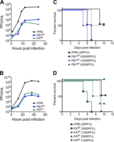
Given the stoichiometric disruption at the RNA and protein level caused by the SP mutations, we hypothesized that the PB1SP and PASP viruses would be attenuated. Single (MOI, 1) and multicycle (MOI, 0.01) replication assays in MDCK cells (ATCC, Manassas, VA) revealed a 2 to 3 log difference between PR8 and either SP virus (Fig. 3A and B). Given our observations in vitro, we set out to determine their phenotype in BALB/c mice (Jackson Laboratory, Bar Harbor, ME). As expected, PB1SP and PASP viruses were significantly attenuated in vivo albeit to a different degree (Fig. 3C and D, respectively). The mouse 50% lethal dose (mLD50) of PB1SP is ∼5 × 10³ PFU while that of PASP is ∼3.3 × 10² PFU, indicating that they are ∼200- and ∼10-fold more attenuated than PR8 (mLD50, ∼ 3 × 101 PFU), respectively (P < 0.001). Interestingly, compared against each other, there is a significant difference between the two SP viruses (P = 0.003).
Fig 3.
In vitro and in vivo characterization of PB1SP and PASP viruses reveals a significant attenuation. (A and B) The replication kinetics of the SP viruses was evaluated in three independent experiments in single (MOI, 1) (A) and multicycle (MOI, 0.01) (B) replication assays in MDCK cells. Error bars represent standard errors. Supernatants were harvested at the indicated time points postinfection, and titers were determined by plaque assays. (C and D) The ability of these viruses to cause disease was evaluated in BALB/c mice (n = 10 per group) infected with several doses of each SP or PR8 virus.
PB1SP and PASP viruses induce increased levels of type I interferon (IFN) due to an increased production of immunostimulatory RNAs.
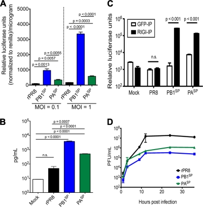
Because of our observations with mice (Fig. 3), we hypothesized that the innate immune response by the host might be accountable for the virus attenuation. Since induction of type I IFN is one of the first lines of defense employed by the immune system by inducing an antiviral state (7), and successful influenza A virus infection is dependent on antagonizing this induction (32), we explored the possibility that the SP viruses are inducing more type I IFN than PR8. To test our hypothesis, we used an MDCK cell line that expresses firefly luciferase driven by the IFN-β promoter. The luciferase activity of cells infected with PB1SP, PASP, and PR8 viruses at MOIs of 0.1 and 1.0 was measured 24 h postinfection (2). In all cases, infection with the SP viruses resulted in a significant increase of reporter activity compared to that with PR8 (Fig. 4A).
Fig 4.
PB1SP and PASP viruses induce type I IFN production. (A) Stable MDCK-IFN-β reporter cells were infected with wild-type PR8, PB1SP, or PASP viruses at two different MOIs (0.1 and 1.0). At 24 h postinfection, luciferase activity was determined per microgram of protein. The experiment was repeated three times. (B) Human DCs derived from PBMCs from three healthy donors were infected at an MOI of 1 with the indicated viruses or left uninfected as a control. At 24 h later, the supernatants were harvested and the type I IFN-α2 levels were determined by ELISA. (C) The amount of immunostimulatory RNAs bound to RIG-I was determined as previously described (1). Error bars represent standard errors. (D) Single-cycle replication curves were conducted in Vero cells to determine the effect of the SP mutations in the replication of the PB1SP and PASP viruses in the absence of type I IFN.
To investigate whether these findings also apply to human dendritic cells (hDCs) exposed to the mutant influenza viruses, peripheral blood mononuclear cells (PBMCs) were isolated from three different healthy donors and derived into conventional DCs in vitro as previously described (8). DCs were infected with SP and PR8 viruses at an MOI of 1. At 24 h postinfection, IFN-α2 levels in the supernatants were determined by enzyme-linked immunosorbent assay (ELISA). Similar to our results obtained with the MDCK reporter cell line, both PB1SP and PASP viruses induced significantly higher levels of type I IFN-α2 than PR8 (Fig. 4B). The amount of type I IFN production in all our assays was significantly higher for PB1SP than for PASP viruses, which correlated with the higher level of attenuation of the former compared to the latter in vivo (Fig. 3C and D). As a result of the stoichiometric disruption we hypothesized that the SP viruses would produce a larger amount of immunostimulatory RNAs. To explore this, the amount of immunostimulatory RNA bound to retinoic acid-inducible gene I (RIG-I) was determined as previously described (1). Briefly, HEK293 cells were infected with PR8, PB1SP, and PASP or left untreated at an MOI of 1 for 1 h. The viruses were subsequently removed, and the cells were transfected with a hemagglutinin (HA)-tagged RIG-I expression plasmid using Lipofectamine 2000 (Invitrogen, Carlsbad, CA). The infections were allowed to progress for 24 h, at which time point the cells were collected and lysed. RIG-I or control (green fluorescent protein [GFP]) immunoprecipitations were performed using an anti-HA antibody or an anti-GFP antibody, respectively. The RNA precipitated was extracted using TRIzol (Invitrogen, Carlsbad, CA) and transfected into a reporter cell line in equimolar amounts between viruses. Twenty-four hours later, firefly luciferase activity was determined. As expected, infection with both SP viruses resulted in a significant increase in the amount of immunostimulatory RNA molecules (Fig. 4C).
Further, the effect of the SP mutations in the replication of both recombinant viruses was also evaluated in an IFN-deficient system to dissect the contribution of an induced antiviral state and a defect in replication by doing single-cycle replication curves in Vero cells (Fig. 4D). Although the differences in Vero cells are reduced compared to those shown in Fig. 3A, there is still a significant reduction in both PB1SP and PASP growth curves in the absence of type I IFN. These results suggest that the attenuation of these viruses is not exclusively due to an increased production of type I IFN (Fig. 4A and B) due to the production of immunostimulatory RNAs (Fig. 4C), but also due to an inherent defect in replication.
SP viruses produce increased levels of noninfectious viral particles.
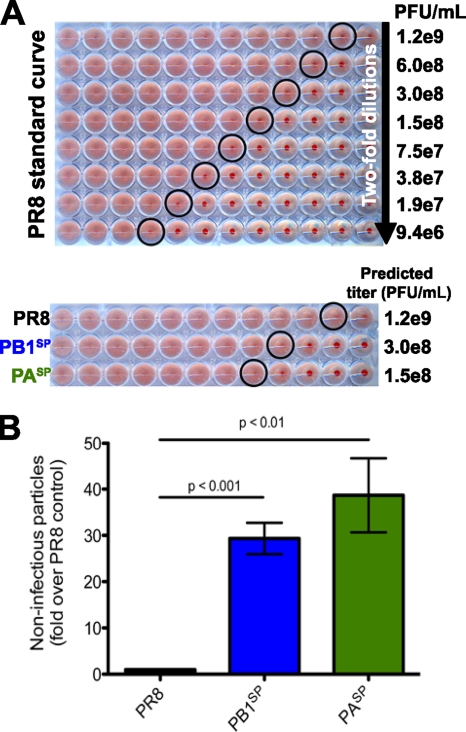
We also speculated that the relatively higher levels in PB1 or PA viral RNA and protein may have a detrimental effect in the production of infectious particles due to the change in the optimal ratio of viral components (i.e., overrepresentation of either PB1 or PA in PB1SP or PASP, respectively). To answer this question, PB1SP, PASP, and PR8 viruses were diluted over 5 orders of magnitude (10−2 to 10−7) and inoculated into the allantois of day 8 embryonated eggs (Charles River Laboratories, Wilmington, MA). After a 2-day incubation at 37°C, eggs were placed at 4°C overnight, and the allantoic fluid was harvested. A hemagglutination (HA) standard curve was generated and compared to the actual titer determined by plaque assays (PFU/ml) for PR8. Using this standard curve, we were able to extrapolate an approximate titer (PFU/ml) based on the HA units of the SP viruses (Fig. 5A). Both SP viruses have a significantly higher number of noninfectious viruses, 25- to 36-fold for PB1SP and 30- to 46-fold for PASP compared to PR8 (Fig. 5B). In order to investigate whether this increase in noninfectious virus particles was due to improper viral RNA packaging, PR8, PB1SP, and PASP viral particles were purified over a sucrose gradient, and RNA was extracted and used in RNA gels and in quantitative PCR (Q-PCR) to determine the frequency of each vRNA species packaged. Both methodologies resulted in slight differences that were not significantly different from results for PR8 (data not shown). Thus, the increased level of noninfectious viral particles likely reflects either an increased release of empty (lacking all viral RNA) particles or budding of mixed populations that are lacking at least one segment.
Fig 5.
Introduction of the SP mutations results in increased shedding of noninfectious viral particles. (A) PR8 virus stocks were used to generate a standard curve by HA. In parallel, the same stocks were used in plaque assays to correlate HA units with PFU/ml for the PR8. HA assays were performed in SP and control viruses to determine their HA values, and the same stocks were also used in plaque assays to estimate the number of noninfectious particles. Given the limited sensitivity of HA assays, four independent viral preparations were tested. (B) Histograms represent the differences between HA units and PFU obtained for the four independent viral preparations for PR8 and PB1SP or PASP. Error bars represent standard deviations.
In conclusion, by generating viruses that encode the G3A/C8U mutations we have demonstrated (i) that these mutations are associated with increased influenza virus promoter activity during virus infection, as previously suggested (25, 31), (ii) that it is possible to increase the expression of specific proteins encoded by influenza viruses during infection, (iii) that disruption of the stoichiometry at the viral RNA and/or protein levels increases the number of immunostimulatory RNAs and in consequence the induction of type I IFN, (iv) that introduction of the SP mutations results in an increase in the ratio of noninfectious versus infectious virus particles, and (v) that the SP mutations attenuate the ability of PB1SP and PASP to replicate efficiently in an IFN-deficient system. It might be interesting to insert these mutations in other influenza virus genes in order to investigate the phenotype of viruses expressing increased levels of other viral proteins.
ACKNOWLEDGMENTS
We are grateful for the comments and suggestions provided by Elena Carnero and Balaji Manicassamy and the excellent technical support provided by Osman Lizardo and Richard Cadagan.
This study was supported by CRIP (Center for Research in Influenza Pathogenesis), an NIAID-funded Center of Excellence for Influenza Research and Surveillance (CEIRS), HHSN266200700010C, to A.G.-S.
Footnotes
Published ahead of print 7 March 2012
REFERENCES
- 1. Baum A, Sachidanandam R, Garcia-Sastre A. 2010. Preference of RIG-I for short viral RNA molecules in infected cells revealed by next-generation sequencing. Proc. Natl. Acad. Sci. U. S. A. 107:16303–16308 [DOI] [PMC free article] [PubMed] [Google Scholar]
- 2. Bergmann M, Muster T. 1995. The relative amount of an influenza A virus segment present in the viral particle is not affected by a reduction in replication of that segment. J. Gen. Virol. 76(Pt 12):3211–3215 [DOI] [PubMed] [Google Scholar]
- 3. Catchpole AP, Mingay LJ, Fodor E, Brownlee GG. 2003. Alternative base pairs attenuate influenza A virus when introduced into the duplex region of the conserved viral RNA promoter of either the NS or the PA gene. J. Gen. Virol. 84:507–515 [DOI] [PubMed] [Google Scholar]
- 4. Chen W, et al. 2001. A novel influenza A virus mitochondrial protein that induces cell death. Nat. Med. 7:1306–1312 [DOI] [PubMed] [Google Scholar]
- 5. Cianci C, Tiley L, Krystal M. 1995. Differential activation of the influenza virus polymerase via template RNA binding. J. Virol. 69:3995–3999 [DOI] [PMC free article] [PubMed] [Google Scholar]
- 6. Crow M, Deng T, Addley M, Brownlee GG. 2004. Mutational analysis of the influenza virus cRNA promoter and identification of nucleotides critical for replication. J. Virol. 78:6263–6270 [DOI] [PMC free article] [PubMed] [Google Scholar]
- 7. Durbin JE, et al. 2000. Type I IFN modulates innate and specific antiviral immunity. J. Immunol. 164:4220–4228 [DOI] [PubMed] [Google Scholar]
- 8. Fernandez-Sesma A, et al. 2006. Influenza virus evades innate and adaptive immunity via the NS1 protein. J. Virol. 80:6295–6304 [DOI] [PMC free article] [PubMed] [Google Scholar]
- 9. Flick R, Hobom G. 1999. Interaction of influenza virus polymerase with viral RNA in the ‘corkscrew’ conformation. J. Gen. Virol. 80(Pt 10):2565–2572 [DOI] [PubMed] [Google Scholar]
- 10. Flick R, Neumann G, Hoffmann E, Neumeier E, Hobom G. 1996. Promoter elements in the influenza vRNA terminal structure. RNA 2:1046–1057 [PMC free article] [PubMed] [Google Scholar]
- 11. Fodor E, et al. 1999. Rescue of influenza A virus from recombinant DNA. J. Virol. 73:9679–9682 [DOI] [PMC free article] [PubMed] [Google Scholar]
- 12. Fodor E, Palese P, Brownlee GG, Garcia-Sastre A. 1998. Attenuation of influenza A virus mRNA levels by promoter mutations. J. Virol. 72:6283–6290 [DOI] [PMC free article] [PubMed] [Google Scholar]
- 13. Fodor E, Pritlove DC, Brownlee GG. 1995. Characterization of the RNA-fork model of virion RNA in the initiation of transcription in influenza A virus. J. Virol. 69:4012–4019 [DOI] [PMC free article] [PubMed] [Google Scholar]
- 14. Fodor E, Pritlove DC, Brownlee GG. 1994. The influenza virus panhandle is involved in the initiation of transcription. J. Virol. 68:4092–4096 [DOI] [PMC free article] [PubMed] [Google Scholar]
- 15. Hsu MT, Parvin JD, Gupta S, Krystal M, Palese P. 1987. Genomic RNAs of influenza viruses are held in a circular conformation in virions and in infected cells by a terminal panhandle. Proc. Natl. Acad. Sci. U. S. A. 84:8140–8144 [DOI] [PMC free article] [PubMed] [Google Scholar]
- 16. Ishihama A, Nagata K. 1988. Viral RNA polymerases. CRC Crit. Rev. Biochem. 23:27–76 [DOI] [PubMed] [Google Scholar]
- 17. Kim HJ, Fodor E, Brownlee GG, Seong BL. 1997. Mutational analysis of the RNA-fork model of the influenza A virus vRNA promoter in vivo. J. Gen. Virol. 78(Pt 2):353–357 [DOI] [PubMed] [Google Scholar]
- 18. Klumpp K, Ruigrok RW, Baudin F. 1997. Roles of the influenza virus polymerase and nucleoprotein in forming a functional RNP structure. EMBO J. 16:1248–1257 [DOI] [PMC free article] [PubMed] [Google Scholar]
- 19. Krug RM. 1981. Priming of influenza viral RNA transcription by capped heterologous RNAs. Curr. Top. Microbiol. Immunol. 93:125–149 [DOI] [PubMed] [Google Scholar]
- 20. Lamb RA, Choppin PW. 1983. The gene structure and replication of influenza virus. Annu. Rev. Biochem. 52:467–506 [DOI] [PubMed] [Google Scholar]
- 21. Leahy MB, Dobbyn HC, Brownlee GG. 2001. Hairpin loop structure in the 3′ arm of the influenza A virus virion RNA promoter is required for endonuclease activity. J. Virol. 75:7042–7049 [DOI] [PMC free article] [PubMed] [Google Scholar]
- 22. Leahy MB, Pritlove DC, Poon LL, Brownlee GG. 2001. Mutagenic analysis of the 5′ arm of the influenza A virus virion RNA promoter defines the sequence requirements for endonuclease activity. J. Virol. 75:134–142 [DOI] [PMC free article] [PubMed] [Google Scholar]
- 23. Lee MK, et al. 2003. A single-nucleotide natural variation (U4 to C4) in an influenza A virus promoter exhibits a large structural change: implications for differential viral RNA synthesis by RNA-dependent RNA polymerase. Nucleic Acids Res. 31:1216–1223 [DOI] [PMC free article] [PubMed] [Google Scholar]
- 24. Luo GX, Luytjes W, Enami M, Palese P. 1991. The polyadenylation signal of influenza virus RNA involves a stretch of uridines followed by the RNA duplex of the panhandle structure. J. Virol. 65:2861–2867 [DOI] [PMC free article] [PubMed] [Google Scholar]
- 25. Neumann G, Hobom G. 1995. Mutational analysis of influenza virus promoter elements in vivo. J. Gen. Virol. 76(Pt 7):1709–1717 [DOI] [PubMed] [Google Scholar]
- 26. Robb NC, Smith M, Vreede FT, Fodor E. 2009. NS2/NEP protein regulates transcription and replication of the influenza virus RNA genome. J. Gen. Virol. 90:1398–1407 [DOI] [PubMed] [Google Scholar]
- 27. Robertson JS. 1979. 5′ and 3′ terminal nucleotide sequences of the RNA genome segments of influenza virus. Nucleic Acids Res. 6:3745–3757 [DOI] [PMC free article] [PubMed] [Google Scholar]
- 28. Solorzano A, et al. 2000. Reduced levels of neuraminidase of influenza A viruses correlate with attenuated phenotypes in mice. J. Gen. Virol. 81:737–742 [DOI] [PubMed] [Google Scholar]
- 29. Steidle S, et al. 2010. Glycine 184 in nonstructural protein NS1 determines the virulence of influenza A virus strain PR8 without affecting the host interferon response. J. Virol. 84:12761–12770 [DOI] [PMC free article] [PubMed] [Google Scholar]
- 30. Tiley LS, Hagen M, Matthews JT, Krystal M. 1994. Sequence-specific binding of the influenza virus RNA polymerase to sequences located at the 5′ ends of the viral RNAs. J. Virol. 68:5108–5116 [DOI] [PMC free article] [PubMed] [Google Scholar]
- 31. Vreede FT, Gifford H, Brownlee GG. 2008. Role of initiating nucleoside triphosphate concentrations in the regulation of influenza virus replication and transcription. J. Virol. 82:6902–6910 [DOI] [PMC free article] [PubMed] [Google Scholar]
- 32. Wang X, et al. 2000. Influenza A virus NS1 protein prevents activation of NF-kappaB and induction of alpha/beta interferon. J. Virol. 74:11566–11573 [DOI] [PMC free article] [PubMed] [Google Scholar]
- 33. Wise HM, et al. 2009. A complicated message: identification of a novel PB1-related protein translated from influenza A virus segment 2 mRNA. J. Virol. 83:8021–8031 [DOI] [PMC free article] [PubMed] [Google Scholar]


