Abstract
Side branch wiring is not infrequently used to protect side branch flow after main vessel stenting. Rarely, it becomes difficult to retrieve the jailed wire behind the stent and therefore it may even be detached and remained in the circulation. This article presents a report of such a case and reviews treatment options.
Keywords: Jailed wire, Stent, Retrieval
Introduction
Wires in side branches may remain and detach behind the stent during main vessel stenting. Several treatment options have been put forward.1–3
Case Report
The patient was a 65 year old male who developed extensive anterior wall myocardial infarction seven years earlier while in excellent medical condition. He had no traditional risk factors except cigarette smoking. Coronary angiography at that time revealed severe (90%) stenosis of left anterior descending artery (LAD) mid-portion, significant stenosis of LAD distal portion and significant stenosis of origin of diagonal branch. Plain balloon angioplasty of diagonal branch and distal LAD and predilation and stenting of LAD mid portion were planned for another session. Two 0.014 guidewires were inserted in LAD and diagonal branches. Plain balloon angioplasty of diagonal branch was done using a 2.0-20 balloon (Voyager, Guidant, Indianapolis, IN, USA). Then, distal lesion of LAD was opened by a 3.0-15 balloon (Voyager, Guidant, Indianapolis, IN, USA) using nominal atmosphere pressure. The proximal lesion of LAD was primarily predilated with a 3.0-14 balloon (Voyager, Guidant, Indianapolis, IN, USA) and then stented with a 3.0-15 stent (Cypher, Cordis Corp., Miami, Florida, USA) at a pressure of 16 atmospheres. In the final angiogram both LAD and diagonal branches were completely open. However, while the guidewires were being pulled back from the coronary arteries at the end of the procedure, diagonal guidewire was trapped and its distal radiopaque segment was separated and remained behind the LAD stent. Since the patient's homodynamic condition was stable and the retained guidewire did not impair the blood flow in LAD or diagonal branch, no further action was made and the patient was sent to post-catheterization unit. The patient was discharged home the day after.
After discharge the patient was followed regularly and he did not show any signs of ischemia. Therefore, no further actions were performed.
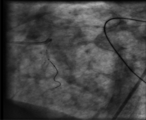
In September 2006, the patient was hospitalized because of inferior wall myocardial infarction for which tissue-type plasminogen activator (tPA) was administered with good results. In coronary angiography, right coronary artery was found to have significant stenosis at mid-portion which was addressed by percutaneous coronary intervention (PCI). In coronary angiography of left system, left anterior descending and diagonal branches were completely patent. The retained broken jailed wire was still in place with no displacement in comparison with previous coronary angiography (Figure 1). The patient has remained in stable cardiac condition until now.
Figure 1.
Retained jailed wire seven years after PCI.
Discussion
Broken retained guidewires in coronary arteries are very infrequently encountered in medical practice and since they are reported only in case reviews, no evidence-based approach has been put forward regarding their optimal management. The first cases of broken guidewires were reported in 1980 while the main treatment option was surgery.1 Despite production of more flexible and high quality guidewires, the incidence of this complication has not been reduced. With technical improvements, physicians began to tackle with more complex cases, so the risk of wire entrapment remains the same or is even increased. Remaining of segments of guidewires (especially their metallic part) may predispose thrombus formation with its well-known sequels, i.e. systemic emboli and coronary thrombosis. Hardware retained in the lumen of a coronary vessel will generally serve as a nidus for endothelial injury and platelet deposition, putting the vessel at risk for acute thrombosis.2 Some situations increase the risk of guidewire rupture during its retrieval from behind the stent. First of all, guidewire detachment (especially its radiopaque portion) is more common with so-called hydrophilic wires, so these types of guidewires should not be used as a jailed wire in treatment of bifurcation lesions. Secondly, this condition is more common when the guidewire is jailed between overlapping parts of stents. Stenting of calcified and tortuous parts of vessels also increases shear force during wire retrieval and therefore the chance of wire detachment.3
Based on available literature, treatment options for this complication depend on the site of entrapment and clinical sequels of the foreign body and are divided to conservative management, interventional techniques, and surgery.
Conservative management is appropriate when risks of intervention outweigh its benefits in a particular patient. It is logical to leave a radiopaque part of a guidewire in a small side branch which had no clinical sequels (this segment of guidewire is less thrombogenic than its metallic peers). Several reports have confirmed short- and long-term safety and efficacy of this method in this group of patients making sure that a small part of a guidewire has been retained in a not so big side branch.
Unfortunately, the total length of retained guidewire is not always discernable in fluoroscopy1 and its distal radiopaque segment may be the peak of an iceberg with its metallic part remaining in the main branch and even in aortic bulb or aorta. In this case, the wire must be retrieved by either interventional or surgical routs since remaining of thrombogenic proximal part of the guidewire may provoke thrombotic sequels.
Interventional techniques include retrieval of segments of guidewire by manufactured snares, homemade snares, paired guidewires knotted together, Dotter basket snares, FilterWires, and retrieval forceps.1–12 Both proximal part and radiopaque distal part have been reported to be successfully captured by snares, depending on visibility and accessibility of each part. Choice of the retrieval device and its size depends on spatial orientation and anatomical position of the guidewire. If endovascular retrieval of retained guidewire was impossible or it is thought that aggressive maneuvers for its retrieval may impose further endothelial damage, surgery will become an alternative option with unproven safety and efficacy. It must be especially taken into account if for some reasons revascularization of patient seems incomplete by interventional procedures.13
In our patient, the radiopaque portion of the guidewire was retained in a not so big side branch. In addition, careful evaluation of cine films showed no part of guide wire in LAD and aorta and it seemed that the guidewire was ruptured exactly near its exit point behind LAD stent. Therefore, it was reasonable to leave it in place without further action. Coronary angiography seven years later proved safety of this approach at least for that period of time.
Conflict of Interests
Authors have no conflict of interests.
References
- 1.Doring V, Hamm C. Delayed surgical removal of a guide-wire fragment following coronary angioplasty. Thorac Cardiovasc Surg. 1990;38(1):36–7. doi: 10.1055/s-2007-1013988. [DOI] [PubMed] [Google Scholar]
- 2.Hartzler GO, Rutherford BD, McConahay DR. Retained percutaneous transluminal coronary angioplasty equipment components and their management. Am J Cardiol. 1987;60(16):1260–4. doi: 10.1016/0002-9149(87)90604-7. [DOI] [PubMed] [Google Scholar]
- 3.Van Gaal WJ, Porto I, Banning AP. Guide wire fracture with retained filament in the LAD and aorta. Int J Cardiol. 2006;112(2):e9–11. doi: 10.1016/j.ijcard.2006.01.040. [DOI] [PubMed] [Google Scholar]
- 4.Sezgin AT, Gullu H, Ermis N. Guidewire entrapment during jailed wire technique. J Invasive Cardiol. 2006;18(8):391–2. [PubMed] [Google Scholar]
- 5.Ghosh PK, Alber G, Schistek R, Unger F. Rupture of guide wire during percutaneous transluminal coronary angioplasty. Mechanics and management. J Thorac Cardiovasc Surg. 1989;97(3):467–9. [PubMed] [Google Scholar]
- 6.Vrolix M, Vanhaecke J, Piessens J, De Geest H. Anunusual case of guide wire fracture during percutaneous transluminal coronary angioplasty. Cathet Cardiovasc Diagn. 1988;15(2):99–102. doi: 10.1002/ccd.1810150208. [DOI] [PubMed] [Google Scholar]
- 7.Khambekar S, Hudson I, Kovac J. Percutaneous coronary intervention to anomalous right coronary artery and retained piece of guidewire in the coronary vasculature. J Interv Cardiol. 2005;18(3):201–4. doi: 10.1111/j.1540-8183.2005.04097.x. [DOI] [PubMed] [Google Scholar]
- 8.Cafri C, Rosenstein G, Ilia R. Fracture of a coronary guidewire during graft thrombectomy with the X-sizer device. J Invasive Cardiol. 2004;16(5):263–5. [PubMed] [Google Scholar]
- 9.Ozkan M, Yokusoglu M, Uzun M. Retained percutaneous transluminal coronary angioplasty guidewire in coronary circulation. Acta Cardiol. 2005;60(6):653–4. doi: 10.2143/AC.60.6.2004939. [DOI] [PubMed] [Google Scholar]
- 10.Kim CK, Beom Park C, Jin U, Ju Cho E. Entrapment of guidewire in the coronary stent during percutaneous coronary intervention. Thorac Cardiovasc Surg. 2006;54(6):425–6. doi: 10.1055/s-2006-923805. [DOI] [PubMed] [Google Scholar]
- 11.Patel T, Shah S, Pandya R, Sanghvi K, Fonseca K. Broken guidewire fragment: a simplified retrieval technique. Catheter Cardiovasc Interv. 2000;51(4):483–6. doi: 10.1002/1522-726x(200012)51:4<483::aid-ccd24>3.0.co;2-f. [DOI] [PubMed] [Google Scholar]
- 12.Ojeda Delgado JL, Jimenez Mena M, Barrios Alonso V, Pena Tizon J, Fernandez Sanchez-Villaran E, Hernandez Madrid A, et al. Guide-wire rupture as a complication of coronary angioplasty. Apropos 2 cases and a review of the literature. Rev Esp Cardiol. 1992;45(2):141–4. [PubMed] [Google Scholar]
- 13.Balbi M, Bezante GP, Brunelli C, Rollando D. Guide wire fracture during percutaneous transluminal coronary angioplasty: possible causes and management. Interact Cardiovasc Thorac Surg. 2010;10(6):992–4. doi: 10.1510/icvts.2009.227678. [DOI] [PubMed] [Google Scholar]


