Abstract
Fecal impaction (FI) is a common cause of lower gastrointestinal tract obstruction lagging behind stricture for diverticulitis and colon cancer. It is the result of chronic or severe constipation and most commonly found in the elderly population. Early recognition and diagnosis is accomplished by way of an adequate history and physical examination in conjunction with an acute abdominal series. Prompt identification and treatment minimizes the risks of complications such as bowel obstruction leading to aspiration, stercoral ulcers, perforation, and peritonitis. Treatment options include gentle proximal softening in the absence of complete bowel obstruction, distal washout, and manual extraction. Surgical resection of the involved colon or rectum is reserved for cases of FI complicated by ulceration and perforation leading to peritonitis. Recurrence is common, and can be managed by increasing dietary fiber content to 30 gm/day, increased water intake, and discontinuation of medications that can contribute to colonic hypomotility.
Keywords: fecal impaction, constipation, stercoral perforation, inspissated stool syndrome
Objectives: On completion of this article, the reader should be able to summarize the management of fecal impaction.
Fecal impaction (FI) is a common gastrointestinal (GI) disorder and a source of significant patient discomfort with potential for major morbidity especially in the elderly population.1 FI is defined as the inability to evacuate large hard inspissated concreted stool or bezoar lodged in the lower GI tract. It is most commonly found in the rectum.2,3 It is also known as coprostasis or inspissated stool syndrome.4 Despite a multimillion dollar laxative industry in our bowel-conscious society, fecal impaction remains prevalent mostly in the pediatric and elderly population. The incidence of fecal impaction increases with age and dramatically impairs the quality of life in the elderly.5 Read et al noted that 42% of patients in a geriatric ward had FI.6 FI is also commonly found in patients with neuropsychiatric disorders such as Alzheimer disease, Parkinson disease, dementia, and severe stroke, as well as in spinal cord injury patients.
Etiology and Pathophysiology
The etiologic factors responsible for FI are similar to those responsible for constipation. FI is seen as an acute complication of chronic and untreated constipation. An extensive list of factors contributing to the development of fecal impaction is listed in Table 1.5,7 Three of the most important risk factors are colonic hypomotility and inadequate dietary fiber and water intake; hence, the population with the highest risk are the elderly and neuropsychiatric patients. An increase in fiber intake to 30 grams per day coupled with adequate hydration helps prevent constipation and fecal impaction by poorly digested fiber. Lack of mobility due to aging or spinal cord injury may also cause fecal impaction related to reduction of colonic mass movement and an inability to use abdominal muscles to assist in defecation. Medications known to retard GI motility include opiate analgesics, anticholinergic agents, calcium channel blockers, antacids, and iron preparations.5 However, paradoxically, laxative abuse is associated with constipation and fecal impaction. The laxative-dependent patient is unable to produce a normal response to colonic distention and progressively requires higher doses to achieve a bowel movement.6,8,9 Congenital and acquired conditions of the colon and rectum, including megacolon, Hirschsprung disease, Chagas disease, intra- and extraluminal obstruction from adhesion, and anastomotic stricture can also cause fecal impaction.4,10 In addition to these etiologic factors, anatomic and functional abnormalities of the anorectum should be considered and excluded such as pelvic floor prolapse.1,11
Table 1. Etiologies of Fecal Impaction1.
| Chronic constipation |
| Metabolic (hypothyroidism, diabetes mellitus, uremia, porphyria, hypercalcemia) |
| Dietary (inadequate fluid and fiber intake) |
| Medications (opiates, antipsychotics, iron preparations, calcium channel blockers) |
| Neurogenic (spinal cord injury, multiple sclerosis, Parkinson disease) |
| Psychiatric illness |
| Anatomic anorectal abnormalities |
| Megarectum (Chagas disease, Hirschsprung disease) |
| Anorectal stenosis (strictures) |
| Neoplasm |
| Functional anorectal abnormalities |
| Increased rectal compliance (irritation, endometriosis) |
| Abnormal rectal sensation (Crohns disease) |
| Pelvic floor dysfunction (rectocele, enterocele, nonrelaxing puborectalis) |
Clinical Presentation and Evaluation
The typical presenting symptoms of fecal impaction are similar to those found in intestinal obstruction from any cause, including abdominal pain and distention, nausea, vomiting, and anorexia.10 These are summarized in Table 2.5 A retrospective review by Gurll and Steer revealed that 39% of patients with fecal impaction had a history of prior impactions.12 These symptoms result from hardened stool impacted in the rectum or distal sigmoid colon with subsequent obstruction. Additional complications such as stercoral ulceration leading to perforation, rectovaginal fistula, megacolon, and colonic perforation may ensue.13 Elderly or institutionalized patients with dementia or psychosis may present with increased agitation, confusion, autonomic dysreflexia—marked by hemodynamic instability, paradoxical diarrhea, and fecal incontinence.10
Table 2. Symptoms Associated with Fecal Impaction.
| Constipation |
| Rectal discomfort |
| Anorexia |
| Nausea |
| Vomiting |
| Abdominal pain |
| Paradoxic diarrhea |
| Fecal incontinence |
| Urinary frequency |
| Urinary overflow incontinence |
| Confusion |
| Agitation |
| Worsening psychosis |
Following a complete history highlighting bowel habits, GI symptoms, surgical history, medical history, and medications, a physical examination (PE) is then performed. A PE would reveal a mild tachycardia, with normal blood pressure signifying moderate to severe dehydration or pain. However, in cases of perforation, a fever could be present as well. A focused abdominal exam would often reveal abdominal distension with tympany and mild diffuse abdominal tenderness with a palpable malleable, tubular structure usually in the left lower quadrant indicating a stool filled rectosigmoid. Signs of perforation (tachycardia, fever, tenderness or peritoneal signs) are usually absent.4,14 Although most impactions occur in the rectal vault, the absence of palpable stool does not rule out a FI.1,10,15
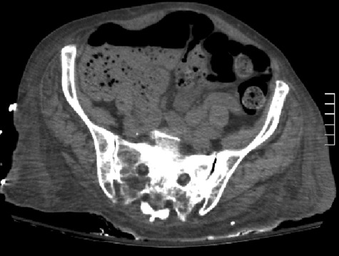
Laboratory work-up should include a complete blood count with a differential (CBC), basic metabolic panel (BMP). However, liver function tests and pancreatic enzymes are not necessary; they may be ordered if the pain is located in the epigastric area. The WBC often shows a mild leukocytosis, with or without a left shift. The BMP would show dehydration, hyponatremic, hypokalemic metabolic alkalosis with paradoxical aciduria as a result of poor oral intake and persistent nausea and vomiting. An acute abdominal series can be used as the first-line imaging modality used to search for intraluminal feces or signs of obstruction. In the advent of the faster, easily available computed tomography (CT) scan, more patients are diagnosed by the presence of large fecal matter in the colon and rectum with or without signs of colonic perforation (Fig. 1). The presence of bowel obstruction as evidenced by dilated small bowel or colon with air-fluid levels contraindicates attempts at proximal softening or washout using oral solutions.
Figure 1.
A computed tomography scan showing fecal impaction more prominent in the right colon.
Treatment
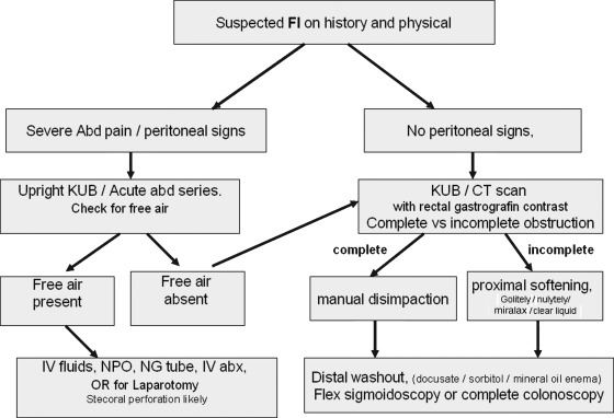
Treatment is aimed at relieving the major complaint and correcting the underlying pathophysiology to prevent recurrence. FI in the rectum will often require digital fragmentation and mechanical removal (see Fig. 2).1
Figure 2.
Suggested treatment algorithm for management of fecal impaction. FI, fecal incontinence; KUB, kidney ureter bladder; IV, intravenous; CT, computed tomography; OR, operating room; abd, abdomen; abx, antibiotics.
Manual Disimpaction
If hardened stool is palpable in the rectum, it may require manual fragmentation or disimpaction. A lubricated, gloved index finger is inserted into the rectum and the hardened stool is gently broken up using a scissoring motion. The finger is then moved in a circular manner, bent slightly and removed, extracting stool with it. This maneuver is repeated until the rectum is cleared of hardened stool. Manual disimpaction may be aided by the use of an anal retractor (i.e., Hill-Ferguson retractor).10,15,16,17,18
Distal Softening or Washout
Softening of hardened stool and stimulation of evacuation with enemas and suppositories is often helpful. A variety of enema solutions is available for use: each has characteristics that may be useful in selected patients. Most enema solutions contain water and an osmotic agent. One such combination contains water, docusate sodium syrup (Colace®; Purdue Pharma L.P., Stamford, CT), and sorbitol. Docusate sodium is a surface active agent that helps soften the stool as it mixes with water.12,19,20,21 Sorbitol is a sugar alcohol that acts as an osmotic agent. Rectally administered solutions mechanically soften the impacted stool and the additional volume gently stimulates the rectum to evacuate.
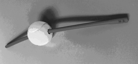
During enema administration, the patient is placed in the left lateral decubitus position with the buttocks close to the edge of the bed and the left leg straight and the right leg bent at knee toward the chest (Sims’ position) with a plastic bag under the hips. The enema is given using a 30-French red rubber catheter or three-way Foley that is passed through a rubber ball (i.e., tennis ball; Fig. 3). The ball allows the administrator to maintain a seal against the patient's anus for prolonged enema contact. Balloon-tipped catheters are not used as they may damage the distal rectum or anal sphincter and generally do not maintain an adequate seal.1,3,14,17 The pressure and volume of enema administration must be appropriate. Enema pressure is controlled by the height of the solution reservoir. Limiting the reservoir height to 3 feet above the anus maintains an adequate pressure limit. The volume and rate of fluid administration is guided by the size of the patient's rectum and the degree of fullness symptoms. Administration of smaller volumes (1–2 L) may be more beneficial than a single large volume enema. A slower rate of enema administration produces less patient discomfort, aids in mixing of solution, and allows instillation of a larger volume.1 The patient's sensation of fullness is a helpful guide during enema instillation. Volumes or rates that produce patient discomfort are avoided.15,22
Figure 3.
A 30-French catheter passed via two small holes in a tennis ball held against the anus used for enema administration.
Once administration is complete, a few minutes are allowed for the solution to mix with and soften the stool. Gentle massaging of the lower abdomen may aid in mixing the combination. The patient then voluntarily evacuates the enema–stool mixture.1 Additional gentle abdominal manipulation often helps in evacuation. Ambulatory patients can evacuate more efficiently by using a commode. This process is repeated until the symptoms are relieved and returns are clear.15,22
Proximal Softening or Washout
Oral lavage with polyethylene glycol solutions containing electrolytes (GoLYTELY® or NuLYTELY®, Braintree Laboratories, Braintree, MA; CoLyte®, Meda Pharmaceuticals, Somerset, NJ) may be used to soften or washout proximal stool.6 Such solutions without electrolytes (Miralax®, Merck & Co., Whitehouse Station, NJ) have also been used. This technique is contraindicated when a complete bowel obstruction exists. The volume and rate of oral lavage is dependent on patient size. To treat childhood fecal impaction, Youssef and coworkers recommend 1 to 1.5 g/kg/d of polyethylene glycol solution (PEG 3350; Miralax®).11 For adults, oral regimens vary from 1 to 2 L of PEG with electrolytes or 17 g of PEG 3350 in 4 to 8 oz of water every 15 minutes until the patient begins passing stool or eight glasses have been consumed.22 Development of nausea, vomiting, or significant abdominal discomfort prompts cessation of fluid intake. A recent Cochrane Review evaluated 10 randomized controlled trials comparing lactulose to PEG. The study involving 868 participants with age range from 3 months to 70 years showed higher weekly stool frequency and improved abdominal pain scores for patients using PEG.23
Other osmotic laxatives such as oral magnesium citrate have also been used for proximal lavage. Thirty to 60 mL of magnesium citrate orally with 4 oz of clear liquids every 4 to 8 hours is a common regimen. Magnesium- and phosphate-containing solutions must be used with extreme caution in patients with renal insufficiency and congestive heart failure.
Special Situations
Barium Impaction
Following barium radiographic studies (barium enema and upper GI studies), the barium may be retained in colon and become impacted with stool. Barium is not water soluble, and as such will become inspissated in the colon once the water is absorbed. Anatomic or functional abnormalities of the lower GI tract can predispose to such impactions.1 The presence of inspissated stool is quite common in a rectal stump. The rectal stump is diverted from the stream of stool, thus reducing the water content in the rectum. The stool and barium left behind becomes dehydrated and impacted thereby making an end to end anastomosis (EEA) more challenging.16,18
Patients undergoing barium studies should ingest additional fluids following the examination to prevent a barium impaction. Use of a laxative such as milk of magnesia may also be beneficial. Medical advice should be sought if no bowel movement occurs within 48 hours of the radiologic examination or development of symptoms of FI.1,15
The presence of a barium impaction is readily apparent on plain films. An anteroposterior or lateral abdominal film will reveal the amount and location of the retained barium. The absence of signs of perforation (contrast extravasation or free air) or bowel obstruction should also be confirmed. Perforation generally requires operative management. In the absence of perforation or obstruction, removal of barium impaction should proceed as outlined earlier.
Anorectal Surgery
Fecal impaction following anorectal surgery is a rare but serious complication. Buls and Goldberg reported a 0.4% incidence of impaction after operative hemorrhoidectomy.24 Fecal impaction occurring after anorectal surgery is multifactorial. Opiates used for pain relief in the postoperative period have significant constipating action. Anal canal edema and sphincter spasm also compound the problem. A patient's fear of pain associated with bowel movements may lead to restriction of bowel movements resulting in hardened, impacted stool. The presence of a significant impaction is suggested by a history of infrequent bowel movements and perineal pressure and pain.1
Mild impactions are relieved with the gentle administration of a retention enema. Posthemorrhoidectomy patients with significant impactions often require disimpaction under anesthesia. An anal block can be administered in the operating room or the endoscopy suite in combination with conscious sedation. 0.5% or 1% Xylocaine with or without epinephrine is injected around the anus and into the anal sphincter complex. The fecal impaction may be gently digitally removed, once the local anesthetic takes effect.
After removal of the impaction, the patient should be placed on additional stool softeners and laxatives and advised on the importance of regular bowel movements. A rectal foreign body or bezoars can be removed using the single or multiple Foley technique. The three-way Foley is passed into the rectum and gently inflated and pulled back to retrieve rectal masses. This technique has the potential risk of causing perforation in an already thin-walled rectum.18
Posttreatment Evaluation and Prevention
Once the impaction has been adequately treated, possible etiologies are explored. A total colonic evaluation by flexible sigmoidoscopy, a colonoscopy, or a barium enema in cases where a colonoscopy is not feasible, should be done after the FI resolves.18,19 A colonoscopy should be performed to reveal anatomic abnormalities (stricture or malignancy). Endocrine and metabolic screening, and thyroid function tests are also indicated. In the absence of an anatomic abnormality, a bulking agent (psyllium, methylcellulose) or an osmotic agent such as PEG (Miralax®) is administered to produce soft regular bowel movements. Other risk factors such as depression, immobility, lack of exercise, and inadequate access to toilet facilities should also be corrected.1,5,10
Conclusion
In summary, fecal impaction is a common GI problem in the elderly. Early identification and treatment minimizes patient discomfort and potential risk for aspiration, stercoral ulceration, or perforation. Treatment options include digital disimpaction and proximal or distal washout (Fig. 2). Following treatment, possible etiologies should be found and preventive therapy such as increased dietary fiber and water intake can be instituted to avoid recurrence.1
Acknowledgments
A previous version of this article entitled, “Fecal Impaction” was written by Farhad Araghizadeh, M.D., and published in Clinics in Colon and Rectal Surgery.1
References
- 1.Araghizadeh F. Fecal impaction. Clin Colon Rectal Surg. 2005;18(2):116–119. doi: 10.1055/s-2005-870893. [DOI] [PMC free article] [PubMed] [Google Scholar]
- 2.Tobias N, Mason D, Lutkenhoff M, Stoops M, Ferguson D. Management principles of organic causes of childhood constipation. J Pediatr Health Care. 2008;22(1):12–23. doi: 10.1016/j.pedhc.2007.01.001. [DOI] [PubMed] [Google Scholar]
- 3.Wald A. Management and prevention of fecal impaction. Curr Gastroenterol Rep. 2008;10(5):499–501. doi: 10.1007/s11894-008-0091-y. [DOI] [PubMed] [Google Scholar]
- 4.Tracey J. Fecal impaction: not always a benign condition. J Clin Gastroenterol. 2000;30(3):228–229. doi: 10.1097/00004836-200004000-00004. [DOI] [PubMed] [Google Scholar]
- 5.De Lillo A R, Rose S. Functional bowel disorders in the geriatric patient: constipation, fecal impaction, and fecal incontinence. Am J Gastroenterol. 2000;95(4):901–905. doi: 10.1111/j.1572-0241.2000.01926.x. [DOI] [PubMed] [Google Scholar]
- 6.Read N W, Abouzekry L, Read M G, Howell P, Ottewell D, Donnelly T C. Anorectal function in elderly patients with fecal impaction. Gastroenterology. 1985;89(5):959–966. doi: 10.1016/0016-5085(85)90194-5. [DOI] [PubMed] [Google Scholar]
- 7.Beck D E. Surgical management of constipation. Clin Colon Rectal Surg. 2005;18(2):81–84. doi: 10.1055/s-2005-870888. [DOI] [PMC free article] [PubMed] [Google Scholar]
- 8.Creason N, Sparks D. Fecal impaction: a review. Nurs Diagn. 2000;11(1):15–23. doi: 10.1111/j.1744-618x.2000.tb00381.x. [DOI] [PubMed] [Google Scholar]
- 9.Gallagher P F, O’Mahony D, Quigley E M. Management of chronic constipation in the elderly. Drugs Aging. 2008;25(10):807–821. doi: 10.2165/00002512-200825100-00001. [DOI] [PubMed] [Google Scholar]
- 10.Wrenn K. Fecal impaction. N Engl J Med. 1989;321(10):658–662. doi: 10.1056/NEJM198909073211007. [DOI] [PubMed] [Google Scholar]
- 11.Youssef N N, Peters J M, Henderson W, Shultz-Peters S, Lockhart D K, Di Lorenzo C. Dose response of PEG 3350 for the treatment of childhood fecal impaction. J Pediatr. 2002;141(3):410–414. doi: 10.1067/mpd.2002.126603. [DOI] [PubMed] [Google Scholar]
- 12.Gurll N, Steer M. Diagnostic and therapeutic considerations for fecal impaction. Dis Colon Rectum. 1975;18(6):507–511. doi: 10.1007/BF02587220. [DOI] [PubMed] [Google Scholar]
- 13.Schwartz J, Rabinowitz H, Rozenfeld V, Leibovitz A, Stelian J, Habot B. Rectovaginal fistula associated with fecal impaction. J Am Geriatr Soc. 1992;40(6):641. doi: 10.1111/j.1532-5415.1992.tb02121.x. [DOI] [PubMed] [Google Scholar]
- 14.Roy A K, Ghildiyal J P. Impaction of feces in a loop of sigmoid colon: a rare cause of incarceration of inguinal hernia in children. Int J Surg. 2008;6(6):e7–e8. doi: 10.1016/j.ijsu.2006.08.005. [DOI] [PubMed] [Google Scholar]
- 15.Eitan A, Bickel A, Katz I M. Fecal impaction in adults: report of 30 cases of seed bezoars in the rectum. Dis Colon Rectum. 2006;49(11):1768–1771. doi: 10.1007/s10350-006-0713-0. [DOI] [PubMed] [Google Scholar]
- 16.Arana-Arri E, Cortés H, Cabriada V, Lekerika N, García-Verdugo A, Shengelia-Shapiro L. Giant faecaloma causing perforation of the rectum presented as a subcutaneous emphysema, pneumoperitoneum and pneumomediastinum: a case report. Eur J Emerg Med. 2007;14(6):351–353. doi: 10.1097/MEJ.0b013e3282004952. [DOI] [PubMed] [Google Scholar]
- 17.Candy D C, Edwards D, Geraint M. Treatment of faecal impaction with polyethelene glycol plus electrolytes (PGE + E) followed by a double-blind comparison of PEG + E versus lactulose as maintenance therapy. J Pediatr Gastroenterol Nutr. 2006;43(1):65–70. doi: 10.1097/01.mpg.0000228097.58960.e6. [DOI] [PubMed] [Google Scholar]
- 18.Ratnapala D N, Borrowdale R C, Lambrianides A L. On-table technique for removing faecalomas from the rectal stump before restoration of intestinal continuity. ANZ J Surg. 2008;78(7):623. doi: 10.1111/j.1445-2197.2008.04593.x. [DOI] [PubMed] [Google Scholar]
- 19.Beck D E. Bowel preparation for colonoscopy. Clin Colon Rectal Surg. 2010;23(1):10–13. doi: 10.1055/s-0030-1247851. [DOI] [PMC free article] [PubMed] [Google Scholar]
- 20.Lohlun J, Margolis M, Gorecki P, Schein M. Fecal impaction causing megarectum-producing colorectal catastrophes. A report of two cases. Dig Surg. 2000;17(2):196–198. doi: 10.1159/000018833. [DOI] [PubMed] [Google Scholar]
- 21.Onders R P Mittendorf E A Utility of laparoscopy in chronic abdominal pain Surgery 20031344549–552., discussion 552–554 [DOI] [PubMed] [Google Scholar]
- 22.Di Palma J A, Smith J R, Cleveland M. Overnight efficacy of polyethylene glycol laxative. Am J Gastroenterol. 2002;97(7):1776–1779. doi: 10.1111/j.1572-0241.2002.05840.x. [DOI] [PubMed] [Google Scholar]
- 23.Lee-Robichaud H, Thomas K, Morgan J, Nelson R L. Lactulose versus polyethylene glycol for chronic constipation. Cochrane Database Syst Rev. 2010;(7):CD007570. doi: 10.1002/14651858.CD007570.pub2. [DOI] [PubMed] [Google Scholar]
- 24.Buls J G, Goldberg S M. Modern management of hemorrhoids. Surg Clin North Am. 1978;58(3):469–478. doi: 10.1016/s0039-6109(16)41530-6. [DOI] [PubMed] [Google Scholar]


