Abstract
Programmed cell death (apoptosis) is essential for the development and homeostasis of metazoans. The central step in the execution of programmed cell death is the activation of caspases. In C. elegans, the core cell death regulators EGL-1(a BH3 domain-containing protein), CED-9 (Bcl-2), and CED-4 (Apaf-1) act in an inhibitory cascade to activate the CED-3 caspase. Here we have identified an additional component eif-3.K (eukaryotic translation initiation factor 3 subunit k) that acts upstream of ced-3 to promote programmed cell death. The loss of eif-3.K reduced cell deaths in both somatic and germ cells, whereas the overexpression of eif-3.K resulted in a slight but significant increase in cell death. Using a cell-specific promoter, we show that eif-3.K promotes cell death in a cell-autonomous manner. In addition, the loss of eif-3.K significantly suppressed cell death-induced through the overexpression of ced-4, but not ced-3, indicating a distinct requirement for eif-3.K in apoptosis. Reciprocally, a loss of ced-3 suppressed cell death induced by the overexpression of eif-3.K. These results indicate that eif-3.K requires ced-3 to promote programmed cell death and that eif-3.K acts upstream of ced-3 to promote this process. The EIF-3.K protein is ubiquitously expressed in embryos and larvae and localizes to the cytoplasm. A structure-function analysis revealed that the 61 amino acid long WH domain of EIF-3.K, potentially involved in protein-DNA/RNA interactions, is both necessary and sufficient for the cell death-promoting activity of EIF-3.K. Because human eIF3k was able to partially substitute for C. elegans eif-3.K in the promotion of cell death, this WH domain-dependent EIF-3.K-mediated cell death process has potentially been conserved throughout evolution.
Introduction
Programmed Cell death is an evolutionarily conserved cellular process that eliminates unnecessary, damaged, or harmful cells [1], [2]. Inappropriate regulation of this process can lead to developmental disorders, tumorigenesis, or degenerative pathologies in C. elegans, flies, mice, or humans [3].
Molecular and genetic studies in C. elegans have led to the identification and characterization of the evolutionarily conserved genes egl-1, ced-3, ced-4, and ced-9, which constitute the core cell death pathway [4]–[6]. The proteins encoded by these genes act in an inhibitory cascade. EGL-1(a BH3-containing protein) promotes cell death by antagonizing the cell death inhibitory function of CED-9, a homolog of BCL-2 [5], [7]. CED-9 inhibits cell death by antagonizing the Apaf-1-like protein CED-4, which promotes death by activating CED-3 [8], [9]. CED-3 belongs to a cysteine protease family known as caspase [10]. It has been proposed that the binding of EGL-1 to CED-9 on the mitochondrial outer membrane transmits a pro-apoptotic signal that results in the CED-4-dependent activation of the cytoplasmic CED-3 caspase, thereby triggering apoptosis [8], [11]. Recent structural evidence suggests that eight CED-4 molecules form a funnel-shaped structure with four-fold symmetry, with each unit being defined by an asymmetric CED-4 dimer [12]. The cavity of this octameric structure provides space for two CED-3 molecules and facilitates their autocatalytic activation. Additionally, the auto-activation of the CED-3 zymogen is negatively regulated by the CED-3 orthologs CSP-2 and CSP-3, which lack caspase activity [13], [14], revealing that the regulation of CED-3 activity during programmed cell death is complex.
Additional factors that regulate the cell killing process during C. elegans development have been reported. MAC-1, an AAA family ATPase, can bind to CED-4 in vitro and prevent programmed cell death [15]. ICD-1 and TFG-1, which are similar to human βNAC and TRK-fused gene, respectively, suppress CED-4-dependent, but CED-3-independent, cell death [16], [17]. In contrast to these cell-death inhibitors, WAN-1, which is a mitochondrial adenine nucleotide translocator and is associated with CED-4 and CED-9 in vitro, can induce ectopic cell death dependently on the core cell death proteins [18]. It is not clear whether additional component(s) may exist to promote the cell killing process upstream of CED-3. Moreover, some cell death effectors that act downstream of (or in parallel to) CED-3, such as CED-8 [19] and WAH-1 [20], or are CED-3 substrates, such as DCR-1 [21], are important for the timing or progression of programmed cell death.
The eukaryotic translation initiation factor 3 (eIF3) plays essential roles in the initiation of translation [22]. The mammalian eIF3 complex contains 10–13 subunits, including five highly conserved core subunits and five to eight less conserved non-core subunits [23], [24]. The 28 kDa human eIF3k protein was originally identified as a non-core subunit of the eIF3 complex [25]. An in vitro reconstitution experiment showed that eIF3k is not required for the formation of the active eIF3 complex [26]. Interestingly, eIF3k is conserved among metazoans, including C. elegans, D. melanogaster, M. musculus, and H. sapiens, but is absent in S. cerevisiae, suggesting a specialized role for eif-3.K in multicellular organisms [25], [27]. In addition, human eIF3k is associated with dynein [27], cyclin D3 [28], the 5-HT7 receptor [29], and keratin K18 [30], suggesting the involvement of eIF3k in processes that are unrelated to translation. Recently, we reported an apoptosis-promoting function for eIF3k in simple epithelial cells [30]. Upon apoptotic stimuli, keratin K18 is cleaved by caspase 3, resulting in the collapse of K8/K18 intermediate filaments into apoptotic bodies and the sequestration of caspase 3 in kerain-containing inclusions [31]. eIF3k binds to keratin inclusions, which in turn leads to the release of keratin-associated caspase into the cytosol to facilitate the execution of apoptosis [30]. Keratin K8/K18 is the major intermediate filament in epithelial cells [31]. It is not clear whether eIF3k may potentiate apoptosis in other cell types, such as neurons or muscle cells, where intermediate filaments other than keratin are present. In addition, it is unclear whether the apoptosis-promoting function of eIF3k has been conserved throughout evolution.
In this work, we characterized the function of eif-3.K in C. elegans and showed that its apoptosis-promoting function has indeed been conserved throughout evolution. Furthermore, we identified a new function for the WH domain of EIF-3.K in the promotion of programmed cell death.
Materials and Methods
Strains
All strains were maintained at 20°C on NGM (nematode growth medium) agar seeded with Escherichia coli OP50 bacteria as previously described [32]. The wild-type strain was the Bristol strain N2. The following mutations were used: linkage group (LG) I, ced-1(e1735) [33], csp-3(tm2486) [14]; LGII, icd-1(tm2873) [17] mIn1[dpy-10(e128)mIs14]; LGIII, ced-7(n1996) [33], ced-4(n1162, n2273) [4], [34], ced-6(n2095) [33]; LGIV, ced-5(n1812), ced-2(n1994) [33], ced-3(n717, n2427) [4], [35], csp-2(tm3077) [13]; LGV, eif-3.K(gk126) (C. elegans knockout consortium); unc-76(e911) [36]; nuc-1(e1392) [37]; LGX, nIs106 [38]. The following integrated lines were used: nIs50[Pmec-7ced-3A] [35], bzIs8[Pmec-4GFP] [39] and smIs1[Pmec-7acCED-3; Pmec-3GFP] [40].
Cell Death Assays
Cell corpse numbers in embryos or germline of indicated mutants were scored as previously described [41]. Extra surviving cells in the anterior pharynx were scored at the late L3 or early L4 larval stage, as previously described [41]. To assay extra surviving cells in the ventral cord, the integrated nIs106 (Plin-11gfp) transgene was utilized [38]. The nIs106 transgene was crossed to ced-2 or ced-7 single mutants or ced-2; eif-3.K or ced-7; eif-3.K double mutants. The extra Pn.aap cells in the P2, P9–P12-derived regions of the transgenic mutants were scored at the L4 stage by the fluorescence microscopy as previously described [38]. The TUNEL assay was carried out using an in situ cell-death detection kit (Roche) as previously described [42]. To assay the UV-C radiation-induced cell death in the germline, adult worms (24 h post the L4 stage) were exposed to 254 nm UV-C light at150 J/m2 using a Stratalinker UV crosslinker (Stratagene, model 2400) as previously described [43], and the cell corpses in the gonadal arms were scored 24 hours after the treatment.
Molecular Biology
To determine the 5′ end of eif-3.K mRNA, we performed an RT-PCR experiment using nested primers 5′-GATGAGACACTTGGCGAGAG-3′ and 5′-CTTGTTTTCATTGACCATAGC-3′ in combination with either the SL1 primer or SL2 primer and sequenced the resulting product. The sequence confirmed the 5′ end of the eif-3.K coding sequence shown on the Wormbase and revealed that eif-3.K mRNA was trans-spliced to either SL1 or SL2. To generate the eif-3.K cDNA construct, the full-length eif-3.K coding region was amplified by RT-PCR using primers 5′-ATGTCGTTCGAGAAACTG-3′ and 5′-GTAAGTTGGGGGCAACTGAGAAATT-3′ and subsequently inserted into the pSTBlue vector (Novagen) at the EcoRV site. To generate Phspeif-3.K, the eif-3.K cDNA was inserted into heat shock vectors pPD49.78 and pPD49.83 (different tissue specificity). To generate Plet-858eif-3.K or Pmec-4eif-3.K, eif-3.K was inserted to the pPD118.25 plasmid containing Plet-858 [44] or the pPD95.77 plasmid containing Pmec-4 [45], respectively. We generated mutant eif-3.K cDNA encoding truncated EIF-3.K protein without the HAM domain (amino acids 23–120) or WH domain (amino acids 148–208) by inverse PCR and inserted the mutant cDNA into the vector containing Plet-858 to generate Plet-858eif-3.KΔHAM or Plet-858eif-3.KΔWH, respectively. eif-3.KΔWH cDNA was also inserted into vectors pPD49.78 and pPD49.83 to yield Phspeif-3.KΔWH. To generate Plet-858WH or PhspWH, the cDNA corresponding to the WH domain (amino acids 148–208) was inserted into the vector containing Plet-858 or Phsp, respectively. The egl-1 [5] or ced-4 cDNA [34] was cloned into pPD52.102 (Andy Fire) to generate Pmec-7egl-1 or Pmec-7ced-4, respectively.
Transgenic Animals
Germline transformation experiments were performed as previously described [46]. For the rescue experiment or structure function analysis of EIF-3.K, the indicated constructs (50 µg/ml) were injected into eif-3.K(gk126) animals with the coinjection marker pTG96 plasmid. The pTG96 plasmid contains sur-5::GFP that is expressed in almost all somatic cells [47].
To overexpress egl-1, ced-4, or eif-3.K in the touch neurons, Pmec-7egl-1, Pmec-7ced-4, or Pmec-7eif-3.K (50 µg/ml) was injected into unc-76(e911); bzIs8 animals with the coinjection marker p76–16B (100 µg/ml), which rescues the unc-76 phenotype [36].To overexpress ced-3 in touch neurons in the bzIs8 transgenic worms, nIs50 carrying the integrated transgene Pmec-7ced-3 (ced-3A line) [35] was crossed to the bzIs8 strain to generate nIs50; bzIs8 double transgenic worms. To express acCED-3 in touch neurons, the integrated transgene smIs1 [40] carrying both Pmec-7acCED-3 and Pmec-3gfp was used [40]. To coexpress eif-3.K and ced-3 in touch neurons Pmec-7eif-3.K (50 µg/ml) was injected into bzIs8; nIs50 animals with the coinjection marker Pmyo-2gfp (2 µg/ml), which expresses GFP in the pharynx [48].
Heat Shock Experiments
To overexpress the wild-type or mutant eif-3.K cDNA or human eIF3k cDNA, young adults carrying the respective transgene were allowed to lay eggs overnight, and the laid embryos were cultured at 20°C (non-heat shock) or at 33°C (heat shock) for 1 hr, which was followed by a 20°C recovery for at least 1.5 hrs. The embryos were scored for cell corpses at the comma and 1.5-fold stages under DIC optics.
Antibodies, Immunostaining and Immunoblotting
To generate anti-EIF-3.K antibodies, the eif-3.K cDNA corresponding to 45–240 amino acids was cloned into the pGEX-4T expression vector. GST- EIF-3.K(45–240) fusion protein was expressed in E. coli and further purified using 10% SDS-PAGE. GST-EIF-3.K(45–240) protein was excised from the gel and used to immunize rabbits. Immune serum was further purified by EIF-3.K-conjugated Affi-Gel as described by the manufacturer’s manual (Bio-Rad).
For immunostaining, embryos and worms were collected off plates and treated with hypochlorite (10 N NaOH and NaOCl) to enrich embryos. Embryos were then washed with ddH2O for three times and fixed in fixation buffer (2% paraformaldehyde, 90% methanol, 10% EGTA, 1 M spermine, 100 mM spermidine, and 0.5 M PIPES) overnight at -80°C as described by Guenther and Garriga [49]. After fixation, embryos were thawed, washed with Tris-Triton buffer (100 mM Tris-HCl pH7.4, 1% Triton X-100, and 1 mM EDTA) and blocked with 5% bovine serum albumin in PBS. Treated embryos were incubated with purified antibodies against EIF-3.K overnight at 4°C. After washing with wash buffer (1X PBS, 1% BSA, 0.5% Triton X-100, 0.05% NaN3, and 1 mM EDTA), embryos were then incubated with rhodamine-conjugated donkey secondary antibodies against rabbit (Jackson Immune Research Laboratories). After incubation for 2 hr at room temperature, antibodies were washed off using wash buffer three times for 5 min each, with DAPI included in the first wash. For MitoTracker® staining, embryos were collected from worms grown in the dark on NGM agar plates containing MitoTracker® Red 580 (1 µg/mL, Molecular Probes). Stained embryos were mounted with VECTASHIELD® mounting medium H-1000 (Vector Laboratories) and observed using confocal laser scanning microscopy (Leica TCS SP2 Confocal Spectral Microscope).
For western blot analysis, total protein extracts of indicated genotypes were resolved by 12% SDS-PAGE and transferred to nitrocellulose membranes. The blot was incubated with affinity-purified EIF-3.K antibodies (1∶2500) and monoclonal anti-α tubulin antibodies (Abcam). ECL detection system (pierce) was used for detection.
Bacteria-mediated RNAi
Induction RNA interference (RNAi) experiments were carried out using a bacterial feeding protocol [50]. L4 larvae were transferred to the control (pPD129.36) or indicated RNAi plates and cultured at 20°C. F1 embryos laid approximately 48 hours later were picked for phenotypic analysis. The eif-3.K RNAi clone was obtained from the Ahringer RNAi library.
Results
The Loss of eif-3.K Causes Reduced Cell Death in Both Somatic and Germline Cells
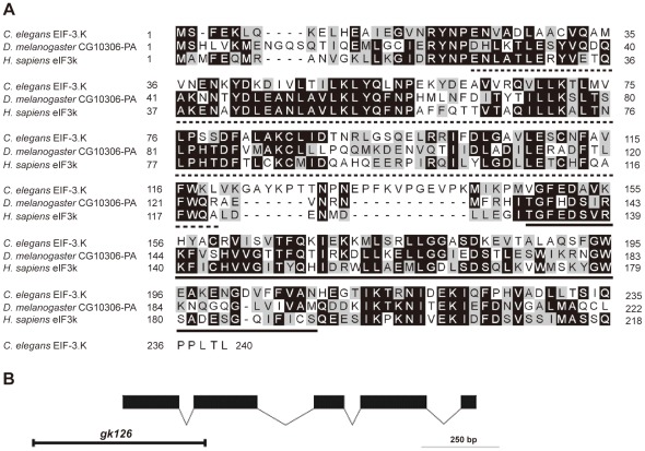
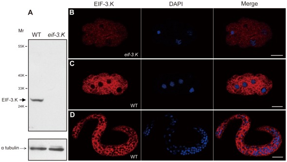
We obtained the cDNA for eif-3.K through reverse transcription polymerase chain reaction (RT-PCR) using total RNA from mixed-stage worms. The predicted full length amino acid sequence of EIF-3.K is 35% identical and 57% similar to that of human eIF3k (Figure 1A). We next characterized the eif-3.K mutant allele gk126, which was isolated by the C. elegans Gene Knockout Consortium. This allele contains a 538 base pair (bp) long deletion from 119 bp upstream of the start ATG codon to the second exon in the eif-3.K locus (Figure 1B). No EIF-3.K protein was detected in the eif-3.K(gk126) mutant by western blotting or immunostaining analyses using purified anti-EIF-3.K antibodies (Figure 2), suggesting that the gk126 allele is null. Because eif-3.K(gk126) and eif-3.K(RNAi) mutant worms were viable and had normal development and growth rates (Table S1), we concluded that eif-3.K is not an essential component of the general translation machinery in C. elegans.
Figure 1. eif-3.K has been conserved throughout evolution.
(A) An alignment between the EIF-3.K protein sequence and the H. sapiens eIF3k and D. melanogaster CG10306-PA protein sequences. Amino acids shaded in black are identical, and amino acids shaded in gray are similar. The predicted HAM and WH domains are underlined with dotted and black lines. (B) The gene structure of eif-3.K was deduced by comparing the coding sequence and the genomic DNA sequence. Boxes represent exons and lines between boxes represent introns. Solid boxes indicate the eif-3.K open reading frame. Open boxes indicate the untranslated region. The transcription direction is from left to right. The deleted region present in the gk126 allele is indicated.
Figure 2. EIF-3.K protein expression.
(A) Western blot analysis of EIF-3.K protein expression. Affinity-purified anti-EIF-3.K antibodies were used to probe a blot of embryonic extracts from wild-type and eif-3.K(gk126) worms (above). Equal loading of the two extracts was confirmed by anti-α tubulin antibodies (below). The sizes of molecular weight markers and the positions of EIF-3.K and α tubulin are indicated. (B–D) Images of an eif-3.K(gk126) mutant early embryo (B), a wild-type early embryo (C) and a wild-type newly hatched larva (D) that were co-stained with anti-EIF-3.K antibodies and DAPI. Merged images are also shown. Scale bar = 10 µm.
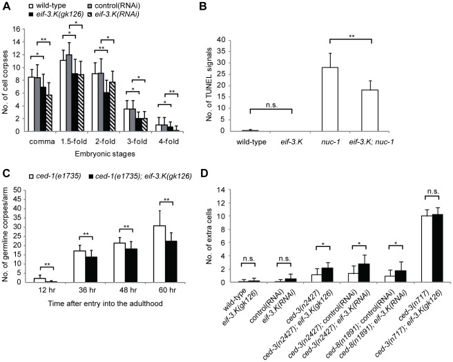
We next examined whether eif-3.K(RNAi) or eif-3.K(gk126) embryos have defective programmed cell death. A time course analysis of embryonic cell corpses using differential interference contrast (DIC) microscopy showed that eif-3.K(RNAi) or eif-3.K(gk126) embryos had fewer cell corpses than wild-type embryos throughout embryogenesis (Figure 3A). To determine whether this decrease in cell corpse number corresponded with a reduction in cell death or was simply due to abnormal corpse morphology, we further analyzed the embryos using the TUNEL (Terminal deoxynucleotidyl transferase dUTP nick end labeling) assay. The degradation of DNA in dying cells is a hallmark of apoptosis and can be detected in situ using TUNEL staining [42], [51]. As shown previously [42], wild-type embryos had very few TUNEL-positive corpses (Figure 3B); however, embryos lacking the nuc-1 gene, which codes for a protein similar to DNAse II that is involved in DNA degradation [42], had many more TUNEL-positive corpses (Figure 3B). The eif-3.K embryos, like the wild-type embryos, had few TUNEL-positive corpses; however, eif-3.K; nuc-1 double mutant embryos had fewer TUNEL-positive corpses than nuc-1 single mutant embryos, indicating that apoptotic.
Figure 3. The loss of eif-3.K results in reduced programmed cell deaths.
(A) The inactivation of eif-3.K by RNAi or by genetic deletion reduced cell corpse numbers throughout embryogenesis. Cell corpses of the indicated genotype or RNAi treatment were scored at the comma, 1.5-fold, 2-fold, 3-fold and 4-fold embryonic stages. The eif-3.K(gk126) embryos were compared to the wild-type embryos, and the eif-3.K(RNAi) embryos were compared to control(RNAi) embryos at each stage. All comparisons were performed using the unpaired t test (*P<0.05, **P<0.001). Data are presented as the mean ± standard deviation. Error bars represent S.D. Greater than 20 embryos per stage were analyzed. (B) The loss of eif-3.K reduces TUNEL staining in nuc-1 embryos. The number of cells exhibiting TUNEL staining was determined in embryos of the indicated genotypes at the 1.5-fold stage. The eif-3.K(gk126) embryos were compared to the wild-type embryos, and the nuc-1(e1392)single mutants were compared to the eif-3.K(gk126); nuc-1(e1392) double mutants. Comparisons were performed using the unpaired t test (*P<0.05, **P<0.001). Data are presented as the mean ± standard deviation. Error bars represent S.D. Greater than 20 embryos of each genotype were analyzed. n.s. indicates no significant difference. (C) A loss of eif-3.K reduces cell corpse number in the germline. Cell corpses in the germline of the ced-1 single mutants (white columns) and eif-3.K; ced-1 double mutants (black columns) were counted at the indicated times after entry into the adulthood. The y axis represents the average number of cell corpses scored in each gonadal arm. The eif-3.K(gk126); ced-1(e1735) double mutants were compared to the ced-1(e1735) single mutants at the same developmental stage using the unpaired t test (*P<0.05, **P<0.001). Data are presented as the mean ± standard deviation for >20 gonadal arms. Error bars represent S.D. (D) A loss of eif-3.K increases the number of extra surviving cells in weak ced-3 mutants. Cells that failed to undergo programmed cell deaths in the anterior pharynx were scored in the indicated animals. All comparisons were performed using the unpaired t test (*P<0.05, **P<0.001). Data are presented as the mean ± standard deviation for >20 larvae. Error bars represent S.D. n.s. indicates no significant difference.
DNA degradation is compromised in the eif-3.K mutants. This result, together with the observed decrease in cell corpse number (Figure 3A), indicates that apoptosis is compromised in eif-3.K mutants during embryogenesis.
Like somatic cells, germline cells also undergo apoptosis in C. elegans [52]. Because few germ cell corpses can be observed in the wild-type adult gonad at any given time due to the prompt removal of cell corpses by the gonadal sheath cells [52], we utilized ced-1(e1735) mutant worms, in which cell corpses are not efficiently removed and therefore accumulate, to increase our chances of detecting cell corpses. We found that ced-1(e1735); eif-3.K(gk126) double mutants had significantly fewer germ cell corpses than ced-1(e1735) single mutants at all adult stages (Figure 3C). Therefore, eif-3.K is also important for programmed cell death in germline cells.
The Loss of eif-3.K Enhances Cell Survival in Sensitized Mutants
We next examined whether a loss of eif-3.K function could prevent cell death and result in an accumulation of surviving cells. Two assays were used to score the surviving cells in various regions of the animal [53]. First, superfluous surviving cells that were present in the anterior pharynx were scored using DIC optics. As was previously shown [54], in the presence of a strong loss-of-function mutation in the pro-apoptotic gene ced-3(n717), which blocks nearly all cell deaths, resulted in approximately 10 additional surviving cells in the anterior pharynx (Figure 3D). Animals harboring the weak ced-3(n2427) mutation had only 1.2 additional surviving cells (Figure 3D). We found that the eif-3.K(RNAi) or eif-3.K(gk126) single mutant animals had 0.2 or 0.5 extra surviving cells, similar to the wild-type animals (Figure 3D), indicating that the loss of eif-3.K could not detectably block apoptosis in these cells; however, the eif-3.K(RNAi) or eif-3.K(gk126) mutation did enhance cell survival in the weak ced-3(n2427) mutant animals. The ced-3(n2427); eif-3.K(RNAi) or ced-3(n2427); eif-3.K(gk126) double mutants had 2.7 or 2.1 additional surviving cells in the anterior pharynx (Figure 3D). This is significantly more than ced-3(n2427), eif-3.K(RNAi) or eif-3.K(gk126) single mutants. Moreover, the RNAi-mediated inactivation of eif-3.K also significantly enhanced cell survival in the worms lacking the ced-8 gene (Figure 3D), which controls the timing of programmed cell death [19]. These results show that the loss of eif-3.K enhances cell survival in sensitized mutants.
We further analyzed the identities of the surviving cells in these mutants. The extraneous surviving cells observed in ced-3(n2427) single mutants and ced-3(n2427); eif-3.K(gk126) double mutants appeared similar and included sisters of muscle cells m1and m2 and neurons I1, I2, and MC (Figure S1). It is possible that these cells are more likely to survive than others when the apoptotic machinery is compromised. Consistent with this hypothesis, m1 and m2 sister cells were occasionally observed to survive in the wild-type or eif-3.K(gk126) animals (Figure S1).We also compared the identities of surviving cells in the ced-3(n2427) animals that were treated with either the eif-3.K or control RNAi. Compared to the control RNAi, eif-3.K RNAi enhanced the survival of the niece of the epithelial cell e1, the sister of the neuron I1 and those cells that were also enhanced by the eif-3.K(gk126) mutation in the ced-3(n2427) single mutants, including sisters of m1, m2, I1, I2, and MC cells (Figure S1). Because the eif-3.K null allele did not enhance the total number of extra surviving cells in the strong ced-3(n717) mutants (Figure 3D), eif-3.K likely functions with ced-3 in the same genetic pathway to promote most, if not all, programmed cell death. Additionally, because the identities of apoptotic cells can be inferred from the cell fates of their differentiated sister cells [55], our observations suggest that eif-3.K exerts a cell death-promoting function in multiple cell types, including neuron, muscle and epithelial cells during development.
Secondly, we scored superfluous surviving cells in the ventral cord in larvae. In contrast to the extra surviving cells we observed in the anterior pharynx, which are generated during embryogenesis [56], extra surviving cells in the ventral cord are generated during larval development [57]. In strong ced-3(n717) mutants, five cells P2.aap and P9–P12.aap in the ventral cord survive [38]. These Pn.aap cells are known to differentiate into VC motor neuron-like cells and express the Plin-11gfp reporter (Table 1) [38]. We scored extra surviving Pn.aap cells using the Plin-11gfp transgene as a marker and found that only 2% of eif-3.K(gk126) worms exhibited extra Pn.aap cells (Table 1). However, the eif-3.K(gk126) mutation increased the average number of extra surviving Pn.aap cells in ced-3(n2427) mutants from 2.6 to 3.6 (Table 1). A previous study showed that strong mutations in genes essential for the removal of apoptotic cells, such as ced-2 or ced-7, block cell death, albeit at low efficiency, as apoptotic cell removal is involved in the death of cells [38], [58]. The frequency of extra Pn.aap cell survival in these mutants can be enhanced by a weak mutation in the core programmed cell death genes ced-3, ced-4, or egl-1 [38]. Therefore, we tested whether the loss of eif-3.K enhanced the frequency of superfluous Pn.aap cell survival in strong ced-2 or ced-7 mutants. We found that the eif-3.K(gk126) mutation increased the frequency of Pn.aap cell survival in ced-2 (n1994) or ced-7(n1996) mutants from 27% to 100% and 83% to 93%, respectively (Table 1). In addition, the average number of extra surviving Pn.aap cells in ced-2(n1994) or ced-7(n1996) mutants also increased from 0.3 to 2.4 and 1.6 to 2.1, respectively (Table 1). These observations support the idea that the cell death machinery is compromised in the eif-3.K mutants. The observed decreased cell death in eif-3.K mutants (Figures 3A–3C) as well as the enhanced cell survival observed in the anterior pharynx (Figure 3D) and ventral cord (Table 1) of sensitized mutants shows that eif-3.K is a positive mediator of programmed cell death.
Table 1. The loss of eif-3.K enhances cell survival in the ventral cord of sensitized mutants.
| Transgene | Genotype | Average number of extra Pn.aap cellsa | Animals with surviving Pn.aap cellsb (%) |
| Plin-11gfp | wild-type | 0.0 | 0 |
| Plin-11gfp | ced-3(n717) | 5.0 | 100 |
| Plin-11gfp | ced-3(n2427) | 2.6 | 99 |
| Plin-11gfp | eif-3.K(gk126) | 0.0 | 2 |
| Plin-11gfp | ced-3(n2427); eif-3.K(gk126) | 3.6 | 100 |
| Plin-11gfp | ced-2(n1994) | 0.3 | 27 |
| Plin-11gfp | ced-2(n1994); eif-3.K(gk126) | 2.4 | 100 |
| Plin-11gfp | ced-7(n1996) | 1.6 | 83 |
| Plin-11gfp | ced-7(n1996); eif-3.K(gk126) | 2.1 | 93 |
aAverage numbers of fluorescent cells caused by expression from Plin-11gfp in P2, 9, 10, 11, and 12-derived regions were determined using DIC microscopy equipped with an ultraviolet light source. Greater than 20 larvae of each genotype were analyzed.
The percentages of animals that had at least one fluorescent cell in the P2, 9, 10, 11, and 12-derived regions were determined.
The Loss of eif-3.K Partially Suppresses the Ectopic Cell Deaths Induced by the Overexpression of egl-1 or ced-4
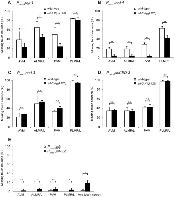
We next tested whether eif-3.K genetically interacts with the core programmed cell death genes ced-3, ced-4, and egl-1. Previous studies have shown that cell-specific expression of these three genes under the control of the Pmec-7 promoter, which is expressed in six touch neurons (AVM, ALMR/L, PVM, and PLMR/L), promotes these neurons to undergo programmed cell death [5], [35]. We tested if the programmed cell death of these cells required the activity of eif-3.K. To facilitate the scoring of touch neurons, the Pmec-4gfp reporter in the integrated transgene bzIs8, which labels the six touch neurons with GFP, was used as a cell viability marker [39]. We found that the eif-3.K(gk126) mutation partially suppressed the apoptosis of the touch neurons that was induced by the overexpression of egl-1 or ced-4 (Figures 4A and 4B). For example, the overexpression of egl-1 or ced-4 resulted in the death of approximately 50% or 29% of the PVM neurons, respectively. The loss of eif-3.K reduced cell death to 24% and 3%, respectively. A significant reduction in cell death was also observed in other touch neurons, except for PLMR/L with egl-1 overexpression (Figure 4A and 4B). Therefore, the efficient apoptosis of touch neurons induced by the overexpression of egl-1 or ced-4 requires eif-3.K. This result showed that eif-3.K functions downstream of or in parallel to egl-1 or ced-4 to promote cell death.
Figure 4. The loss of eif-3.K partially suppresses cell death induced by the overexpression of egl-1and ced-4, but not ced-3.
(A–D) The percentage of animals missing specific touch neurons are shown for the wild-type (white columns) or eif-3.K(gk126) (black columns) embryos carrying the Pmec-7egl-1 (A), Pmec-7ced-4 (B), Pmec-7ced-3 (C), or Pmec-7acCED-3 (D) transgenes. The eif-3.K(gk126) transgenic worms were compared to the analogous wild-type transgenic worms. Comparisons were performed using the unpaired t test (*P<0.05, **P<0.001). Data are presented as the mean ± standard deviation. Error bars represent S.D. n.s. indicates no significant difference. More than 100 animals were scored for each strain. (A)The percentage of animals missing specific touch neurons or missing at least one touch neuron are shown for wild-type control Pmec-7gfp transgenic animals (white columns) or Pmec-7eif-3.K transgenic animals (black columns). More than 100 animals were scored for each strain.
In contrast, eif-3.K(gk126) failed to suppress the apoptosis of touch neurons induced by ced-3 overexpression. The overexpression of ced-3 resulted in the death of approximately 34% of the PVM neurons in the wild-type animals, similar to the percentage (40%) of cell death observed in the eif-3.K(gk126) mutants (Figure 4C). When activated CED-3 (acCED-3) was expressed in touch neurons via the Pmec-7acCED-3 transgene [40], 40% and 43% of the PVM neurons were killed in the wild-type and eif-3.K(gk126) worms, respectively (Figure 4D). This result showed that eif-3.K also fails to inhibit apoptosis caused by the overexpression of activated CED-3. Similar results were observed in other touch neurons expressing either the Pmec-7ced-3 or the Pmec-7acCED-3 transgene (Figures 4C and 4D).
We next examined if overexpression of ced-3 using the heat shock promoter Phsp was able to rescue the cell death defects caused by the eif-3.K(gk126) mutation. Heat shock-induced ced-3 overexpression rescued the defect at the comma and 1.5-fold stages, but it also slightly elicited ectopic cell killing at the comma stage (Table 2). The eif-3.K(gk126) embryos carrying the Phspced-3 transgene had approximately 7.0 cell corpses at the comma stage under non-heat shock condition. The heat shock-induced overexpression of ced-3 in the transgenic embryos at the same developmental stage increased the cell corpse number to 10.7, which was significantly more than the 8.4 cell corpses that were observed in the wild-type embryos carrying the control Phspgfp transgene under the same conditions (Table 2). This result supports the model that ced-3 acts downstream of eif-3.K to execute programmed cell death.
Table 2. Overexpression of eif-3.K or ced-3 in cell death-defective mutants.
| No. of Cell Corpsesb | ||||
| Transgene | Genotype | Heat shocka | comma | 1.5-fold |
| Phspgfp | wild-type | – | 8.0±0.9 | 10.8±1.2 |
| Phspgfp | wild-type | + | 8.4±1.1 | 11.1±1.5 |
| Phspgfp | ced-3(n717) | – | 0.0±0.0 | 0.0±0.0 |
| Phspgfp | ced-3(n717) | + | 0.0±0.0 | 0.0±0.2 |
| Phspgfp | ced-4(n1162) | – | 0.0±0.0 | 0.0±0.2 |
| Phspgfp | ced-4(n1162) | + | 0.1±0.3 | 0.1±0.1 |
| Phspgfp | eif-3.K(gk126) | – | 6.7±2.2 | 9.0±2.1 |
| Phspgfp | eif-3.K(gk126) | + | 6.8±2.0 | 8.9±1.9 |
| Phspced-3 | wild-type | – | 7.8±1.1 | 10.8±1.6 |
| Phspced-3 c | wild-type | + | 11.2±2.1** | 12.8±2.2** |
| Phspced-3 | eif-3.K(gk126) | – | 7.0±1.8 | 8.9±2.0 |
| Phspced-3 c | eif-3.K(gk126) | + | 10.7±0.6** | 11.6±1.7** |
| Phspeif-3.K | wild-type | – | 8.5±2.1 | 11.2±2.4 |
| Phspeif-3.K | wild-type | + | 9.4±1.7 | 13.4±2.9* |
| Phspeif-3.K | ced-3(n717) | – | 0.0±0.0 | 0.0±0.2 |
| Phspeif-3.K | ced-3(n717) | + | 0.2±0.4 | 0.4±0.5* |
| Phspeif-3.K | ced-4(n1162) | – | 0.1±0.3 | 0.0±0.2 |
| Phspeif-3.K | ced-4(n1162) | + | 0.2±0.4 | 0.1±0.2 |
Transgenic animals were subjected to heat-shock (+) or left at 20°C (−).
Transgenic embryos were scored for the number of cell corpses 1.5 hrs after heat shock (see Materials and methods). Data are presented as the mean ± standard deviation from two independent stably transmitting lines.
Greater than 20 embryos were analyzed from each line except for 1.5-fold embryos carrying Phspced-3 (n≥8) after heat shock due to high lethality.
The transgenic embryos after heat shock were compared to the corresponding transgenic embryos without heat shock. All comparisons were performed using the unpaired t test (*P<0.05, **P<0.001).
eif-3.K Acts Upstream of ced-3 in the Promotion of Programmed Cell Death
To determine whether eif-3.K promotes programmed cell death, we tested if the overexpression of eif-3.K caused cells that would normally live to undergo programmed cell death by overexpressing eif-3.K under the control of the heat shock promoter Phsp in the wild-type animals. The overexpression of eif-3.K, but not of the control gfp, slightly but significantly increased the cell corpse number at the 1.5-fold stage, despite a lack of significant ectopic killing at the comma stage (Table 2). Nonetheless, this ectopic killing at the 1.5-fold stage supports for a cell death-promoting function for eif-3.K. In addition, this ectopic killing was significantly suppressed by the strong ced-3(n717) or ced-4(n1162) mutations (Table 2). This result, in combination with the reciprocal experiment in which the loss of eif-3.K suppressed the efficient apoptosis of touch neurons in the presence of ced-4 overexpression (Figure 4B), suggests a mutual requirement for eif-3.K and ced-4. Additionally, because the loss of eif-3.K failed to suppress the efficient apoptosis of touch neurons in the presence of ced-3 overexpression, our result suggests a unidirectional requirement of eif-3.K for ced-3 to achieve effective ectopic cell death under overexpression conditions.
eif-3.K Promotes Cell Death in a Cell-Autonomous Fashion
To determine whether eif-3.K promotes programmed cell death in a cell-autonomous fashion, we tested if the Pmec-7eif-3.K transgene, in which eif-3.K is expressed under the Pmec-7 promotor in touch neurons, could trigger touch neuron apoptosis in the wild-type animals. Although the overexpression of eif-3.K transgene resulted in a low frequency of individual touch neuron apoptosis, approximately 19.2% of trasngenic worms had at least one missing touch neuron (Figure 4E). In contrast, only 1.9% of the wild-type animals carrying the control Pmec-7gfp transgene had one missing touch neuron (Figure 4E). This result not only reinforced the cell death-promoting function for eif-3.K but also showed that eif-3.K executes this function in a cell-autonomous fashion.
The Loss of eif-3.K Significantly Reduces Ectopic Cell Deaths in icd-1 Mutants
The inactivation of icd-1 (inhibitor of cell death-1) by RNAi results in ectopic cell death that can be blocked by the loss of ced-4 but not ced-3, revealing that the cell death in icd-1(RNAi) embryos is ced-4-dependent but ced-3-indpendent [17]. The observation that eif-3.K is required for cell death induced by the overexpression of ced-4 but not ced-3 (Figures 4B–4D) prompted us to test whether eif-3.K could suppress ectopic cell death resulting from the loss of icd-1. Like the icd-1(RNAi) embryos described previously [17], icd-1(tm2873) embryos had additional cell corpses compared to the wild-type embryos at the comma and 1.5-fold stages (Figure 5A). Although the eif-3.K(gk126) mutation significantly reduced the cell corpse number in icd-1(tm2873) embryos at both stages, the cell corpse number was not reduced to the extent observed in the eif-3.K(gk126) mutants alone (Figure 5B). Instead, the cell corpse number for the double mutant was between that observed in the eif-3.K(gk126) and icd-1(tm2873) single mutants. This result is consistent with the model that eif-3.K acts in parallel with icd-1 to promote cell death; however, we cannot rule out the possibility that icd-1 may in part prevent programmed cell death in an eif-3.K-dependent manner.
Figure 5. eif-3.K partially suppresses the ectopic cell deaths caused by the loss of icd-1.
(A) A quantification of the cell corpses present in the wild-type and icd-1(tm2873) embryos at the comma (left) and 1.5-fold (right) stages. (B) A quantification of the cell corpses present in the eif-3.K(gk126) and icd-1(tm2873) single mutant and icd-1(tm2873); eif-3.K(gk126) double mutant embryos at the comma (left) and 1.5-fold (right) stages. The y axis shows the percentage of embryos and the x axis shows the cell corpse number. More than 20 embryos for each genotype at each stage were scored.
EIF-3.K is Widely Expressed throughout Embryogenesis and Localized to the Cytoplasm
To determine the localization pattern of EIF-3.K, we raised antibodies against a recombinant EIF-3.K protein (see Experimental Procedures). Using affinity-purified EIF-3.K antibodies and western blot analysis, we detected a band of apparent molecular mass 27 kDa from wild-type worm extracts by western blot analysis (Figure 2A). This protein was absent in extracts from the eif-3.K(gk126) mutants (Figure 2A), confirming that the 27 kDa protein is the product of the eif-3.K gene. We used the purified EIF-3.K antibodies to stain embryos and larvae. EIF-3.K was widely expressed in embryos and larvae and was localized to the cytoplasm (Figures 2B–2D). EIF-3.K did not appear to be associated with mitochondria, where several cell death regulators such as CED-9, WAH-1, and WAN-1 are located [7], [18], [20], because EIF-3.K did not co-localize with MitoTracker Red, a marker of mitochondria (Figure S2).
The WH Domain of EIF-3.K is Necessary and Sufficient for its Cell Death-Promoting Activity
EIF3.K contains two distinct domains, the HAM (HEAT Analogous Repeats) and WH (Winged Helix) domains, which have been implicated in protein-protein and protein-RNA interactions, respectively [59]. To test the importance of these domains for EIF-3.K function, we deleted the region corresponding to the HAM or WH domains, respectively (eif-3.KΔHAM or eif-3.KΔWH constructs). We then tested the ability of the mutant construct to rescue the cell death defects in eif-3.K(gk126) embryos by expressing the mutant construct under the control of the ubiquitous let-858 promoter Plet-858 [44]. To our surprise, the HAM domain, comprising more than one-third of the EIF-3.K protein, was dispensable for eif-3.K activity, as Plet-858eif-3.KΔHAM completely rescued the cell death defect in the eif-3.K(gk126) embryos (Table 3). In contrast, Plet-858eif-3.KΔWH failed to rescue the defect (Table 3), suggesting an essential role for the WH domain in the cell death-promoting function of eif-3.K. Because the expression level of the Plet-858eif-3.K transgene was lower than that of the endogenous eif-3.K, as detected by western blotting or immunostaining analysis (data not shown), the stronger heat shock promoter Phsp was subsequently used to increase the expression of the mutant eif-3.K construct in an effort to confirm our results. The heat shock-induced expression of the wild-type and mutant eif-3.K genes resulted in slightly higher protein expression levels (Figure S3). The overexpressed proteins exhibited a similar localization as the endogenous EIF-3.K protein, suggesting that these proteins localize normally (Figure S3). Similar to the results obtained using the Plet-858 promoter, expression under the heat shock promoters revealed that eif-3.KΔHAM, but not eif-3.KΔWH, rescued the eif-3.K mutant phenotype at the comma and 1.5-fold stages (Table 3). These data show that the WH domain, but not the HAM domain, is necessary for the cell death-promoting function of EIF-3.K.
Table 3. Structure and function analysis of eif-3.K.
| No. of Cell Corpsesb | ||||
| Transgene | Genotype | Heat shocka | comma | 1.5-fold |
| none | wild-type | – | 8.4±0.9 | 11.3±1.0 |
| none | eif-3.K(gk126) | – | 6.9±2.1 | 9.0±2.3 |
| Plet-858eif-3.KΔHAM | eif-3.K(gk126) | – | 7.8±1.1* | 11.0±1.0** |
| Plet-858eif-3.KΔWH | eif-3.K(gk126) | – | 6.5±1.2 | 9.1±1.3 |
| Plet-858WH | eif-3.K(gk126) | – | 8.1±0.7* | 11.0±1.2** |
| Phspgfp | eif-3.K(gk126) | – | 6.7±2.2 | 9.0±2.1 |
| Phspgfp | eif-3.K(gk126) | + | 6.8±2.0 | 8.9±1.9 |
| Phspeif-3.K | eif-3.K(gk126) | – | 6.9±2.0 | 8.8±1.9 |
| Phspeif-3.K | eif-3.K(gk126) | + | 8.8±1.2** | 11.3±1.1** |
| Phspeif-3.K | wild-type | – | 8.5±2.1 | 11.2±2.4 |
| Phspeif-3.K | wild-type | + | 9.4±1.7 | 13.4±2.9** |
| Phspeif-3.KΔWH | eif-3.K(gk126) | – | 6.4±2.1 | 8.7±1.8 |
| Phspeif-3.KΔWH | eif-3.K(gk126) | + | 6.2±1.1 | 8.6±0.8 |
| Phspeif-3.KΔHAM | eif-3.K(gk126) | – | 6.5±2.0 | 8.8±2.0 |
| Phspeif-3.KΔHAM | eif-3.K(gk126) | + | 8.3±1.1** | 11.2±1.9** |
| PhspWH | eif-3.K(gk126) | – | 6.5±2.1 | 9.0±1.8 |
| PhspWH | eif-3.K(gk126) | + | 8.3±0.9** | 10.9±1.0* |
| PhspWH | wild-type | – | 8.4±2.0 | 11.0±2.4 |
| PhspWH | wild-type | + | 10.1±2.2* | 13.1±2.1* |
| PhspeIF3k(human) | eif-3.K(gk126) | – | 6.5±1.9 | 8.6±1.7 |
| PhspeIF3k(human) | eif-3.K(gk126) | + | 8.7±0.8** | 10.4±0.6** |
aTransgenic animals were subjected to heat-shock (+) or left at 20°C (−).
Transgenic embryos were scored for the number of cell corpses 1.5 hrs after heat shock.
(see Materials and methods).
Data are presented as the mean ± standard deviation from two independent stably transmitting lines. Greater than 20 embryos were analyzed from each line.
For the Plet-858 expressing transgene, eif-3.K mutant embryos carrying the transgene were compared to eif-3.K mutant without the transgene.
For the Phsp expressing transgene, the transgenic embryos after heat shock were compared to the corresponding transgenic embryos without heat shock. All comparisons were performed using the unpaired t test (*P<0.05, **P<0.001).
We next tested if the WH domain of EIF-3.K is sufficient to rescue the cell-death defect caused by the eif-3.K mutation. We expressed the WH domain alone using either the Plet-858 or Phsp promoters in the transgenes Plet-858WH or PhspWH, respectively. We found that either promoter rescued the cell death defect in the eif-3.K mutants (Table 3). Moreover, when the PhspWH transgene was expressed in the wild-type animals, it induced ectopic cell deaths. Furthermore, superfluous cell corpses were observed at the comma and 1.5-fold stages (Table 3). Therefore, the WH domain is both necessary and sufficient for the cell death-promoting activity of eif-3.K.
Human eIF3k can Partially Substitute for C. elegans EIF-3.K
The human eIF3k mediates apoptosis in simple epithelial cells, likely by binding to keratin K18 via its HAM domain [30]; however, the HAM domain of C. elegans EIF-3.K appears dispensable for its function in cell death. We tested whether the expression of human eIF3k by Phsp was able to rescue the cell death defect caused by the eif-3.K mutation. We found that human eIF3k partially rescued the defective apoptosis in the eif-3.K(gk126) mutants (Table 3). This result indicates that the pro-apoptotic function of EIF-3.K has been conserved through evolution from C. elegans to humans and that the mechanisms by which human eIF3k and C. elegans EIF-3.K promote apoptosis may, in part, be similar.
Discussion
eIF3 is the largest and most complex translation initiation factor, consisting of thirteen subunits in both C. elegans and humans [22]. The RNAi-based or genetic inactivation of ten eIF3 subunits, including eif-3.A (egl-45), eif-3.B, eif-3.C, eif-3.D, eif-3.E, eif-3.F, eif-3.G, eif-3.H, eif-3.J, or cif-1(eif-3.M), in C. elegans can cause sterility, embryonic lethality or gross developmental defects [60]–[63]. In contrast, the eif-3.K null mutant is viable and healthy (Table S1), suggesting that eif-3.K is not essential for general translation initiation. Similarly, human eIF3k is dispensable for the formation of an active eIF3 complex in vitro [26]. We have previously shown that human eIF3k promotes apoptosis in cultured simple epithelial cells [30]. In this work, we provide evidence that eif-3.K has a cell death-promoting function at an organismal level and that this function has been conserved through evolution.
In C. elegans, the loss of eif-3.K caused reduced programmed cell death (Figures 3A–3C) and enhanced cell survival in sensitized mutants (Figure 3D and Table 1). In contrast, the overexpression of eif-3.K by the heat shock promoter or a touch neuron-specific promoter resulted in ectopic cell death (Table 2 and Figure 4E). These results demonstrate that eif-3.K promotes programmed cell death. Our results also show that eif-3.K is essential for the efficient cell death that is induced by the overexpression of egl-1 or ced-4, but not ced-3, as the loss of eif-3.K partially suppresses the cell death that is induced by the overexpression of egl-1 or ced-4 only (Figures 3A–3D). In addition, the observation that ced-3 overexpression can rescue the cell death-defective phenotype of eif-3.K mutants and that the ced-3 strong mutation can suppress cell death caused by heat shock-induced eif-3.K overexpression (Table 2) further reinforces the notion that eif-3.K requires ced-3 to promote programmed cell death. Furthermore, the wide range in the identity and type of extraneous surviving cells that are affected by the eif-3.K mutation (Figure S1) suggests that eif-3.K may be involved in the majority of programmed cell death. This is consistent with the ubiquitous expression of EIF-3.K in embryos and larvae (Figures 2C and 2D). In addition to physiological cell deaths, DNA damage caused by genotoxic stress such as UV or IR radiation also induces cell deaths in the germline [43], [64]. We found that the eif-3.K(gk126) mutation significantly reduced UV-induced cell deaths in the germline (Figure S5), indicating that eif-3.K also mediates DNA damage-induced cell death.
During C. elegans development, the activity of the executioner caspase CED-3 is under both positive and negative regulation. Previous studies have shown that CED-4 facilitates the auto-cleavage of pro-CED-3 to generate the active CED-3 caspase during the promotion of cell death [12], [65], [66], while the CED-3 paralogs CSP-2 and CSP-3 associate with the CED-3 zymogen and inhibits its auto-activation, thereby protecting cells from inappropriate apoptosis [13], [14]. Our observation that neither EIF-3.K nor its WH domain bind to CED-3 or CED-4 in a yeast 2-hybrid system (Figure S4) suggests that EIF-3.K may not promote cell death through a direct association with either protein. In addition, since CED-3 and CED-4 are the only known proteins involved in CED-3 activation from pro-CED-3 [9], [12], EIF-3.K likely does not affect this activation process directly. It is possible that EIF-3.K may promote programmed cell death after CED-4-induced CED-3 activation. Human eIF3k has been proposed to promote apoptosis by facilitating the release of active caspases from an inhibitory compartment of intermediate filament-containing inclusions into the cytosol, thereby allowing the released caspase better access to its cytosolic substrates [30]. Although the mechanism by which eIF3k may affect the release of caspases from intermediate filament-containing inclusions is not clear, the binding of eIF3k to intermediate filaments is known to be important for the release process [30]. Similarly, C. elegans EIF-3.K may promote programmed cell death by affecting the distribution of active CED-3, thus facilitating the substrate cleavage and the subsequent execution of cell death. Alternatively, EIF-3.K might promote programmed cell death in parallel with CED-4 by antagonizing CSP-2 or CSP-3, thus facilitating CED-3 auto-activation from the zymogen in germline or somatic cells, respectively [13], [14]. To test the latter possibility, we used bzIs8 (Pmec-4gfp ), which labels six touch neurons, as marker to monitor the survival of touch neurons and tested the effect of the eif-3.K mutation on the missing cell phenotype of the csp-3(lf) animals. As previously shown [14], in csp-3(lf) animals six touch neurons were lost randomly at a frequency from 2% to 10% (Table S2) and 24% of animals lost at least one touch neuron (Table S2). The eif-3.K(gk126) mutation strongly suppressed this missing cell defect in csp-3 mutants (Table S2). In addition, loss of csp-2 resulted in increased germline cell deaths [13], and this phenotype can also be suppressed by the eif-3.K mutation (Figure S6). These results suggest that EIF-3.K may promote cell death downstream of or in parallel to csp-2 or csp-3 in the germline and somatic cell deaths, respectively.
Human eIF3k co-localizes with keratin and requires keratin for its apoptosis-promoting function in simple epithelial cells [30]. Upon apoptotic stimuli, keratin K18 is cleaved by caspase 3 at VEVD238 of the L1-2 linker region or DALD397 of the C terminal (tail) domain, resulting in a collapse of keratin filaments [67]. C. elegans contains eleven genes that encode cytoplasmic intermediate filaments, including ifa-1, mua-6, ifa-3, ifa-4, ifb-1, ifb-2, ifc-1, ifc-2, ifd-1, ifd-2, and ifp-1 [68]. No detectable change in either the localization or the level of the EIF-3.K protein was observed by immunostaining analysis in mua-6(rh85) or ifb-1(ju71) mutant embryos or in embryos treated with ifa-1, mua-6, ifa-4, ifb-1, ifc-1, or ifd-1 interfering RNAs (data not shown). Potential CED-3 cleavage sites (DXXD) are found in IFA-1 (DAED), IFC-2 (DNRD), IFD-1(DNRD and DVDD), and IFP-1 (DSVD). The RNAi-mediated inactivation of ifa-1, but not ifc-2, ifd-1, or ifp-1, reduced the number of cell corpses at the comma stage. For example, the ifa-1(RNAi) worms had on average 6.9±1.8 cell corpses, which is similar to the number of cell corpses observed in eif-3.K mutants; however, whether IFA-1, IFC-2, IFD-1, or IFP-1 are direct targets of the CED-3 caspase or are involved in programmed cell death needs to be evaluated. It is not yet clear whether EIF-3.K localizes to intermediate filaments or mediates programmed cell death through intermediate filaments in C. elegans, similar to human eIF3k. Previously, the pro-apoptotic function of human eIF3k was identified and assayed in simple epithelial cells [30] in which keratin K8/K18 is the major intermediate filament. It will be interesting to determine whether human eIF3k, like C. elegans eIF3.K, can promote apoptosis in muscle or neuron cells, as human eIF3k is widely expressed in many tissues, including the brain and muscle [27], where no or very little keratin is expressed [69]. In addition, because eIF3k is also present in D. melanogaster, an organism that lacks intermediate filaments, it will be interesting to see if eIF3k plays a role in apoptosis in D. melanogaster as well.
C. elegans EIF-3.K and human eIF3k both contain two conserved domains, the WH and HAM domains. The HAM domain, but not the WH domain, of human eIF3k interacts with keratin 18 in a yeast two-hybrid system and thus may be important for eIF3k localization to keratin [30]; however, in C. elegans, the HAM domain is dispensable for the cell death-promoting function of EIF-3.K and the WH domain alone is sufficient to promote cell death (Table 3). This result suggests that the WH domain may promote programmed cell death by an IF-independent mechanism. The WH domain has been implicated in DNA or RNA binding [59], but how it may promote programmed cell death needs further study. The result that human eIF3k can partially rescue the cell death defect in the eif-3.K mutants suggests that the eIF3k family may promote apoptosis through a conserved mechanism, which may be dependent upon the WH domain.
Supporting Information
The identification of extraneous surviving cells in the mutants. The y axis represents the percentage of animals with specific superfluous surviving cells (x axis). The extra surviving cells are named after their sister or niece cells, such as “e1 sister cell” and “I2 niece cell”. M4, MC, NSM, I1 and I2 are neurons. e1 is an epithelial cell, and m1 and m2 are muscle cells. L: left, R: right. The identities of extraneous surviving cells were determined as previously described [41]. More than 20 worms for each genotype were scored.
(TIF)
EIF-3.K is not associated with mitochondria. A wild-type embryo was co-stained with anti-EIF-3.K antibodies (A) and MitoTracker (B). The merged image is shown in C. Scale bar = 10 µm.
(TIF)
Deletion of the WH domain does not affect the expression pattern or stability of EIF-3.K. The wild-type embryo (A) and eif-3.K mutant embryo (B) with no transgene, and the eif-3.K mutant embryos carrying the transgene Phspeif-3.K (C), Phspeif-3. KΔHAM (D), or Phspeif-3. KΔWH (E) were heat shocked and co-stained with anti-EIF-3.K antibodies (red) and DAPI (blue). Representative images of anti-EIF-3.K antibody staining (upper panel) and merged images of anti-EIF-3.K antibody and DAPI staining (lower panel) are shown. Scale bar = 10 µm.
(TIF)
Neither EIF-3.K nor the WH domain alone interacts with CED-3 or CED-4 in a yeast 2-hybrid assay. Pairs of constructs expressing the indicated fusion proteins were transformed into the yeast strain MaV203. The resulting transformants were streaked on SC-Trp-Leu-His or SC-Trp-Leu plates containing 30 mM 3 AT. Growth on the SC-Trp-Leu-His+30 mM 3 AT plate indicates an interaction between the fusion proteins. The E2F1and RB pair was used as positive control [70]. “-” in the lower panel indicates no insert was present in the AD fusion construct.
(TIF)
Loss of eif-3.K reduced DNA damage-induced apoptosis. Apoptotic germ cell corpses were scored in the wild-type (black columns) and eif-3.K(gk126) (white columns) young adult worms 24 hr following exposure to 150 J/m2 UV-C radiation. The eif-3.K(gk126) mutants were compared to the wild-type using the unpaired t test (**P<0.001). More than 20 gonadal arms were scored for each genotype.
(TIF)
Loss of eif-3.K suppressed the increased cell death phenotype of csp-2 mutants in the germline. Germ cell corpses were scored in ced-6(n2095) (white columns), ced-6(n2095); csp-2(tm3077) (gray columns), ced-6(n2095); eif-3.K(gk126) (black columns), ced-6(n2095); csp-2(tm3077); eif-3.K(gk126) (slashed columns) worms 48 hours after entering adulthood. The y axis represents the average number of cell corpses scored in each gonadal arm. The data were compared using the unpaired t test (*P<0.05, **P<0.001). More than 20 gonadal arms of each genotype were scored.
(TIF)
eif-3.K is not essential for embryonic or larval development.
(DOC)
The missing cell defect in csp-3 mutants was suppressed by loss of eif-3.K .
(DOC)
Acknowledgments
We thank the Caenorhabditis Genetic Center (CGC) for providing strains, TC5 Bio-Image Tools, College of Life Science, NTU for image assistance, and the C. elegans core facility (supported by the National Science Council in Taiwan) for technical and material support.
Footnotes
Competing Interests: The authors have declared that no competing interests exist.
Funding: This work was supported in part by a National Taiwan University grant (10R70602A3) and the National Science Council (Taiwan).The funders had no role in study design, data collection and analysis, decision to publish, or preparation of the manuscript. No additional external funding received for this study.
References
- 1.Raff MC, Barres BA, Burne JF, Coles HS, Ishizaki Y, et al. Programmed cell death and the control of cell survival: lessons from the nervous system. Science. 1993;262:695–700. doi: 10.1126/science.8235590. [DOI] [PubMed] [Google Scholar]
- 2.Jacobson MD, Weil M, Raff MC. Programmed cell death in animal development. Cell. 1997;88:347–354. doi: 10.1016/s0092-8674(00)81873-5. [DOI] [PubMed] [Google Scholar]
- 3.Fuchs Y, Steller H. Programmed cell death in animal development and disease. Cell. 2011;147:742–758. doi: 10.1016/j.cell.2011.10.033. [DOI] [PMC free article] [PubMed] [Google Scholar]
- 4.Ellis HM, Horvitz HR. Genetic control of programmed cell death in the nematode C. elegans. Cell. 1986;44:817–829. doi: 10.1016/0092-8674(86)90004-8. [DOI] [PubMed] [Google Scholar]
- 5.Conradt B, Horvitz HR. The C. elegans protein EGL-1 is required for programmed cell death and interacts with the Bcl-2-like protein CED-9. Cell. 1998;93:519–529. doi: 10.1016/s0092-8674(00)81182-4. [DOI] [PubMed] [Google Scholar]
- 6.Hengartner MO, Ellis RE, Horvitz HR. Caenorhabditis elegans gene ced-9 protects cells from programmed cell death. Nature. 1992;356:494–499. doi: 10.1038/356494a0. [DOI] [PubMed] [Google Scholar]
- 7.Hengartner MO, Horvitz HR. C. elegans cell survival gene ced-9 encodes a functional homolog of the mammalian proto-oncogene bcl-2. Cell. 1994;76:665–676. doi: 10.1016/0092-8674(94)90506-1. [DOI] [PubMed] [Google Scholar]
- 8.Chen F, Hersh BM, Conradt B, Zhou Z, Riemer D, et al. Translocation of C. elegans CED-4 to nuclear membranes during programmed cell death. Science. 2000;287:1485–1489. doi: 10.1126/science.287.5457.1485. [DOI] [PubMed] [Google Scholar]
- 9.Yang X, Chang HY, Baltimore D. Essential role of CED-4 oligomerization in CED-3 activation and apoptosis. Science. 1998;281:1355–1357. doi: 10.1126/science.281.5381.1355. [DOI] [PubMed] [Google Scholar]
- 10.Yuan J, Shaham S, Ledoux S, Ellis HM, Horvitz HR. The C. elegans cell death gene ced-3 encodes a protein similar to mammalian interleukin-1 beta-converting enzyme. Cell. 1993;75:641–652. doi: 10.1016/0092-8674(93)90485-9. [DOI] [PubMed] [Google Scholar]
- 11.Pourkarimi E, Greiss S, Gartner A. Evidence that CED-9/Bcl2 and CED-4/Apaf-1 localization is not consistent with the current model for C. elegans apoptosis induction. Cell Death Differ. 2011;19:406–15. doi: 10.1038/cdd.2011.104. [DOI] [PMC free article] [PubMed] [Google Scholar]
- 12.Qi S, Pang Y, Hu Q, Liu Q, Li H, et al. Crystal structure of the Caenorhabditis elegans apoptosome reveals an octameric assembly of CED-4. Cell. 2010;141:446–457. doi: 10.1016/j.cell.2010.03.017. [DOI] [PubMed] [Google Scholar]
- 13.Geng X, Zhou QH, Kage-Nakadai E, Shi Y, Yan N, et al. Caenorhabditis elegans caspase homolog CSP-2 inhibits CED-3 autoactivation and apoptosis in germ cells. Cell Death Differ. 2009;16:1385–1394. doi: 10.1038/cdd.2009.88. [DOI] [PMC free article] [PubMed] [Google Scholar]
- 14.Geng X, Shi Y, Nakagawa A, Yoshina S, Mitani S, et al. Inhibition of CED-3 zymogen activation and apoptosis in Caenorhabditis elegans by caspase homolog CSP-3. Nat Struct Mol Biol. 2008;15:1094–1101. doi: 10.1038/nsmb.1488. [DOI] [PMC free article] [PubMed] [Google Scholar]
- 15.Wu D, Chen PJ, Chen S, Hu Y, Nunez G, et al. C. elegans MAC-1, an essential member of the AAA family of ATPases, can bind CED-4 and prevent cell death. Development. 1999;126:2021–2031. doi: 10.1242/dev.126.9.2021. [DOI] [PubMed] [Google Scholar]
- 16.Chen L, McCloskey T, Joshi PM, Rothman JH. ced-4 and proto-oncogene tfg-1 antagonistically regulate cell size and apoptosis in C. elegans. Curr Biol. 2008;18:1025–1033. doi: 10.1016/j.cub.2008.06.065. [DOI] [PMC free article] [PubMed] [Google Scholar]
- 17.Bloss TA, Witze ES, Rothman JH. Suppression of CED-3-independent apoptosis by mitochondrial betaNAC in Caenorhabditis elegans. Nature. 2003;424:1066–1071. doi: 10.1038/nature01920. [DOI] [PubMed] [Google Scholar]
- 18.Shen Q, Qin F, Gao Z, Cui J, Xiao H, et al. Adenine nucleotide translocator cooperates with core cell death machinery to promote apoptosis in Caenorhabditis elegans. Mol Cell Biol. 2009;29:3881–3893. doi: 10.1128/MCB.01509-08. [DOI] [PMC free article] [PubMed] [Google Scholar]
- 19.Stanfield GM, Horvitz HR. The ced-8 gene controls the timing of programmed cell deaths in C. elegans. Mol Cell. 2000;5:423–433. doi: 10.1016/s1097-2765(00)80437-2. [DOI] [PubMed] [Google Scholar]
- 20.Wang X, Yang C, Chai J, Shi Y, Xue D. Mechanisms of AIF-mediated apoptotic DNA degradation in Caenorhabditis elegans. Science. 2002;298:1587–1592. doi: 10.1126/science.1076194. [DOI] [PubMed] [Google Scholar]
- 21.Nakagawa A, Shi Y, Kage-Nakadai E, Mitani S, Xue D. Caspase-dependent conversion of Dicer ribonuclease into a death-promoting deoxyribonuclease. Science. 2010;328:327–334. doi: 10.1126/science.1182374. [DOI] [PMC free article] [PubMed] [Google Scholar]
- 22.Hinnebusch AG. eIF3: a versatile scaffold for translation initiation complexes. Trends Biochem Sci. 2006;31:553–562. doi: 10.1016/j.tibs.2006.08.005. [DOI] [PubMed] [Google Scholar]
- 23.Phan L, Zhang X, Asano K, Anderson J, Vornlocher HP, et al. Identification of a translation initiation factor 3 (eIF3) core complex, conserved in yeast and mammals, that interacts with eIF5. Mol Cell Biol. 1998;18:4935–4946. doi: 10.1128/mcb.18.8.4935. [DOI] [PMC free article] [PubMed] [Google Scholar]
- 24.Browning KS, Gallie DR, Hershey JW, Hinnebusch AG, Maitra U, et al. Unified nomenclature for the subunits of eukaryotic initiation factor 3. Trends Biochem Sci. 2001;26:284. doi: 10.1016/s0968-0004(01)01825-4. [DOI] [PubMed] [Google Scholar]
- 25.Mayeur GL, Fraser CS, Peiretti F, Block KL, Hershey JW. Characterization of eIF3k: a newly discovered subunit of mammalian translation initiation factor elF3. Eur J Biochem. 2003;270:4133–4139. doi: 10.1046/j.1432-1033.2003.03807.x. [DOI] [PubMed] [Google Scholar]
- 26.Masutani M, Sonenberg N, Yokoyama S, Imataka H. Reconstitution reveals the functional core of mammalian eIF3. EMBO J. 2007;26:3373–3383. doi: 10.1038/sj.emboj.7601765. [DOI] [PMC free article] [PubMed] [Google Scholar]
- 27.Karki S, Ligon LA, DeSantis J, Tokito M, Holzbaur EL. PLAC-24 is a cytoplasmic dynein-binding protein that is recruited to sites of cell-cell contact. Mol Biol Cell. 2002;13:1722–1734. doi: 10.1091/mbc.02-02-0011. [DOI] [PMC free article] [PubMed] [Google Scholar]
- 28.Shen X, Yang Y, Liu W, Sun M, Jiang J, et al. Identification of the p28 subunit of eukaryotic initiation factor 3(eIF3k) as a new interaction partner of cyclin D3. FEBS Lett. 2004;573:139–146. doi: 10.1016/j.febslet.2004.07.071. [DOI] [PubMed] [Google Scholar]
- 29.De Martelaere K, Lintermans B, Haegeman G, Vanhoenacker P. Novel interaction between the human 5-HT7 receptor isoforms and PLAC-24/eIF3k. Cell Signal. 2007;19:278–288. doi: 10.1016/j.cellsig.2006.06.012. [DOI] [PubMed] [Google Scholar]
- 30.Lin YM, Chen YR, Lin JR, Wang WJ, Inoko A, et al. eIF3k regulates apoptosis in epithelial cells by releasing caspase 3 from keratin-containing inclusions. J Cell Sci. 2008;121:2382–2393. doi: 10.1242/jcs.021394. [DOI] [PubMed] [Google Scholar]
- 31.Oshima RG. Apoptosis and keratin intermediate filaments. Cell Death Differ. 2002;9:486–492. doi: 10.1038/sj.cdd.4400988. [DOI] [PubMed] [Google Scholar]
- 32.Brenner S. The genetics of Caenorhabditis elegans. Genetics. 1974;77:71–94. doi: 10.1093/genetics/77.1.71. [DOI] [PMC free article] [PubMed] [Google Scholar]
- 33.Ellis RE, Jacobson DM, Horvitz HR. Genes required for the engulfment of cell corpses during programmed cell death in Caenorhabditis elegans. Genetics. 1991;129:79–94. doi: 10.1093/genetics/129.1.79. [DOI] [PMC free article] [PubMed] [Google Scholar]
- 34.Yuan J, Horvitz HR. The Caenorhabditis elegans cell death gene ced-4 encodes a novel protein and is expressed during the period of extensive programmed cell death. Development. 1992;116:309–320. doi: 10.1242/dev.116.2.309. [DOI] [PubMed] [Google Scholar]
- 35.Shaham S, Horvitz HR. Developing Caenorhabditis elegans neurons may contain both cell-death protective and killer activities. Genes Dev. 1996;10:578–591. doi: 10.1101/gad.10.5.578. [DOI] [PubMed] [Google Scholar]
- 36.Bloom L, Horvitz HR. The Caenorhabditis elegans gene unc-76 and its human homologs define a new gene family involved in axonal outgrowth and fasciculation. Proc Natl Acad Sci U S A. 1997;94:3414–3419. doi: 10.1073/pnas.94.7.3414. [DOI] [PMC free article] [PubMed] [Google Scholar]
- 37.Sulston JE. Post-embryonic development in the ventral cord of Caenorhabditis elegans. Philos Trans R Soc Lond B Biol Sci. 1976;275:287–297. doi: 10.1098/rstb.1976.0084. [DOI] [PubMed] [Google Scholar]
- 38.Reddien PW, Cameron S, Horvitz HR. Phagocytosis promotes programmed cell death in C. elegans. Nature. 2001;412:198–202. doi: 10.1038/35084096. [DOI] [PubMed] [Google Scholar]
- 39.Harbinder S, Tavernarakis N, Herndon LA, Kinnell M, Xu SQ, et al. Genetically targeted cell disruption in Caenorhabditis elegans. Proc Natl Acad Sci U S A 94. 1997;13128–13133 doi: 10.1073/pnas.94.24.13128. [DOI] [PMC free article] [PubMed] [Google Scholar]
- 40.Parrish J, Li L, Klotz K, Ledwich D, Wang X, et al. Mitochondrial endonuclease G is important for apoptosis in C. elegans. Nature. 2001;412:90–94. doi: 10.1038/35083608. [DOI] [PubMed] [Google Scholar]
- 41.Ledwich D, Wu YC, Driscoll M, Xue D. Analysis of programmed cell death in the nematode Caenorhabditis elegans. Methods Enzymol. 2000;322:76–88. doi: 10.1016/s0076-6879(00)22009-0. [DOI] [PubMed] [Google Scholar]
- 42.Wu YC, Stanfield GM, Horvitz HR. NUC-1, a caenorhabditis elegans DNase II homolog, functions in an intermediate step of DNA degradation during apoptosis. Genes Dev. 2000;14:536–548. [PMC free article] [PubMed] [Google Scholar]
- 43.Stergiou L, Doukoumetzidis K, Sendoel A, Hengartner MO. The nucleotide excision repair pathway is required for UV-C-induced apoptosis in Caenorhabditis elegans. Cell Death Differ 14, 2007;1129–1138 doi: 10.1038/sj.cdd.4402115. [DOI] [PubMed] [Google Scholar]
- 44.Kelly WG, Xu S, Montgomery MK, Fire A. Distinct requirements for somatic and germline expression of a generally expressed Caernorhabditis elegans gene. Genetics. 1997;146:227–238. doi: 10.1093/genetics/146.1.227. [DOI] [PMC free article] [PubMed] [Google Scholar]
- 45.Lai CC, Hong K, Kinnell M, Chalfie M, Driscoll M. Sequence and transmembrane topology of MEC-4, an ion channel subunit required for mechanotransduction in Caenorhabditis elegans. J Cell Biol. 1996;133:1071–1081. doi: 10.1083/jcb.133.5.1071. [DOI] [PMC free article] [PubMed] [Google Scholar]
- 46.Mello C, Fire A. DNA transformation. Methods Cell Biol. 1995;48:451–482. [PubMed] [Google Scholar]
- 47.Gu T, Orita S, Han M. Caenorhabditis elegans SUR-5, a novel but conserved protein, negatively regulates LET-60 Ras activity during vulval induction. Mol Cell. 1998;Biol18:4556–4564. doi: 10.1128/mcb.18.8.4556. [DOI] [PMC free article] [PubMed] [Google Scholar]
- 48.Okkema PG, Fire A. The Caenorhabditis elegans NK-2 class homeoprotein CEH-22 is involved in combinatorial activation of gene expression in pharyngeal muscle. Development. 1994;120:2175–2186. doi: 10.1242/dev.120.8.2175. [DOI] [PubMed] [Google Scholar]
- 49.Guenther C, Garriga G. Asymmetric distribution of the C. elegans HAM-1 protein in neuroblasts enables daughter cells to adopt distinct fates. Development. 1996;122:3509–3518. doi: 10.1242/dev.122.11.3509. [DOI] [PubMed] [Google Scholar]
- 50.Kamath RS, Martinez-Campos M, Zipperlen P, Fraser AG, Ahringer J. Effectiveness of specific RNA-mediated interference through ingested double-stranded RNA in Caenorhabditis elegans. Genome Biol. 2001;2:RESEARCH0002.1–10. doi: 10.1186/gb-2000-2-1-research0002. [DOI] [PMC free article] [PubMed] [Google Scholar]
- 51.Gavrieli Y, Sherman Y, Ben-Sasson SA. Identification of programmed cell death in situ via specific labeling of nuclear DNA fragmentation. J Cell Biol. 1992;119:493–501. doi: 10.1083/jcb.119.3.493. [DOI] [PMC free article] [PubMed] [Google Scholar]
- 52.Gumienny TL, Lambie E, Hartwieg E, Horvitz HR, Hengartner MO. Genetic control of programmed cell death in the Caenorhabditis elegans hermaphrodite germline. Development. 1999;126:1011–1022. doi: 10.1242/dev.126.5.1011. [DOI] [PubMed] [Google Scholar]
- 53.Schwartz HT. A protocol describing pharynx counts and a review of other assays of apoptotic cell death in the nematode worm Caenorhabditis elegans. Nat Protoc. 2007;2:705–714. doi: 10.1038/nprot.2007.93. [DOI] [PubMed] [Google Scholar]
- 54.Shaham S, Reddien PW, Davies B, Horvitz HR. Mutational analysis of the Caenorhabditis elegans cell-death gene ced-3. Genetics. 1999;153:1655–1671. doi: 10.1093/genetics/153.4.1655. [DOI] [PMC free article] [PubMed] [Google Scholar]
- 55.Avery L, Horvitz HR. A cell that dies during wild-type C. elegans development can function as a neuron in a ced-3 mutant. Cell. 1987;51:1071–1078. doi: 10.1016/0092-8674(87)90593-9. [DOI] [PMC free article] [PubMed] [Google Scholar]
- 56.Sulston JE, Schierenberg E, White JG, Thomson JN. The embryonic cell lineage of the nematode Caenorhabditis elegans. Dev Biol. 1983;100:64–119. doi: 10.1016/0012-1606(83)90201-4. [DOI] [PubMed] [Google Scholar]
- 57.Sulston JE, Horvitz HR. Post-embryonic cell lineages of the nematode, Caenorhabditis elegans. Dev Biol. 1977;56:110–156. doi: 10.1016/0012-1606(77)90158-0. [DOI] [PubMed] [Google Scholar]
- 58.Kinchen JM, Cabello J, Klingele D, Wong K, Feichtinger R, et al. Two pathways converge at CED-10 to mediate actin rearrangement and corpse removal in C. elegans. Nature. 2005;434:93–99. doi: 10.1038/nature03263. [DOI] [PubMed] [Google Scholar]
- 59.Wei Z, Zhang P, Zhou Z, Cheng Z, Wan M, et al. Crystal structure of human eIF3k, the first structure of eIF3 subunits. J Biol Chem. 2004;279:34983–34990. doi: 10.1074/jbc.M405158200. [DOI] [PubMed] [Google Scholar]
- 60.Luke-Glaser S, Roy M, Larsen B, Le Bihan T, Metalnikov P, et al. CIF-1, a shared subunit of the COP9/signalosome and eukaryotic initiation factor 3 complexes, regulates MEL-26 levels in the Caenorhabditis elegans embryo. Mol Cell Biol. 2007;27:4526–4540. doi: 10.1128/MCB.01724-06. [DOI] [PMC free article] [PubMed] [Google Scholar]
- 61.Kamath RS, Fraser AG, Dong Y, Poulin G, Durbin R, et al. Systematic functional analysis of the Caenorhabditis elegans genome using RNAi. Nature. 2003;421:231–237. doi: 10.1038/nature01278. [DOI] [PubMed] [Google Scholar]
- 62.Sonnichsen B, Koski LB, Walsh A, Marschall P, Neumann B, et al. Full-genome RNAi profiling of early embryogenesis in Caenorhabditis elegans. Nature. 2005;434:462–469. doi: 10.1038/nature03353. [DOI] [PubMed] [Google Scholar]
- 63.Simmer F, Moorman C, van der Linden AM, Kuijk E, van den Berghe PV, et al. Genome-wide RNAi of C. elegans using the hypersensitive rrf-3 strain reveals novel gene functions. PLoS Biol. 2003;1:77–84. doi: 10.1371/journal.pbio.0000012. [DOI] [PMC free article] [PubMed] [Google Scholar]
- 64.Gartner A, Milstein S, Ahmed S, Hodgkin J, Hengartner MO. A conserved checkpoint pathway mediates DNA damage–induced apoptosis and cell cycle arrest in C. elegans. Mol Cell 5, 2000;435–443 doi: 10.1016/s1097-2765(00)80438-4. [DOI] [PubMed] [Google Scholar]
- 65.Seshagiri S, Miller LK. Caenorhabditis elegans CED-4 stimulates CED-3 processing and CED-3-induced apoptosis. Curr Biol. 1997;7:455–460. doi: 10.1016/s0960-9822(06)00216-8. [DOI] [PubMed] [Google Scholar]
- 66.Wu D, Wallen HD, Inohara N, Nunez G. Interaction and regulation of the Caenorhabditis elegans death protease CED-3by CED-4 and CED-9. J Biol Chem. 1997;272:21449–21454. doi: 10.1074/jbc.272.34.21449. [DOI] [PubMed] [Google Scholar]
- 67.Caulin C, Salvesen GS, Oshima RG. Caspase cleavage of keratin 18 and reorganization of intermediate filaments during epithelial cell apoptosis. J Cell Biol. 1997;138:1379–1394. doi: 10.1083/jcb.138.6.1379. [DOI] [PMC free article] [PubMed] [Google Scholar]
- 68.Carberry K, Wiesenfahrt T, Windoffer R, Bossinger O, Leube RE. Intermediate filaments in Caenorhabditis elegans. Cell Motil Cytoskeleton. 2009;66:852–864. doi: 10.1002/cm.20372. [DOI] [PubMed] [Google Scholar]
- 69.Lazarides E. Intermediate filaments as mechanical integrators of cellular space. Nature. 1980;283:249–256. doi: 10.1038/283249a0. [DOI] [PubMed] [Google Scholar]
- 70.Vidal M, Brachmann RK, Fattaey A, Harlow E, Boeke JD. Reverse two-hybrid and one-hybrid systems to detect dissociation of protein-protein and DNA-protein interactions. Proc Natl Acad Sci U S A 93, 1996;10315–10320 doi: 10.1073/pnas.93.19.10315. [DOI] [PMC free article] [PubMed] [Google Scholar]
Associated Data
This section collects any data citations, data availability statements, or supplementary materials included in this article.
Supplementary Materials
The identification of extraneous surviving cells in the mutants. The y axis represents the percentage of animals with specific superfluous surviving cells (x axis). The extra surviving cells are named after their sister or niece cells, such as “e1 sister cell” and “I2 niece cell”. M4, MC, NSM, I1 and I2 are neurons. e1 is an epithelial cell, and m1 and m2 are muscle cells. L: left, R: right. The identities of extraneous surviving cells were determined as previously described [41]. More than 20 worms for each genotype were scored.
(TIF)
EIF-3.K is not associated with mitochondria. A wild-type embryo was co-stained with anti-EIF-3.K antibodies (A) and MitoTracker (B). The merged image is shown in C. Scale bar = 10 µm.
(TIF)
Deletion of the WH domain does not affect the expression pattern or stability of EIF-3.K. The wild-type embryo (A) and eif-3.K mutant embryo (B) with no transgene, and the eif-3.K mutant embryos carrying the transgene Phspeif-3.K (C), Phspeif-3. KΔHAM (D), or Phspeif-3. KΔWH (E) were heat shocked and co-stained with anti-EIF-3.K antibodies (red) and DAPI (blue). Representative images of anti-EIF-3.K antibody staining (upper panel) and merged images of anti-EIF-3.K antibody and DAPI staining (lower panel) are shown. Scale bar = 10 µm.
(TIF)
Neither EIF-3.K nor the WH domain alone interacts with CED-3 or CED-4 in a yeast 2-hybrid assay. Pairs of constructs expressing the indicated fusion proteins were transformed into the yeast strain MaV203. The resulting transformants were streaked on SC-Trp-Leu-His or SC-Trp-Leu plates containing 30 mM 3 AT. Growth on the SC-Trp-Leu-His+30 mM 3 AT plate indicates an interaction between the fusion proteins. The E2F1and RB pair was used as positive control [70]. “-” in the lower panel indicates no insert was present in the AD fusion construct.
(TIF)
Loss of eif-3.K reduced DNA damage-induced apoptosis. Apoptotic germ cell corpses were scored in the wild-type (black columns) and eif-3.K(gk126) (white columns) young adult worms 24 hr following exposure to 150 J/m2 UV-C radiation. The eif-3.K(gk126) mutants were compared to the wild-type using the unpaired t test (**P<0.001). More than 20 gonadal arms were scored for each genotype.
(TIF)
Loss of eif-3.K suppressed the increased cell death phenotype of csp-2 mutants in the germline. Germ cell corpses were scored in ced-6(n2095) (white columns), ced-6(n2095); csp-2(tm3077) (gray columns), ced-6(n2095); eif-3.K(gk126) (black columns), ced-6(n2095); csp-2(tm3077); eif-3.K(gk126) (slashed columns) worms 48 hours after entering adulthood. The y axis represents the average number of cell corpses scored in each gonadal arm. The data were compared using the unpaired t test (*P<0.05, **P<0.001). More than 20 gonadal arms of each genotype were scored.
(TIF)
eif-3.K is not essential for embryonic or larval development.
(DOC)
The missing cell defect in csp-3 mutants was suppressed by loss of eif-3.K .
(DOC)


