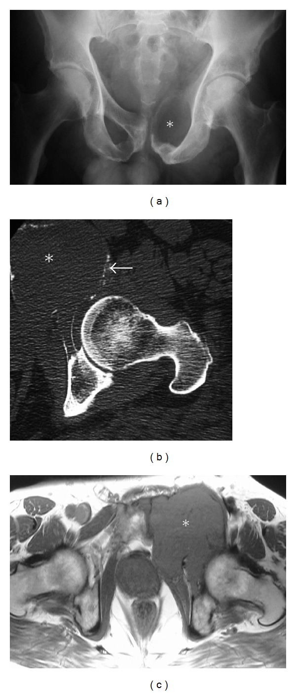
Figure 5.

Plasmacytoma. A 68-year-old male with plasmacytoma. Radiographs (a) of the pelvis showing expansion of the left obturator foramen and an expansile osteolytic lesion involving the superior pubic ramus (asterisk). Axial CT (b) depicts the large lytic mass expanding the superior pubic rami (asterisk). A thin mineralized periphery in keeping with expanded cortex is appreciated on CT (arrow). Axial T1 MR (c) demonstrates the expansile mass (asterisk) displacing the left femoral vessels, indenting the left anterolateral aspect of the prostate and invading the adjacent obturator internus muscle.
