Abstract
Previously, arsenic trioxide showed impressive regression rates of acute promyelocytic leukemia. Here, we investigated molecular determinants of sensitivity and resistance of cell lines of different tumor types towards arsenic trioxide. Arsenic trioxide was the most cytotoxic compound among 8 arsenicals investigated in the NCI cell line panel. We correlated transcriptome-wide microarray-based mRNA expression to the IC50 values for arsenic trioxide by bioinformatic approaches (COMPARE and hierarchical cluster analyses, Ingenuity signaling pathway analysis). Among the identified pathways were signaling routes for p53, integrin-linked kinase, and actin cytoskeleton. Genes from these pathways significantly predicted cellular response to arsenic trioxide. Then, we analyzed whether classical drug resistance factors may also play a role for arsenic trioxide. Cell lines transfected with cDNAs for catalase, thioredoxin, or the anti-apoptotic bcl-2 gene were more resistant to arsenic trioxide than mock vector transfected cells. Multidrug-resistant cells overexpressing the MDR1, MRP1 or BCRP genes were not cross-resistant to arsenic trioxide. Our approach revealed that response of tumor cells towards arsenic trioxide is multi-factorial.
Introduction
Arsenic is a natural semimetal in soil, water and air. It exists as red arsenic (As2S2), yellow arsenic (As2S3), white arsenic (As2O3, arsenic trioxide), phenylarsine oxide (C6H5AsO), and as salts of sodium, potassium and calcium [1]. Since ancient times arsenic was used for medical purposes [2]. Arsenic was appreciated as Fowler's Solution for many diseases in the 18th and 19th century, i.e., syphilis, cancer, ulcers, etc. [3]. In the 20th century, Paul Ehrlich, the founder of modern chemotherapy, found the arsenical salvarsan, which was the standard therapy against syphilis for decades [4]. On the other side, arsenic compounds can be poisonous [5]. The revival of arsenic in modern medicine was initiated by Chinese scientists showing dramatic regression rates of acute promyelocytic leukemia by arsenic trioxide [6]. These findings were subsequently corroborated in clinical studies in the USA [7].
Various molecular determinants of the biological effect of arsenic trioxide have been elucidated. It promotes the degradation of the oncogenic fusion protein of the PML and retinoic acid receptor α (RARα) genes which arises from t(15;17) translocation in acute promyelocytic leukemia, resulting in induction of cellular differentiation [7], [8]. Apoptosis is selectively induced in malignant cells through enhancement of reactive oxygen species and activation of caspases [9]–[12]. Cells can arrest in the G1 or G2/M phases of the cell cycle after treatment with arsenic trioxide [12]. Tumor angiogenesis is targeted by arsenic trioxide through inhibition of vascular epithelial growth factor production [13].
While focusing on mono-specific drugs without adverse effects on normal tissues, it turned out that drug resistance frequently occurs. Subpopulations of cancer cells with specific point mutations in target proteins can survive attacks of mono-specific drugs due to reduced binding affinity to these drugs. They overgrow the entire tumor population resulting in drug-resistant phenotypes, as in the case of Gleevec® resistance [14]. Therefore, it has recently been proposed that multi-target attacking drugs maybe superior by avoiding development of resistance to single mono-specific drugs. The development of multi-kinase inhibitors represents an example for this novel treatment concept.
The aim of the current study was to investigate sensitivity and resistance of tumor cells towards arsenic trioxide. For this reason, we first analyzed transcriptome-wide microarray-based mRNA expression by bioinformatic approaches (COMPARE and hierarchical cluster analyses, Ingenuity signaling pathway analysis) to identify novel molecular determinants for response of the cell line panel of the National Cancer Institute (NCI), USA, towards arsenic trioxide [15].
A second aim was to analyze whether classical determinants of resistance towards established anti-cancer drugs may also play a role in arsenic trioxide resistance. To this end, anti-oxidative stress response genes as well as multidrug resistance transporters have been tested for their influence on arsenic trioxide resistance. A major obstacle of cancer therapy is the development of cross-resistance and even worse multidrug resistance [16]–[17]. The role of the drug transporters P-glycoprotein (Pgp, MDR1, ABCB1) and multidrug resistance related protein 1 (MRP1, ABCC1) has been discussed with contradictory results [18]–[21] and it is unclear whether or not arsenic trioxide is transported by these two multidrug resistance pumps. Therefore, we have readdressed this question. Furthermore, we analyzed the breast cancer resistance protein (BCRP, ABCG2) whose relevance for resistance to arsenic is unknown as yet.
Furthermore, it has been claimed that arsenic trioxide generates reactive oxygen species (ROS) [22]–[24] leading to apoptosis. The role of ROS-detoxifying enzymes for arsenic trioxide has been investigated. Again, conflicting data have been reported [25]–[27]. Since most of these studies only measured enzymatic activities, we used cell lines transfected with cDNAs for catalase or thioredoxin to clarify whether or not these genes confer resistance to arsenic trioxide.
Materials and Methods
Cell Lines
The panel of 60 human tumor cell lines of the Developmental Therapeutics Program of the NCI, USA, consisted of leukemia (CCRF-CEM, HL-60, K-562, MOLT-4, RPMI-8226, SR), melanoma (LOX-IMVI, MALME-3M, M14, SK-MEL2, SK-MEL28, SK-MEL-5, UACC-257, UACC-62), non-small cell lung cancer (A549, EKVX, HOP-62, HOP-92, NCI-H226, NCI-H23, NCI-H322M, NCI-460, NCI-H522), colon cancer (COLO205, HCC-2998, HCT-116, HCT-15, HT29, KM12, SW-620), renal cancer (786-0, A498, ACHN, CAKI-1, RXF-393, SN12C, TK-10, UO-31), ovarian cancer (IGROV1, OVCAR-3, OVCAR-4, OVCAR-5, OVCAR-8, SK-OV-3) cell lines, cell lines of tumors of the central nervous system (SF-268, SF-295, SF-539, SNB-19, SNB-75, U251), prostate carcinoma (PC-2, DU-145), and breast cancer (MCF-7, NCI/ADR-Res, MDA-MB-231, Hs578T, MDA-MB-435, MDA-N, BT-549, T-47D). Their origin and processing have been previously described [28].
Multidrug-Resistant Tumor Cell Lines: Leukemic CCRF-CEM cells were maintained in RPMI 1640 medium (Invitrogen, Eggenstein, Germany) supplemented with 10% fetal calf serum in a humidified 5% CO2 atmosphere at 37°C. Cells were passaged twice weekly. All experiments were performed with cells in the logarithmic growth phase. P-glycoprotein/multidrug resistance gene 1 (MDR1)-expressing CEM/ADR5000 cells were maintained in 5000 ng/ml doxorubicin. The establishment of the resistant subline has been described [29].
The multidrug-resistance gene 1 (MRP1)-expressing HL-60/AR subline was continuously treated with 100 nM daunorubicin. The establishment of this cell line has been reported (Brügger et al., 1999) [30]. Sensitive and resistant cells were kindly provided by Dr. J. Beck (Department of Pediatrics, University of Greifswald, Greifswald, Germany). Breast cancer cells transduced with control vector (MDA-MB-231-pcDNA3) or with cDNA for the breast cancer resistance protein BCRP (MDA-MB-231-BCRP clone 23) were maintained under standard conditions as described above for CCRF-CEM cells. The generation of the cell lines followed a published protocol [31]. The cell lines were continuously maintained in 800 ng/ml gentamicin (Invitrogen, Karlsruhe, Germany). Oxidative stress-related cell lines: The mouse thymic lymphoma-derived WEHI7.2 parental cell line was obtained from Dr. Roger Miesfeld (University of Arizona, Tucson, AZ). Cells were maintained in Dulbecco’s Modified Eagle Medium - low glucose (Invitrogen, Carlsbad, CA) supplemented with 10 % calf serum (Hyclone Laboratories, Logan, UT) at 37°C in a 5 % CO2 humidified environment. Stock cultures were maintained in exponential growth at a density between 0.02 and 2×10⁶ cells/ml. WEHI7.2 cells stably transfected with and overexpressing human bcl-2 (Hb12), constructed and maintained as described in [32], were also obtained from Dr. Miesfeld. Thioredoxin overexpressing cells (THX) were constructed by stably transfecting human thioredoxin into WEHI7.2 cells, then selecting and maintaining clones as described [33]. THX cells express 1.8-fold more thioredoxin than the parental cells [33]. Catalase overexpressing cells were constructed by stably transfecting WEHI7.2 cells with a vector containing rat catalase as described [34]. The CAT38 clone expressing 1.4-fold parental cell catalase activity was selected and maintained in 800 µg/ml G418 (GIBCO-BRL). Hydrogen peroxide resistant cells (200R) were developed by subculturing parental cells in the presence of fresh H2O2 every three days as described [35]. This procedure resulted in a population of cells that is 2.8-fold more resistant to 200 µM H2O2 than the parental cells. 200R cells were maintained in the presence of 200 µM H2O2. Any variant normally grown in the presence of drug was cultured in the absence of drug for one week prior to each experiment.
Drug Response
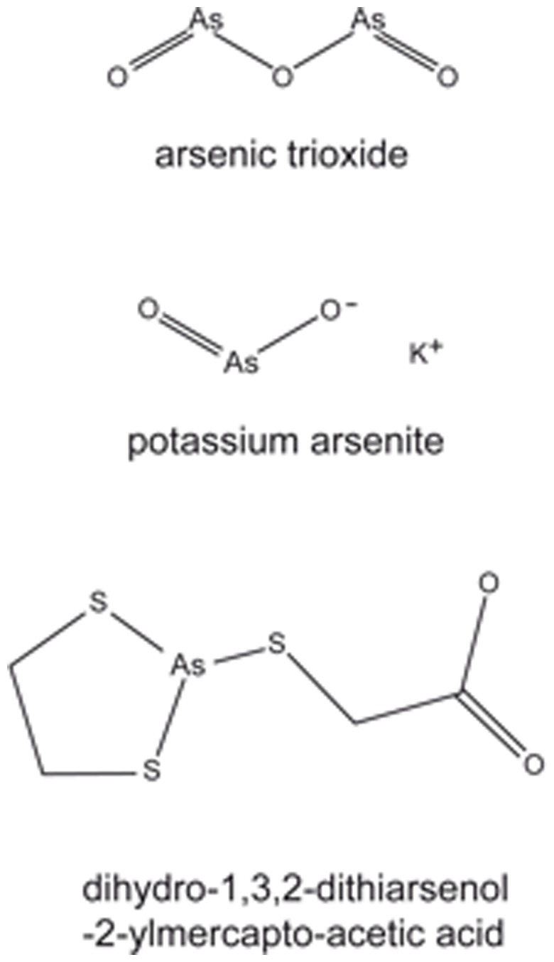
The sulforhodamine B assay for the determination of drug sensitivity in the NCI cell lines has been reported [36]. The inhibition concentration 50% (IC50) values for free and formulated arsenic trioxide (Trisenox) as well as for other arsenic compounds (potassium arsenite, dihydro-1,3,2-dithioarsenol-2-ylmercapto-acetic acid) and standard cytostatic drugs have been deposited in the database of the Developmental Therapeutics Program of the NCI (http://dtp.nci.nih.gov). Their chemical structures are shown in Figure 1 .
Figure 1. Chemical structures of arsenic trioxide, potassium arsenite, and dihydro-1,3,2-dithiarsenol-2ylmercapto-acetic acid.
Growth Inhibition Assay: The in vitro response to drugs was evaluated by means of a growth inhibition assay as described [37]. Aliquots of 5×10⁴ cells/ml were seeded in 24-well plates and drugs were added immediately at different concentrations. Arsenic trioxide was used in different doses to allow calculation of IC50 values. Cells were counted 7 days after treatment with the drugs. The resulting growth data represent the net outcome of cell proliferation and cell death.
MTS assay: The response of WEHI7.2 parental cells and WEHI7.2 cell variants towards arsenic trioxide was measured using the MTS assay (Promega Corp., Madison, WI, USA) as described previously [38]. Briefly, cells were plated at 1.5×10⁴ cells/well in 100 µl medium in a 96-well plate and incubated in the absence or presence of the indicated concentrations of arsenic trioxide for 48 hrs. Relative absorbance was measured by incubating the cells for 3 hrs at 37°C with the MTS solution, prepared and used according to the manufacturer’s protocol (Promega Corp., Madison, WI), and reading at 490 nm using a Microplate Autoreader (Bio-Tek Instruments, Winooski, VT). Response was calculated as percent absorbance of untreated control. The IC50 represent the mean of three independent experiments. The degrees of resistance were calculated by dividing the IC50 of transfected cell lines and multidrug-resistant cell lines, respectively, by the IC50 value of their corresponding mock vector control or parental cell line.
Microarray-Based Bioinformatic and Statistical Analyses
Cell lines of the NCI-60 panel were grown under standard conditions [29]. RNA isolation and microarray hybridization procedures have been described [39]–[40]. The microarray data have been deposited at the website of the NCI Developmental Therapeutics Program (http://dtp.nci.nih.gov). Hierarchical cluster analysis is an explorative statistical method and aims to group at first sight heterogeneous objects into clusters of homogeneous objects. Objects are classified by calculation of distances according to the closeness of between-individual distances. All objects are assembled into a cluster tree (dendrogram). The merging of objects with similar features leads to the formation of a cluster, where the length of the branch indicates the degree of relatedness. The procedure continues to aggregate clusters until there is only one. The distance of a subordinate cluster to a superior cluster represents a criterion for the closeness of clusters as well as for the affiliation of single objects to clusters. Thus, objects with tightly related features appear together, while the separation in the cluster tree increases with progressive dissimilarity. Previously, cluster models have been validated for gene expression profiling and for approaching molecular pharmacology of cancer [39], [41]. Cluster analyses applying the WARD method were done by means of the WinSTAT program (Kalmia Co., Cambridge, USA). Missing values are automatically omitted by the program and the closeness of two joined objects is calculated by the number of data points they contained. In order to calculate distances of all variables included in the analysis, the program automatically standardizes the variables by transforming the data with a mean = 0 and a variance = 1. To visualize the relationships between the IC50 values for arsenic trioxide and mRNA expression levels by cluster analyses, cluster image maps were formed.
For COMPARE analysis, the mRNA expression values of genes of interest and IC50 values for free and formulated arsenic trioxide (Trisenox) of the NCI cell lines were selected from the NCI database (http://dtp.nci.nih.gov). The mRNA expression has been determined by microarray analyses as reported [39]. COMPARE analyses were performed to produce rank-ordered lists of genes expressed in the NCI cell lines. The methodology has been described previously in detail [42]. Briefly, every gene of the NCI microarray database was ranked for similarity of its mRNA expression to the IC50 values for the corresponding compound. To derive COMPARE rankings, a scale index of correlations coefficients (R-values) was created. In the standard COMPARE approach, greater mRNA expression in cell lines correlate with enhanced drug resistance, whereas in reverse COMPARE analyses greater mRNA expression in cell lines indicated drug sensitivity.
The Ingenuity Pathway Analysis software (IPA) (Ingenuity Systems, Mountain View, CA, USA; http://www.ingenuity.com) was utilized to identify networks and pathways of interacting genes and other functional groups in genomic data. Using the IPA Functional Analysis tool we were able to associate biological functions and diseases to the experimental results. Moreover, we used a biomarker filter tool and the Network Explorer for visualizing molecular relationships.
Pearson’s correlation test was used to calculate significance values and rank correlation coefficients as a relative measure for the linear dependency of two variables. This test was implemented into the WinSTAT Program (Kalmia Co.). Pearson’s correlation test determined the correlation of rank positions of values. Ordinal or metric scaling of data is suited for the test and transformed into rank positions. There is no condition regarding normal distribution of the data set for the performance of this test. We used Pearson’s correlation test to correlate microarray-based mRNA expression of candidate genes with the IC50 values for arsenic trioxide.
The Chi2-test was applied to bivariate frequency distributions of pairs of nominal scaled variables. It was used to calculate significance values (P-values) and rank correlation coefficients (R-values) as a relative measure for the linear dependency of two variables. This test was implemented into the WinSTAT program (Kalmia Co.). The Chi2-test determines the difference between each observed and theoretical frequency for each possible outcome, squaring them, dividing each by the theoretical frequency, and taking the sum of the results. Performing the Chi2-test necessitated defining cell lines as being sensitive or resistant to arsenic trioxide. This was done by taking the median IC50 value (log10 = −5.346 M for formulated arsenic trioxide and log10 = −5.467 M for free arsenic trioxide) as a cut-off threshold.
Results
Cross-resistance of Arsenic Compounds in the NCI Cell Line Panel
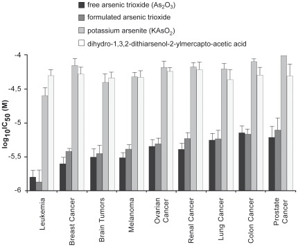
The NCI database contained 9 arsenic-containing compounds, of which five were inactive or only minimally active against the cancer cell lines tested. The four cytotoxic arsenicals were free and formulated arsenic trioxide as well as potassium arsenite, dihydro-1,3,2-dithioarsenol-2-ylmercapto-acetic acid. The inactive or weakly active arsenicals were arsenic(III) 2,3-dimercapto succinic acid, simethyl arsinic acid, lithiume arsenate (Li3AsO4), sodium arsenic tungsten polyoxymetalate hydrate, and arsenic acid (H3AsO4) trilithium salt. These substances have been investigated over a dose range from 10−8 to 10−4 M in 60 tumor cell lines and IC50 values have been calculated thereof. The IC50 values for the four cytotoxic arsenic compounds are shown in Figure 2 . Free and formulated arsenic trioxides were more cytotoxic than the two other arsenicals. Leukemia cell lines were more sensitive than cell lines from other tumor types. Among cell lines of solid cancers, cell lines from brain tumors, melanoma, or breast cancer were most sensitive to free or formulated arsenic trioxide, whereas colon or prostate cancer cell lines were most resistant. Cell lines from lung or kidney cancer showed intermediate sensitivity.
Figure 2. IC50 values of four arsenicals for the NCI cell line panel. Mean values and SEM of IC50 are grouped according to the tumor origin of the cell lines.
We correlated the IC50 values for free and formulated arsenic trioxide with those of other arsenic-containing compounds (potassium arsenite, dihydro-1,3,2-dithioarsenol-2-ylmercapto-acetic acid). As shown in Table 1 , the IC50 values for free and formulated arsenic trioxide were highly correlated (P = 4.14×10⁻¹⁴). Furthermore, the IC50 values for arsenic trioxide of the cell line panel were significantly correlated with the IC50 values for potassium arsenite and dihydro-1,3,2-dithiarsenol-2-ylmercapto-acetic acid, indicating that the cell lines reveal cross-resistance to arsenic-containing drugs.
Table 1. Cross-resistance between arsenic trioxide and other arsenic compounds in the NCI cell line panel.
| free As2O3 | formulated As2O3 | |
| formulated As2O3 | 4.14×10⁻¹⁴* | N/A |
| potassium arsenite (KAsO2) | 1.76×10⁻¹²* | 2.51×10⁻⁹* |
| dihydro-1,3,2-dithiarsenol-2-ylmercapto-acetic acid | 0.04565* | n.s. |
Log10 IC50 values obtained from SRB assays have been subjected to Pearson’s correlation test.
N/A, not applicable; N.S. not significant (P>0.05). * denotes significant correlation.
Gene-hunting Approach
COMPARE and Cluster Analyses of Microarray-Based mRNA Hybridization
We applied a pharmacogenomic approach to explore novel molecular determinants of sensitivity and resistance to arsenic trioxide. We mined the genome-wide mRNA expression database of the NCI and correlated the expression data with the IC50 values for arsenic trioxide. This represents a hypothesis-generating bioinformatic approach, which allows the identification of novel putative molecular determinants of cellular response towards arsenic trioxide.
Standard COMPARE analysis was performed to identify genes, while expression was associated with arsenic trioxide resistance. Vice versa, reverse COMPARE analysis was done to find factors associated with arsenic trioxide sensitivity. Only correlations with a correlation coefficient of R>0.5 (standard COMPARE) or R<−0.55 (reverse COMPARE) were considered (Table S1).
Among the genes identified by this approach were genes from diverse functional groups such as signal transduction (SYDE1, SFN, PPAP2C, EZR, GPRC5A), DNA biosynthesis and transcriptional regulation (UPRT, MED12, SFRS15), adhesion and cytoskeletal organization (PDLIM5, PERP, DSG2, SDC1) and others (ID1, ILKAP, HMIX2, UBA1, ARHGEF6, CYTH1, TXNRD1, CMTM4).
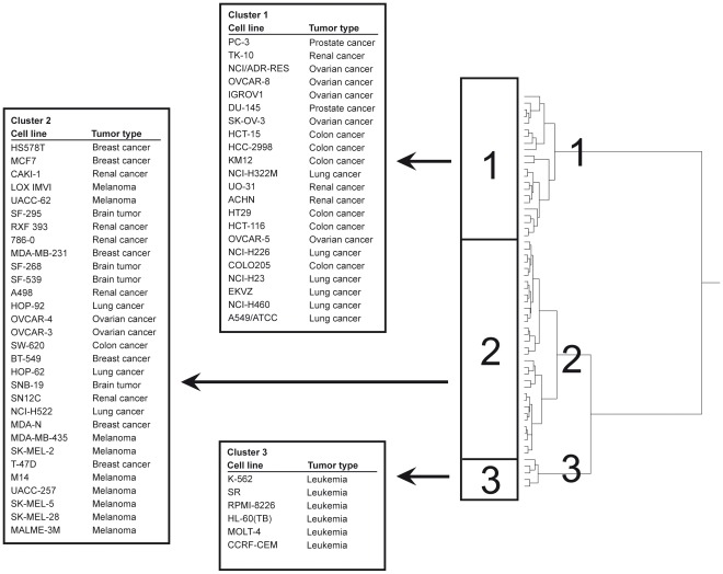
Next, the genes identified by standard and reverse COMPARE analyses were subjected to hierarchical cluster analysis. The dendrogram obtained by this procedure can be divided into three major branches ( Figure 3 ). The distribution of cell lines being sensitive or resistant to formulated arsenic trioxide was significantly different between the branches of the dendrograms. The sensitive/resistant ratio in cluster 1 was 2∶20, 22∶10 in cluster 2 and 6∶0 in cluster 3. The distribution of cell lines among the dendrogram predicted resistance to formulated arsenic trioxide with significance (P = 3.2×10⁻⁶; Chic2-test; Table 2 ). A similar relationship was found for free arsenic trioxide (P = 4.5×10⁻⁶; Chic2-test; Table 2 ).
Figure 3. Hierarchical cluster analysis of microarray-based mRNA gene expression obtained by standard and reverse COMPARE analyses.
The dendrogram shows the clustering of the NCI-60 cell line panel and indicates the degrees of relatedness between cell lines.
Table 2. Separation of clusters of the NCI cell line panel obtained by the hierarchical cluster analysis shown in Figure 3 in comparison to drug sensitivity.
| Partitiona | Cluster 1 | Cluster 2 | Cluster 3 | Chi2 Test | ||
| formulated arsenic trioxide | sensitive | ≤−5.346 | 2 | 22 | 6 | |
| resistant | >−5.346 | 20 | 10 | 0 | 3.32635×10⁻⁶ | |
| free arsenic trioxide | sensitive | ≤−5.467 | 2 | 21 | 6 | |
| resistant | >−5.467 | 20 | 10 | 0 | 4.50505×10⁻⁶ |
The median log10IC50 value (M) for each drug was used as a cut-off to separate tumor cell lines as being "sensitive" or "resistant".
Signaling Pathway Profiling
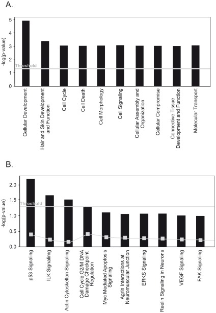
As a next step, we employed a signaling pathway analysis to better understand the biological consequences of arsenic trioxide treatment. The genes identified by microarray and COMPARE analyses were subjected to Ingenuity Pathway Analysis (version 6.5). The genes identified by COMPARE analysis have a function in cellular development, hair and skin development and function, cell cycle, cell death, and cell morphology and others ( Figure 4A ). The top canonical pathways were signaling routes for p53, ILK, and actin cytoskeleton ( Figure 4B ).
Figure 4. Identification of signaling pathways and interaction of gene products associated with cellular response of cancer cells towards formulated arsenic trioxide.
The genes identified by COMPARE analyses (Table S1) were subjected to Ingenuity Pathway Analysis Software. (A) Top 10 categories of biological functions of the candidate genes. (B) Top 10 canonical signaling pathways, which the candidate genes were assigned to.
Candidate Gene Approach
In the second part of our investigation, we analyzed whether classical mechanisms of resistance towards established anti-cancer drugs would also affect response of tumor cells towards arsenic trioxide.
Role of oxidative stress response, damage, or metabolism for resistance to arsenic trioxide
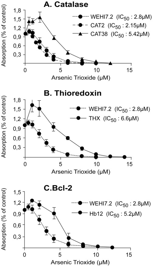
Figure 5 shows the arsenic trioxide response of WEHI7.2 mouse thymic lymphoma cells selected for resistance to H2O2 or stably transfected with catalase, thioredoxin, or bcl-2. The CAT38 clone was 1.94-fold more resistant to arsenic trioxide than the parental WEHI7.2 cells ( Figure 5A ). Thioredoxin-transfected cells were 2.36-fold more resistant to arsenic trioxide than WEHI7.2 cells ( Figure 5B ). WEHI7.2 cells selected for resistance to H2O2 were not resistant to arsenic trioxide (data not shown). Finally, bcl-2-transfected cells were 1.86-fold more resistant to arsenic trioxide than WEHI7.2 cells ( Figure 5C ).
Figure 5. Cytotoxicity of arsenic trioxide on WEHI7.2 cell lines.
Cells stably transfected with expression vectors carrying cDNAs for (A) catalase, (B) thioredoxin, or (C) Bcl-2, and with mock control vector. Values represent the mean (± SEM) of three independent experiments.
Role of ABC-Transporters for Resistance to Arsenic Trioxide
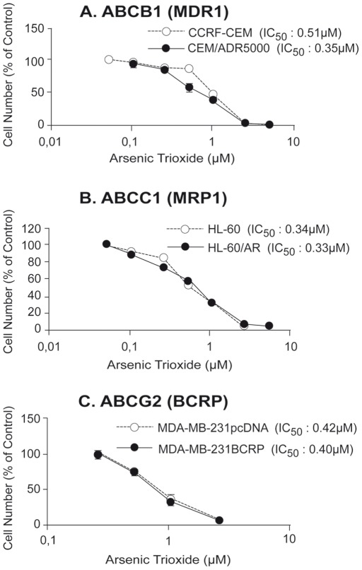
As multidrug resistance (MDR) and MDR-conferring drug transporters of the ABC transporter family are a major cause of failure to many established anti-cancer drugs, we addressed the question, whether cellular response to arsenic trioxide treatment may also be affected by ABC transporters. The role of three ABC transporters has been exemplarily validated using cell lines that selectively overexpress either the ABCB1 (MDR1), ABCC1 (MRP1), or the ABCG2 (BCRP) gene. Based on the IC50 values calculated from the dose response curves shown in Figure 6A , ABCB1 (MDR1)-overexpressing CEM/ADR5000 cells were slightly more sensitive to arsenic trioxide as compared to parental CCRF-CEM cells (degree of increased sensitivity: 0.69). ABCC1(MRP1)-overexpressing HL60/AR cells and ABCG2 (BCRP)-overexpressing MDA-MB-231-BCRP cells were not more resistance to arsenic trioxide than their drug-sensitive counterparts ( Figure 6B and C ).
Figure 6. Cytotoxicity of sensitive and multidrug-resistant tumor cells to arsenic trioxide.
(A) Sensitive CCRF-CEM and multidrug-resistant ABCB1 (MDR1)-overexpressing CEM/ADR5000 cells; (B) sensitive HL60 and multidrug-resistant ABCC1 (MRP1)-overexpressing HL60/AR cells; and (C) sensitive MDA-MB-231-pcDNA and multidrug-resistant ABCG2 (BCRP)-transduced MDA-MB-231-BCRP cells. Values represent the mean (± SEM) of three independent experiments.
Discussion
Gene-hunting Approach
In the present investigation, we analyzed molecular determinants of sensitivity and resistance of cancer tumor cell lines towards arsenic trioxide. In general, there are two ways to reach this goal: (1) gene-hunting and (2) candidate gene approaches. Applying the first approach, we correlated the IC50 values for arsenic trioxide of 60 tumor cell lines with the microarray-based transcriptome-wide mRNA expression levels of this cell line panel [39] by COMPARE analysis. This approach has been successfully used to unravel the mode of action of novel compounds [43]. Cluster and COMPARE analyses are also useful for comparing gene expression profiles with IC50 values for investigational drugs to identify candidate genes for drug resistance [44] and to identify prognostic expression profiles in clinical oncology [45].
We identified genes from diverse functional groups, which were tightly associated with the response of tumor cells to arsenic trioxide, such as genes belonging to p53 signaling and others, most of which have not been associated with cellular response to arsenic trioxide. Interestingly, the oxidative stress response and DNA repair (TXNRD1 and UBA1) appeared in the COMPARE analysis, which speaks DNA damage as mode of action of arsenic trioxide. The gene-hunting approach applied by us delivered several novel candidate genes that may regulate the response of cancer cells to arsenic trioxide. These results merit further investigation to prove the contribution of these genes to arsenic trioxide resistance.
The microarray technology has also been applied by other investigators to analyze genes potentially relevant for cellular response towards arsenic trioxide [46]–[49]. In these studies, the gene expression between untreated and arsenic trioxide-treated cell lines has been compared to identify genes up- or down-regulated upon drug challenge. This approach delivers genes as a response to cytotoxic stress and is different from our approach. In the present investigation, we correlated the basal gene expression of untreated cells in a panel of 60 cell lines with their IC50 values to arsenic trioxide. These two experimental settings refer to two different types of drug resistance. The first approach may unravel genes conferring resistance after drug treatment. This type is called acquired or secondary resistance. In our approach, we identify genes involved in the initial responsiveness of tumor cells to drug treatment. This type is known as inherent or primary resistance. Both types of drug resistance can clinically be observed. As an example, small cell lung cancer frequently responds well to chemotherapy at the beginning of a therapy, but gradually develops resistance during subsequent treatment courses (acquired or secondary resistance). Non-small cell lung cancers do not respond well to chemotherapy even at the beginning of a treatment (inherent or primary resistance). This implies that those tumors express drug resistance mechanisms prior to drug treatment.
It is interesting to note that the microarray analysis in the current study identified genes from functional groups similar to those that previous studies identified as associated with cellular response to arsenic trioxide. These include cell cycle-regulating genes [46−49], transcription factors and cofactors [47], [49], signal transducers [46], [50], DNA repair genes [46], [49] and apoptosis-regulating genes [47]. This indicates that these cellular functions may be of importance for resistance to arsenic trioxide. The appearance of these genes was a clue for the involvement of reactive oxygen species (see above), which was indeed validated by our subsequent experiments. Liu et al. [51] also identified oxidative stress response genes and proteins related to the NRF2 pathway in the NCI-60 cell line panel as possible determinants of response to arsenic trioxide. In our approach, we analyzed not the entire set of significantly correlating genes as Liu and colleagues did, but only the genes with the highest COMPARE ranks. Here, genes related to p53 signaling, cell cycle arrest, DNA repair, and apoptosis provide clues on reactive oxygen species as underlying mechanism. Therefore, the report of Liu et al. and the present investigation do nicely complement each other and strengthen the hypothesis of oxidative stress response as important mechanism for arsenic trioxide’s response in cancer cells.
Additional functional groups of genes, which did not appear in the present investigation, were proteasome degradation, RNA processing calcium signaling, the IFN pathway and protein synthesis [47], [50]. Other arsenic trioxide effects include impairment of the genomic differentiation program in human macrophages [52] and alterations in the expression of multiple micro-RNAs. A more detailed analysis is required to determine the relative importance of the multiple effects in the observed drug response.
Candidate Gene Approach
As a second approach, we analyzed whether several classical drug resistance mechanisms may also play a role for the resistance towards arsenic trioxide. These classical mechanisms did not appear in our COMPARE analyses, although their mRNA expression values were also included into the analysis. This indicates that the above genes identified by COMPARE might be more relevant for response of tumor cells towards arsenic trioxide. Nevertheless, the role of those classical drug resistance mechanisms is worth investigating, because of their generally accepted role for drug resistance to anti-cancer agents.
It has been demonstrated that arsenic trioxide generates ROS (preferentially H2O2 but also O2 •- [22]–[24], [53]) and that the cytotoxic activity of arsenic trioxide is reduced by N-acetylcysteine [27], [54]–[56] and enhanced by buthionine sulfoximine [54], [57], [58]. These results imply that oxidative stress induced by arsenic trioxide is important for cytotoxicity. Therefore, it is surprising that contradictory results have been reported for ROS-detoxifying enzymes. Either increased, decreased, or unchanged enzymatic activities upon cellular challenge with arsenic trioxide have been observed for glutathione S-transferase-pi [25], [26], [58], glutathione peroxidase [25], [59]–[61], glutathione reductase [25], [60], catalase [27], [55], [60], [62], [63], superoxide dismutases [25], [27], [56], [61] and thioredoxin reductase [64]. To clarify the role of ROS-detoxifying enzymes, it may not be sufficient to measure enzymatic activities. Therefore, we have used cell lines transfected with cDNAs for catalase or thioredoxin and treated them with arsenic trioxide. We found that transfection of catalase cDNA or thioredoxin cDNA conferred resistance towards arsenic trioxide. In addition to the glutathione redox system, the thioredoxin system represents another major antioxidant system maintaining the intracellular redox state. Thioredoxin scavenges ROS, regulates antioxidant enzymes, and inhibits proapoptotic proteins [65]. Oxidized thioredoxin is reduced by thioredoxin reductase, which is relevant for arsenic trioxide’s activity as shown in the present investigation.
It is unclear from the literature, whether arsenic trioxide induces apoptosis and whether Bcl-2 is protective. This is further complicated by the conflicting data indicating that arsenic trioxide can up- and down-regulate the apoptosis-regulating bcl-2, bcl-xL, and bax genes depending on the model system [18], [66]–[68]. Therefore, we attempted to clarify the role of the anti-apoptotic bcl-2 gene by treating bcl-2 transfected cells with arsenic trioxide. As expected, we observed that bcl-2 mediated resistance to this compound providing evidence for the importance of the mitochondrial pathway of apoptosis for arsenic trioxide’s cytotoxicity towards cancer cells.
ABC Transporters and Multidrug Resistance
Multidrug resistance (MDR) is based on numerous mechanisms, one of which is the influence of ATP-binding cassette (ABC) transporters. They are involved in the active transport of phospholipids, ions, peptides, steroids, polysaccharides, amino acids, bile acids, pharmaceutical drugs and other xenobiotic compounds [16]. ABCB1 (P-glycoprotein, P-gp, MDR1), ABCC1-C6 (MRP1-6) and ABCG2 (BCRP) confer resistance to cytostatic drugs of tumors and contribute to the failure of tumor [17]. It is still unclear, to which extent human ABC transporters contribute to arsenic trioxide-related drug resistance phenomena.
While some authors found no cross-resistance or even collateral sensitivity of cell lines overexpressing P-glycoprotein (MDR1, ABCB1) [18], [69]–[71] or MRP1 (ABCC1) [18], [20], [21], [72], [73], others claim a role of these ABC transporters in arsenic trioxide resistance [19], [74]. This discussion, i.e. whether arsenic trioxide leads to an induction or repression of these two drug transporters, is controversial [66], [70], [75]–[80]. The role of BCRP (ABCG2), another important multidrug resistance-conferring ABC-transporter has not been addressed as yet. For this reason, we have analyzed multidrug-resistant CBM/ADR5000 cells which specifically overexpress P-glycoprotein, but none of the other ABC transporters [17], [30]. These cells were slightly more sensitive to arsenic trioxide, indicating that P-glycoprotein does not play a major role for resistance to this drug. Furthermore, we have analyzed HL60/AR cells, which have been reported to overexpress MRP1 [81]. In a previous investigation we found that other transporters [82] are also overexpressed in this cell line. Since this cell line did not reveal cross-resistance to arsenic trioxide, we conclude that these ABC transporters are not relevant for resistance towards this drug. Likewise, MDA-MB-231/BCRP cells transfected with a cDNA for BCRP were not cross-resistant to arsenic trioxide. In summary, our data do not support that the ABC-transporters P-gp, MRP1 and BCRP considerably contribute to resistance to arsenic trioxide. This indicates that clinically refractory tumors overexpressing these ABC transporters might still be responsive to arsenic trioxide.
Conclusions
In the present investigation, we analyzed molecular determinants of sensitivity and resistance of cancer tumor cell lines to arsenic trioxide. By the gene-hunting approach, we identified genes, which were not yet known to be linked to responsiveness of cancer cells towards arsenic trioxide-. These genes need to be investigated in more detail in future studies. By the candidate gene approach, we analyzed the role of several classical drug resistance mechanisms for the resistance towards arsenic trioxide-apoptotic bcl-2 gene as well as the thioredoxin reductase gene. ABC transporters were not responsible for resistance to arsenic trioxide (MRP1, BCRP).
Our approach clearly revealed that response of tumor cells towards arsenic trioxide is multi-factorial. At least some of the functional groups of genes are also implicated in clinical responsiveness of tumors towards chemotherapy. Whether the genes identified in the present study also determine clinical responsiveness to arsenic trioxide merits further investigation.
Supporting Information
Genes determining sensitivity or resistance towards formulated arsenic trioxide in the NCI cell line panel as identified by microarray mRNA expression profiling and COMPARE analysis (see Supporting Information).
(DOC)
Footnotes
Competing Interests: The authors have declared that no competing interests exist.
Funding: These authors have no support or funding to report.
References
- 1.Miller WH, Schipper HM, Lee JS, Singer J, Waxman S. Mechanisms of action of arsenic trioxide. Cancer Res. 2002;62:3893–3903. [PubMed] [Google Scholar]
- 2.Klaassen CD. J.G. Hardman, AG Gilman, and LE Limbird (eds.) Goodman and Gilman's The Pharmacological basis of therapeutics, 1649–1672. New York: McGraW-HILL; 1996. Heavy metals and heavy-metal antagonists. [Google Scholar]
- 3.Haller JS. Therapeutic mule: the use of arsenic in the nineteenth century materia medica. Pharmacy in History. 1975;17:87–100. [PubMed] [Google Scholar]
- 4.Chan PC, Huff J. Arsenic carcinogenesis in animals and in humans: mechanistic, experimental, and epidemiological evidence. J Environ Sci Health Part C Environ Carcinog Ecotoxicol Rev. 1997;C15:83–122. [Google Scholar]
- 5.Knowles PC, Benson AA. The biogeochemistry of arsenic. Trends Biochem Sci. 1983;8:178–180. [Google Scholar]
- 6.Shen ZX, Chen GQ, Ni JH, Li XS, Xiong SM, et al. Use of arsenic trioxide (As2O3) in the treatment of acute promyelocytic leukemia (APL): II. Clinical efficacy and pharmacokinetics in relapsed patients. Blood. 1997;89:3354–3360. [PubMed] [Google Scholar]
- 7.Soignet SL, Frankel SR, Douer D, Tallman MS, Kantarjian H, et al. United States multicenter study of arsenic trioxide in relapsed acute promyelocytic leukemia. J Clin Oncol. 2001;19:3852–3860. doi: 10.1200/JCO.2001.19.18.3852. [DOI] [PubMed] [Google Scholar]
- 8.Chen GQ, Zhu J, Shi XG, Ni JH, Zhong HJ, et al. In vitro studies on cellular and molecular mechanisms of arsenic trioxide (As2O3) in the treatment of acute promyelocytic leukemia: As2O3 induces NB4 cell apoptosis with downregulation of Bcl-2 expression and modulation of PML-RARa/PML proteins. Blood. 1996;88:1052–1061. [PubMed] [Google Scholar]
- 9.Huang XJ, Wiernik PH, Klein RS, Gallagher RE. Arsenic trioxide induces apoptosis of myeloid leukemia cells by activation of caspases. Med Oncol. 1999;16:58–64. doi: 10.1007/BF02787360. [DOI] [PubMed] [Google Scholar]
- 10.Anderson KC, Boise LH, Louie R, Waxman S. Arsenic trioxide in multiple myeloma: rationale and future directions. Cancer J. 2002;8:12–25. doi: 10.1097/00130404-200201000-00003. [DOI] [PubMed] [Google Scholar]
- 11.Hayashi T, Hideshima T, Akiyama M, Richardson P, Schlossman RL, et al. Arsenic trioxide inhibits growth of human multiple myeloma cells in the bone marrow microenvironment. Mol Cancer Ther. 2002;1:851–860. [PubMed] [Google Scholar]
- 12.Liu Q, Hilsenbeck S, Gazitt Y. Arsenic trioxide-induced apoptosis in myeloma cells: p53-dependent G1 or G2/M cell cycle arrest, activation of caspase-8 or caspase-9, and synergy with APO2/TRAIL. Blood. 2003;101:4078–4087. doi: 10.1182/blood-2002-10-3231. [DOI] [PubMed] [Google Scholar]
- 13.Roboz GJ, Dias S, Lam G, Lane WJ, Soignet SL, et al. Arsenic trioxide induces dose- and time-dependent apoptosis of endothelium and may exert an antileukemic effect via inhibition of angiogenesis. Blood. 2000;96:1525–1530. [PubMed] [Google Scholar]
- 14.Sawyers CL. Research on resistance to cancer drug Gleevec. Science. 2001;294(5548):1834. doi: 10.1126/science.294.5548.1834b. [DOI] [PubMed] [Google Scholar]
- 15.Efferth T, Kaina B. Microarray-based prediction of cytotoxicity of tumor cells to arsenic trioxide. Cancer Genomics Proteomics. 2004;1:363–370. [PubMed] [Google Scholar]
- 16.Efferth T. The human ATP-binding cassette transporter genes: from the bench to the bedside. Curr Mol Med. 2001;1:45–65. doi: 10.2174/1566524013364194. [DOI] [PubMed] [Google Scholar]
- 17.Gillet JP, Efferth T, Remacle J. Chemotherapy-induced resistance by ATP-binding cassette transporter genes. Biochim Biophys Acta. 2007;1775:237–262. doi: 10.1016/j.bbcan.2007.05.002. [DOI] [PubMed] [Google Scholar]
- 18.Perkins C, Kim CN, Fang G, Bhalla KN. Arsenic induces apoptosis of multidrug-resistant human myeloid leukemia cells that express Bcr-Abl or overexpress MDR, MRP, Bcl-2, or Bcl-x(L). Blood. 2000;95:1014–1022. [PubMed] [Google Scholar]
- 19.Chen X, Zhang M, Liu LX. The overexpression of multidrug resistance-associated proteins and gankyrin contribute to arsenic trioxide resistance in liver and gastric cancer cells. Oncol Rep. 2009;22:73–80. [PubMed] [Google Scholar]
- 20.Seo T, Urasaki Y, Takemura H, Ueda T. Arsenic trioxide circumvents multidrug resistance based on different mechanisms in human leukemia cell lines. Anticancer Res. 2005;25:991–998. [PubMed] [Google Scholar]
- 21.Diaz Z, Mann KK, Marcoux S, Kourelis M, Colombo M, et al. A novel arsenical has antitumor activity toward As2O3-resistant and MRP1/ABCC1-overexpressing cell lines. Leukemia 22: 1853–1863. Erratum in: Leukemia. 2008;23:431. doi: 10.1038/leu.2008.194. [DOI] [PubMed] [Google Scholar]
- 22.Brown E, Yedjou CG, Tchounwou PB. Cytotoxicity and oxidative stress in human liver carcinoma cells exposed to arsenic trioxide (HepG(2). Met Ions Biol Med. 2008;10:583–587. [PMC free article] [PubMed] [Google Scholar]
- 23.Bowling BD, Doudican N, Manga P, Orlow SJ. Inhibition of mitochondrial protein translation sensitizes melanoma cells to arsenic trioxide cytotoxicity via a reactive oxygen species dependent mechanism. Cancer Chemother Pharmacol. 2008;63:37–43. doi: 10.1007/s00280-008-0705-y. [DOI] [PMC free article] [PubMed] [Google Scholar]
- 24.Laparra JM, Vélez D, Barberá R, Farré R, Montoro R. As2O3-induced oxidative stress and cycle progression in a human intestinal epithelial cell line (Caco-2). Toxicol In Vitro. 2008;22:444–449. doi: 10.1016/j.tiv.2007.11.004. [DOI] [PubMed] [Google Scholar]
- 25.Yeh JY, Cheng LC, Ou BR, Whanger DP, Chang LW. Differential influences of various arsenic compounds on glutathione redox status and antioxidative enzymes in porcine endothelial cells. Cell Mol Life Sci. 2002;59:1972–1982. doi: 10.1007/PL00012519. [DOI] [PMC free article] [PubMed] [Google Scholar]
- 26.Wang X, Kong L, Zhao J, Yang P. Arsenic trioxide in the mechanism of drug resistance reversal in MCF-7/ADM cell line of human breast cancer. Zhonghua Zhong Liu Za Zhi. 2002;24:339–343. [PubMed] [Google Scholar]
- 27.Han YH, Kim SH, Kim SZ, Park WH. Apoptosis in arsenic trioxide-treated Calu-6 lung cells is correlated with the depletion of GSH levels rather than the changes of ROS levels. J Cell Biochem. 2008;104:862–878. doi: 10.1002/jcb.21673. [DOI] [PubMed] [Google Scholar]
- 28.Alley MC, Scudiero DA, Monks A, Hursey ML, Czerwinski MJ, et al. Feasibility of drug screening with panels of human tumor cell lines using a microculture tetrazolium assay. Cancer Res 48: 589. 1988;–601 [PubMed] [Google Scholar]
- 29.Kimmig A, Gekeler V, Neumann M, Frese G, Handgretinger R, et al. Susceptibility of multidrug-resistant human leukemia cell lines to human interleukin 2-activated killer cells. Cancer Res. 1990;50:6793–6799. [PubMed] [Google Scholar]
- 30.Brügger D, Herbart H, Gekeler V, Seitz G, Liu C, et al. Functional analysis of P-glycoprotein and multiderug resistance-associated protein-ralated multidrug resistance in AML-blasts. Leuk Res. 1999;23:467–475. doi: 10.1016/s0145-2126(98)00192-1. [DOI] [PubMed] [Google Scholar]
- 31.Doyle LA, Yang W, Abruzzo LV, Krogmann T, Gao Y, et al. A multidrug resistance transporter from human MCF-7 breast cancer cells. Proc Natl Acad Sci USA. 1998;95:15665–15670. doi: 10.1073/pnas.95.26.15665. [DOI] [PMC free article] [PubMed] [Google Scholar]
- 32.Lam M, Dubyak G, Chen L, Nunez G, Miesfeld RL, et al. Evidence that BCL-2 represses apoptosis by regulating endoplasmic reticulum-associated Ca2+ fluxes. Proc Natl Acad Sci USA. 1994;91:6569–6573. doi: 10.1073/pnas.91.14.6569. [DOI] [PMC free article] [PubMed] [Google Scholar]
- 33.Baker A, Payne CM, Briehl MM, Powis G. Thioredoxin, a gene found overexpressed in human cancer, inhibits apoptosis in vitro and in vivo. Cancer Res. 1997;57:5162–5167. [PubMed] [Google Scholar]
- 34.Tome ME, Baker AF, Powis G, Payne CM, Briehl MM. Catalase-overexpressing thymocytes are resistant to glucocorticoid-induced apoptosis and exhibit increased net tumor growth. Cancer Res. 2001;61:2766–2773. [PubMed] [Google Scholar]
- 35.Tome ME, Briehl MM. Thymocytes selected for resistance to hydrogen peroxide show altered antioxidant enzyme profiles and resistance to dexamethasone-induced apoptosis. Cell Death Differ. 2001;8:953–961. doi: 10.1038/sj.cdd.4400904. [DOI] [PubMed] [Google Scholar]
- 36.Rubinstein LV, Shoemaker RH, Paull KD, Simon RM, Tosini S, et al. Comparison of in vitro anticancer-drug-screening data generated with a tetrazolium assay versus a protein assay against a diverse panel of human tumor cell lines. J Natl Cancer Inst. 1990;82:1113–1118. doi: 10.1093/jnci/82.13.1113. [DOI] [PubMed] [Google Scholar]
- 37.Efferth T, Sauerbrey A, Olbrich A, Gebhart E, Rauch P, et al. Molecular modes of action of artesunate in tumor cell lines. Mol Pharmacol. 2003;64:382–394. doi: 10.1124/mol.64.2.382. [DOI] [PubMed] [Google Scholar]
- 38.Efferth T, Briehl MM, Tome ME. Role of antioxidant genes for the activity of artesunate against tumor cells. Int J Oncol. 2003;23:1231–1235. [PubMed] [Google Scholar]
- 39.Scherf U, Ross DT, Waltham M, Smith LH, Lee JK, et al. A gene expression database for the molecular pharmacology of cancer. Nat Genet. 2000;24:236–244. doi: 10.1038/73439. [DOI] [PubMed] [Google Scholar]
- 40.Amundson SA, Do KT, Vinikoor LC, Lee RA, Koch-Paiz CA, et al. Integrating global gene expression and radiation survival parameters across the 60 cell lines of the National Cancer Institute Anticancer Drug Screen. Cancer Res. 2008;68:415–424. doi: 10.1158/0008-5472.CAN-07-2120. [DOI] [PubMed] [Google Scholar]
- 41.Efferth T, Fabry U, Osieka R. Apoptosis and resistance to daunorubicin in human leukemic cells. Leukemia. 1997;11:1180–1186. doi: 10.1038/sj.leu.2400669. [DOI] [PubMed] [Google Scholar]
- 42.Wosikowski K, Schuurhuis D, Johnson K, Paull KD, Myers TG, et al. Identification of epidermal growth factor receptor and erbB2 pathway inhibitors by correlation with gene expression patterns. J Natl Cancer Inst. 1997;89:1505–1515. doi: 10.1093/jnci/89.20.1505. [DOI] [PubMed] [Google Scholar]
- 43.Leteurtre F, Kohlhagen G, Paull KD, Pommier Y. Topoisomerase II inhibition and cytotoxicity of the anthrapyrazoles DuP 937 and DuP 941 (Losoxantrone) in the National Cancer Institute preclinical antitumor drug discovery screen. J Natl Cancer Inst. 1994;86:1239–1244. doi: 10.1093/jnci/86.16.1239. [DOI] [PubMed] [Google Scholar]
- 44.Efferth T, Gebhart E, Ross DD, Sauerbrey A. Identification of gene expression profiles predicting tumor cell response to l-alanosine. Biochem Pharmacol. 2003;66:613–621. doi: 10.1016/s0006-2952(03)00341-1. [DOI] [PubMed] [Google Scholar]
- 45.Efferth T, Olbrich A, Bauer R. mRNA expression profiles for the response of human tumor cell lines to the antimalarial drugs artesunate, arteether, and artemether. Biochem Pharmacol. 2002;64:617–623. doi: 10.1016/s0006-2952(02)01221-2. [DOI] [PubMed] [Google Scholar]
- 46.Ahn WS, Bae SM, Lee KH, Kim YW, Lee JM, et al. Comparison of effects of As2O3 and As4O6 on cell growth inhibition and gene expression profiles by cDNA microarray analysis in SiHa cells. Oncol Rep. 2004;12:573–580. [PubMed] [Google Scholar]
- 47.Zheng PZ, Wang KK, Zhang QY, Huang QH, Du YZ, et al. Systems analysis of transcriptome and proteome in retinoic acid/arsenic trioxide-induced cell differentiation/apoptosis of promyelocytic leukemia. Proc Natl Acad Sci U S A. 2005;102:7653–7658. doi: 10.1073/pnas.0502825102. [DOI] [PMC free article] [PubMed] [Google Scholar]
- 48.Zhao S, Zhang J, Zhang X, Dong X, Sun X. Arsenic trioxide induces different gene expression profiles of genes related to growth and apoptosis in glioma cells dependent on the p53 status. Mol Biol Rep. 2008;35:421–429. doi: 10.1007/s11033-007-9102-6. [DOI] [PubMed] [Google Scholar]
- 49.Burnichon V, Jean S, Bellon L, Maraninchi M, Bideau C, et al. Patterns of gene expressions induced by arsenic trioxide in cultured human fibroblasts. Toxicol Lett. 2003;143:155–162. doi: 10.1016/s0378-4274(03)00171-1. [DOI] [PubMed] [Google Scholar]
- 50.Wang HY, Liu SX, Zhang M. Gene expression profile changes in NB4 cells induced by arsenic trioxide. Acta Pharmacol Sin. 2003;24:646–650. [PubMed] [Google Scholar]
- 51.Liu Q, Zhang H, Smeester L, Zou F, Kesic M, et al. The NRF2-mediated oxidative stress response pathway is associated with tumor cell resistance to arsenic trioxide across the NCI-60 panel. BMC Med Genomics. 2010;3:37. doi: 10.1186/1755-8794-3-37. [DOI] [PMC free article] [PubMed] [Google Scholar]
- 52.Bourdonnay E, Morzadec C, Sparfel L, Galibert MD, Jouneau S, et al. Global effects of inorganic arsenic on gene expression profile in human macrophages. Mol Immunol. 2009;46:649–656. doi: 10.1016/j.molimm.2008.08.268. [DOI] [PubMed] [Google Scholar]
- 53.Woo SH, Park IC, Park MJ, Lee HC, Lee SJ, et al. Arsenic trioxide induces apoptosis through a reactive oxygen species-dependent pathway and loss of mitochondrial membrane potential in HeLa cells. Int J Oncol. 2002;21:57–63. [PubMed] [Google Scholar]
- 54.Nakagawa Y, Akao Y, Morikawa H, Hirata I, Katsu K, et al. Arsenic trioxide-induced apoptosis through oxidative stress in cells of colon cancer cell lines. Life Sci. 2002;70:2253–2269. doi: 10.1016/s0024-3205(01)01545-4. [DOI] [PubMed] [Google Scholar]
- 55.Biswas S, Zhao X, Mone AP, Mo X, Vargo M, et al. Arsenic trioxide and ascorbic acid demonstrate promising activity against primary human CLL cells in vitro. Leuk Res. 2010;34:925–931. doi: 10.1016/j.leukres.2010.01.020. [DOI] [PMC free article] [PubMed] [Google Scholar]
- 56.Han YH, Kim SZ, Kim SH, Park WH. Suppression of arsenic trioxide-induced apoptosis in HeLa cells by N-acetylcysteine. Mol Cells. 2008;26:18–25. [PubMed] [Google Scholar]
- 57.Gartenhaus RB, Prachand SN, Paniaqua M, Li Y, Gordon LI. Arsenic trioxide cytotoxicity in steroid and chemotherapy-resistant myeloma cell lines: enhancement of apoptosis by manipulation of cellular redox state. Clin Cancer Res. 2002;8:566–572. [PubMed] [Google Scholar]
- 58.Yang CH, Kuo ML, Chen JC, Chen YC. Arsenic trioxide sensitivity is associated with low level of glutathione in cancer cells. Br J Cancer. 1999;81:796–799. doi: 10.1038/sj.bjc.6690766. [DOI] [PMC free article] [PubMed] [Google Scholar]
- 59.Jing Y, Dai J, Chalmers-Redman RM, Tatton WG, Waxman S. Arsenic trioxide selectively induces acute promyelocytic leukemia cell apoptosis via a hydrogen peroxide-dependent pathway. Blood. 1999;94:2102–2111. [PubMed] [Google Scholar]
- 60.Allen T, Rana SV. Effect of arsenic (AsIII) on glutathione-dependent enzymes in liver and kidney of the freshwater fish Channa punctatus. Biol Trace Elem Res. 2004;100:39–48. doi: 10.1385/BTER:100:1:039. [DOI] [PubMed] [Google Scholar]
- 61.Li JJ, Tang Q, Li Y, Hu BR, Ming ZY, et al. Role of oxidative stress in the apoptosis of hepatocellular carcinoma induced by combination of arsenic trioxide and ascorbic acid. Acta Pharmacol Sin. 2006;27:1078–1084. doi: 10.1111/j.1745-7254.2006.00345.x. [DOI] [PubMed] [Google Scholar]
- 62.Coe E, Schimmer AD. Catalase activity and arsenic sensitivity in acute leukemia. Leuk Lymphoma. 2008;49:1976–1981. doi: 10.1080/10428190802353617. [DOI] [PubMed] [Google Scholar]
- 63.Giommarelli C, Corti A, Supino R, Favini E, Paolicchi A, et al. Gamma-glutamyltransferase-dependent resistance to arsenic trioxide in melanoma cells and cellular sensitization by ascorbic acid. Free Radic Biol Med. 2009;46:1516–1526. doi: 10.1016/j.freeradbiomed.2009.03.006. [DOI] [PubMed] [Google Scholar]
- 64.Lu J, Chew EH, Holmgren A. Targeting thioredoxin reductase is a basis for cancer therapy by arsenic trioxide. Proc Natl Acad Sci U S A. 2007;104:12288–12293. doi: 10.1073/pnas.0701549104. [DOI] [PMC free article] [PubMed] [Google Scholar]
- 65.Tonissen KF, Di Trapani G. Thioredoxin system inhibitors as mediators of apoptosis for cancer therapy. Mol Nutr Food Res. 2009;53:87–103. doi: 10.1002/mnfr.200700492. [DOI] [PubMed] [Google Scholar]
- 66.Han B, Zhou G, Zhang Q, Zhang J, Wang X, et al. Effect of arsenic trioxide (ATO) on human lung carcinoma PG cell line: ATO induced apoptosis of PG cells and decreased expression of Bcl-2, Pgp. J Exp Ther Oncol. 2004;4:335–342. [PubMed] [Google Scholar]
- 67.Xue DB, Zhang WH, Yun XG, Song C, Zheng B, et al. Regulating effects of arsenic trioxide on cell death pathways and inflammatory reactions of pancreatic acinar cells in rats. Chin Med J (Engl) 2007;120:690–695. [PubMed] [Google Scholar]
- 68.Lee C, Lin Y, Huang M, Liu C, Chow J, et al. Increased cellular glutathione and protection by bone marrow stromal cells account for the resistance of non-acute promyelocytic leukemia acute myeloid leukemia cells to arsenic trioxide in vivo. Leukemia Lymphoma. 2006;47:521–529. doi: 10.1080/10428190500305851. [DOI] [PubMed] [Google Scholar]
- 69.Chan JY, Siu KP, Fung KP. Effect of arsenic trioxide on multidrug resistant hepatocellular carcinoma cells. Cancer Lett. 2006;236:250–258. doi: 10.1016/j.canlet.2005.05.017. [DOI] [PubMed] [Google Scholar]
- 70.Wei H, Su H, Bai D, Zhao H, Ge J, et al. Arsenic trioxide inhibits p-glycoprotein expression in multidrug-resistant human leukemia cells that overexpress the MDR1 gene. Chin Med J (Engl) 2003;116:1644–1648. [PubMed] [Google Scholar]
- 71.Lehmann S, Bengtzen S, Paul A, Christensson B, Paul C. Effects of arsenic trioxide (As2O3) on leukemic cells from patients with non-M3 acute myelogenous leukemia: studies of cytotoxicity, apoptosis and the pattern of resistance. Eur J Haematol. 2001;66:357–364. doi: 10.1034/j.1600-0609.2001.066006357.x. [DOI] [PubMed] [Google Scholar]
- 72.Salerno M, Petroutsa M, Garnier-Suillerot A. The MRP1-mediated effluxes of arsenic and antimony do not require arsenic-glutathione and antimony-glutathione complex formation. J Bioenerg Biomembr. 2002;34:135–145. doi: 10.1023/a:1015180026665. [DOI] [PubMed] [Google Scholar]
- 73.Vernhet L, Allain N, Payen L, Anger JP, Guillouzo A, et al. Resistance of human multidrug resistance-associated protein 1-overexpressing lung tumor cells to the anticancer drug arsenic trioxide. Biochem Pharmacol. 2001;61:1387–1391. doi: 10.1016/s0006-2952(01)00606-2. [DOI] [PubMed] [Google Scholar]
- 74.Seo T, Urasaki Y, Ueda T. Establishment of an arsenic trioxide-resistant human leukemia cell line that shows multidrug resistance. Int J Hematol. 2007;85:26–31. doi: 10.1532/IJH97.05142. [DOI] [PubMed] [Google Scholar]
- 75.Au WY, Chim CS, Wai Lie AK, Pang A, Kwong YL. Real-time quantification of the multidrug resistance-1 gene expression in relapsed acute promyelocytic leukemia treated with arsenic trioxide. Haematologica. 2002;87:1109–1111. [PubMed] [Google Scholar]
- 76.Takeshita A, Shinjo K, Naito K, Matsui H, Shigeno K, et al. P-glycoprotein (P-gp) and multidrug resistance-associated protein 1 (MRP1) are induced by arsenic trioxide (As2O3), but are not the main mechanism of As(2)O(3)-resistance in acute promyelocytic leukemia cells. Leukemia. 2003;17:648–650. doi: 10.1038/sj.leu.2402851. [DOI] [PubMed] [Google Scholar]
- 77.Wang DH, Wei HL, Zhao HS, Hao CY, Min ZH, et al. Arsenic trioxide overcomes apoptosis inhibition in K562/ADM cells by regulating vital components in apoptotic pathway. Pharmacol Res. 2005;52:376–385. doi: 10.1016/j.phrs.2005.05.010. [DOI] [PubMed] [Google Scholar]
- 78.Wang T, Ma LM, Zhang HP, Wang HW, Yang LH, et al. Zhonghua Xue Ye Xue Za Zhi 28: 438–443. [Article in Chinese]; 2007. The effect of arsenic trioxide (As2O3) combined with BSO on K562/ADM cell and its mechanisms. [PubMed] [Google Scholar]
- 79.Liang H, Zhang Y, Zhang JD, Gu J, Ma L, et al. Zhong Xi Yi Jie He Xue Bao 5: 647–650. [Article in Chinese]; 2007. Effects of arsenic trioxide on expressions of vascular endothelial growth factor and P-glycoprotein in multidrug resistant leukemia cell line K562/A02. [DOI] [PubMed] [Google Scholar]
- 80.Xue YW, Han JG, Li BX, Yang BF. [Article in Chinese]; 2007. [Reversal effect and mechanism of arsenic trioxide on multidrug resistance of gastric carcinoma cells SGC7901] Yao Xue Xue Bao 42: 949–953. [PubMed] [Google Scholar]
- 81.Efferth T, Davey M, Olbrich A, Rücker G, Gebhart E, et al. Activity of drugs from traditional Chinese medicine toward sensitive and MDR1- or MRP1-overexpressing multidrug-resistant human CCRF-CEM leukemia cells. Blood Cells Mol Dis. 2002;28:160–168. doi: 10.1006/bcmd.2002.0492. [DOI] [PubMed] [Google Scholar]
- 82.Gillet JP, Efferth T, Steinbach D, Hamels J, de Longueville F, et al. Microarray-based detection of multidrug resistance in human tumor cells by expression profiling of ATP-binding cassette transporter genes. Cancer Res. 2004;64:8987–8993. doi: 10.1158/0008-5472.CAN-04-1978. [DOI] [PubMed] [Google Scholar]
Associated Data
This section collects any data citations, data availability statements, or supplementary materials included in this article.
Supplementary Materials
Genes determining sensitivity or resistance towards formulated arsenic trioxide in the NCI cell line panel as identified by microarray mRNA expression profiling and COMPARE analysis (see Supporting Information).
(DOC)


