Introduction
Capsule endoscopy (CE) has revolutionized the investigation of gastrointestinal tract disease and is a safe device. Capsule retention is the most common complication occurring in 1.4% of the procedures. Patients with pre-existing conditions such as Crohn's disease (CD), have a higher retention rate at 2.6%.1 The retained capsule can be excreted spontaneously or after drug stimulation in a few cases, but the majority will need to be removed by surgical or endoscopic intervention including double-balloon enteroscopy (DBE).1–3
This report presents a case of successful removal of retained CE causing intestinal obstruction by retrograde single-balloon enteroscopy (SBE) in a patient with CD.
Case report
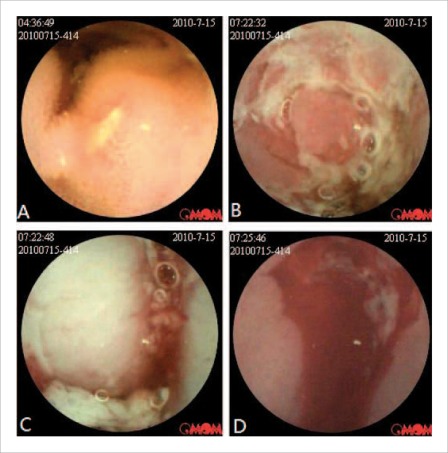
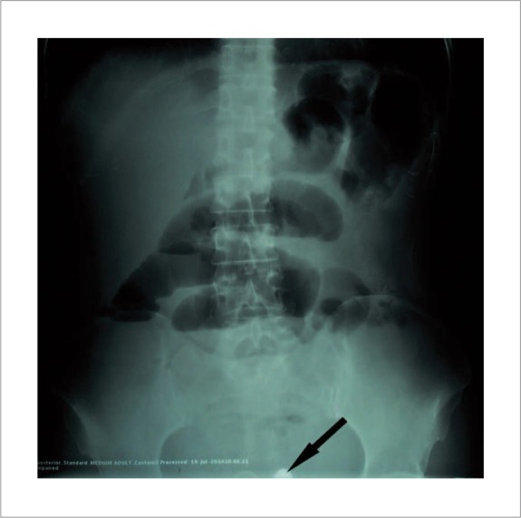
A 47-year-old woman received CE (OMOM capsule, Jinshan Science and Technology Company, Chongqing, China) for the evaluation of a 1 year history of recurrent diarrhea associated with mild abdominal pain. Esophagogastro-duodenoscopy and colonoscopy were negative. CE demonstrated the presence of multiple erosions and ulcers with strictures and active hemorrhage in the small bowel (Fig. 1), with a clinical suspicion of Crohn's disease (CD). The capsule failed to pass after three days and the patient had an acute episode of abdominal pain with abdominal distension. Plain x-ray of the abdomen and CT scan revealed fluid levels suggestive of partial small bowel obstruction (SBO) and the retained capsule (Fig. 2). The patient was admitted and treated conservatively.
Figure 1.
Capsule endoscopy revealed multiple ulcers (A), erosions (B) and strictures (C) with active hemorrhage (D) in small bowel, compatible with Crohn's disease.
Figure 2.
The abdominal plain film showed partial small bowel obstruction and retained capsule (pointed by arrow).
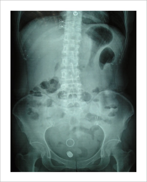
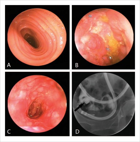
Symptoms improved after three days (Fig. 3) and an antegrade DBE (EN-450P5, Fujinon, Saitama, Japan) was attempted to retrieve the capsule. With the aid of the over tube, the enteroscope was advanced to 250 cm beyond the ligament of Treitz. Multiple small shallow ulcers were found in the upper jejunum and biopsies were taken. At the junction of jejunum and ileum, a significant stricture with cobble stone-like lesions and multiple ulcerations was found. Despite dilation with the over tube, the enteroscope could not be advanced and retrieval of the capsule failed. Fluoroscopy confirmed the capsule was located in the distal small bowel. Contrast injection through the enteroscope revealed a 10 cm stricture of with a narrowed diameter of 0.5 cm (Fig. 4).
Figure 3.
The abdominal plain film showed retained capsule and remission of small bowel obstruction after conservative treatment.
Figure 4.
Antegrade double-balloon enteroscopy revealed normal mucosa in upper jejunum (A), multiple small shallow ulcers (B) and a significant stricture (C). The fluoroscopy showed the retained capsule in the distal intestinal lumen (D).
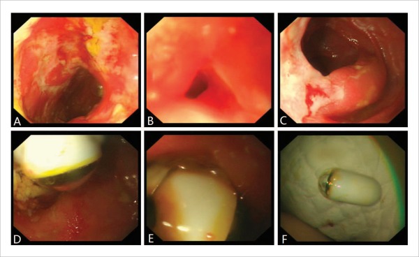
Nausea with vomiting and abdominal distention despite nasogastric decompression persisted on the fourth day, and a retrograde SBE (SIF-Q260, Olympus, Tokyo, Japan) was performed after a low enema (30mL 50% magnesium sulfate, 60mL glycerin and 90mL warm water). After inserting the enteroscope to 10 cm beyond the ileocecal valve, scattered ulcers and a significant stricture were observed. After gentle dilatation of the small bowel stricture, the retained capsule could be seen. It was grasped with a snare and removed by withdrawal of the enteroscope (Fig. 5).
Figure 5.
Retrograde single-balloon enteroscopy revealed scattered ulcers (A) and a significant stricture (B). After dilation, the stricture became accessible (C) and the retained capsule was revealed (D) and retrieved by snare (E, F).
After the procedure, there was complete relief of pain and resolution of bowel obstruction, with passage of flatus and feces. The biopsy results supported the diagnosis of CD. The patient was started on oral prednisone and mesalamine and a follow-up at 4 months showed good clinical response.
Discussion
Capsule retention is the most common complication of CE1, in addition to perforation, aspiration and SBO4–6. Surgical exploration is the most frequent treatment for capsule retention at 58.7%.1 However, balloon-assisted enteroscopy has now been shown to be an effective first-line treatment option that can prevent unnecessary surgery.3,7,8 When capsule retention occurs in patients with CD, medication such as intravenous steroids and infliximab may promote the successful excretion of the retained capsule.9,10 However, there are no proven benefits with steroid treatment in cases with SBO. In our patient, we decided to perform balloon-assisted enteroscopy after partial relief of symptoms to remove the retained capsule.
In general, antegrade enteroscopy is used for retrieval of the retained capsule.3 We reported the first case of removing a retained capsule in the terminal ileum using retrograde SBE after antegrade DBE failed. This approach is especially useful when the retained capsule is located in the distal small bowel.
Retained capsules often cause obstruction proximal to an obstructing lesion such as a Crohn's stricture.11 It is reasonable to adopt the antegrade approach initially rather than a retrograde approach when dealing with the retained capsule.3 However, our case provided additional information to the overall management. First, in some cases, the retained capsule is actually stuck between two major strictures after its spontaneous passage through the first one and it may be necessary to dilate the stricture before scope advancement. Secondly, in a patient with partial SBO, a low enema for bowel prep after resolution of the obstructive symptoms may be sufficient for retrograde enteroscopy.
There are more and more studies discussing the benefit of stricture dilation in the treatment of patients with symptomatic CD.12 However, there is an increased risk of perforation with balloon dilatation, especially in the cases with acute inflammation.12,13 In our case, we only used the over tube to gently dilate the stricture in order to minimize the risk of perforation. Retrograde enteroscopy may offer an alternative option for the removal of retained capsule. Further studies are necessary to determine the proper timing and bowel preparation.
Abbreviations
- CE
capsule endoscopy
- CD
Crohn's disease
- DBE
double-balloon enteroscopy
- SBE
single-balloon enteroscopy
- SBO
small bowel obstruction
Footnotes
Previously published online: www.landesbioscience.com/journals/jig
References
- 1.Liao Z, Gao R, Xu C, Li ZS. Indications and detection, completion, and retention rates of small-bowel capsule endoscopy: a systematic review. Gastrointest Endosc. 2010;71:280–286. doi: 10.1016/j.gie.2009.09.031. [DOI] [PubMed] [Google Scholar]
- 2.Cave D, Legnani P, de Franchis R, Lewis BS ICCE, author. ICCE consensus for capsule retention. Endoscopy. 2005;37:1065–1067. doi: 10.1055/s-2005-870264. [DOI] [PubMed] [Google Scholar]
- 3.Van Weyenberg SJ, Van Turenhout ST, Bouma G, Van Waesberghe JH, Van der Peet DL, Mulder CJ, et al. Double-balloon endoscopy as the primary method for small-bowel video capsule endoscope retrieval. Gastrointest Endosc. 2010;71:535–541. doi: 10.1016/j.gie.2009.10.029. [DOI] [PubMed] [Google Scholar]
- 4.Gonzalez Carro P, Picazo Yuste J, Fernandez Diez S, Perez Roldan F, Roncero Garcia-Escribano O. Intestinal perforation due to retained wireless capsule endoscope. Endoscopy. 2005;37:684. doi: 10.1055/s-2005-861424. [DOI] [PubMed] [Google Scholar]
- 5.Lin OS, Brandabur JJ, Schembre DB, Soon MS, Kozarek RA. Acute symptomatic small bowel obstruction due to capsule impaction. Gastrointest Endosc. 2007;65:725–728. doi: 10.1016/j.gie.2006.11.033. [DOI] [PubMed] [Google Scholar]
- 6.Repici A, Barbon V, De Angelis C, Luigiano C, De Caro G, Hervoso C, et al. Acute small-bowel perforation secondary to capsule endoscopy. Gastrointest Endosc. 2008;67:180–183. doi: 10.1016/j.gie.2007.05.044. [DOI] [PubMed] [Google Scholar]
- 7.Al-toma A, Hadithi M, Heine D, Jacobs M, Mulder C. Retrieval of a video capsule endoscope by using a double-balloon endoscope. Gastrointest Endosc. 2005;62:613. doi: 10.1016/j.gie.2005.04.006. [DOI] [PubMed] [Google Scholar]
- 8.Tanaka S, Mitsui K, Shirakawa K, Tatsuguchi A, Nakamura T, Hayashi Y, et al. Successful retrieval of video capsule endoscopy retained at ileal stenosis of Crohn's disease using double-balloon endoscopy. J Gastroenterol Hepatol. 2006;21:922–923. doi: 10.1111/j.1440-1746.2006.04120.x. [DOI] [PubMed] [Google Scholar]
- 9.O'Donnell S, Qasim A, Ryan BM, O'Connor HJ, Breslin N, O Morain CA. The role of capsule endoscopy in small bowel Crohn's disease. J Crohns Colitis. 2009;3:282–286. doi: 10.1016/j.crohns.2009.07.002. [DOI] [PubMed] [Google Scholar]
- 10.Vanfleteren L, van der Schaar P, Goedhard J. Ileus related to wireless capsule retention in suspected Crohn's disease: emergency surgery obviated by early pharmacological treatment. Endoscopy. 2009;41:E134–E135. doi: 10.1055/s-0029-1214665. [DOI] [PubMed] [Google Scholar]
- 11.Cheifetz AS, Lewis BS. Capsule endoscopy retention: is it a complication? J Clin Gastroenterol. 2006;40:688–691. doi: 10.1097/00004836-200609000-00005. [DOI] [PubMed] [Google Scholar]
- 12.Despott EJ, Gupta A, Burling D, Tripoli E, Konieczko K, Hart A, et al. Effective dilation of small-bowel strictures by double-balloon enteroscopy in patients with symptomatic Crohn's disease. Gastrointest Endosc. 2009;70:1030–1036. doi: 10.1016/j.gie.2009.05.005. [DOI] [PubMed] [Google Scholar]
- 13.Mensink PB, Haringsma J, Kucharzik T, Cellier C, Pérez-Cuadrado E, Mönkemüller K, et al. Complications of double balloon enteroscopy: a multicenter survey. Endoscopy. 2007;39:613–615. doi: 10.1055/s-2007-966444. [DOI] [PubMed] [Google Scholar]


