Abstract
Information on how cells interface with nanomaterials in biological environments has important implications for the practice of nanomedicine and safety consideration of nanomaterials. However, our current understanding of nanobiological interactions is still very limited. Here, we report the direct observation of nanomaterial bio-complex formation (other than protein corona) from nanomaterials dispersed in biologically relevant solutions. We observed highly selective binding of the components of cell culture medium and phosphate buffered saline to ZnO and CuO nanoparticles, independent of protein molecules. Our discoveries may provide new insights into the understanding of how cells interact with nanomaterials.
Nanotechnology has held great promise for revolutionizing biomedicine. Nanomaterials (NMs) are widely being under investigation for bioimaging1, killing tumors2, delivering drugs/genes3,4. The response of biological systems to NMs depends primarily on the surface properties of NMs in a biological environment5,6. For example, there is increasing evidence of rapid formation of protein coronas, as NMs within biological environments acquire a coating of protein molecules7,8,9,10,11, and thus there is some consensus that cellular responses to materials in a biological medium reflect the adsorbed biomolecule layer, rather than the material itself. The concept of the nanoparticle (NP) protein corona is important towards understanding the dynamic surface properties of NMs in biological environments. Unfortunately, the interface issues between NMs and biological systems are too complicated and it is still not very clear how the spontaneous formation of a protein corona influences the materials' interactions with biological systems and contributes to biological outcomes of materials. On the other hand, the increasing use of NMs in industrial and consumer products has aroused global concern regarding their potential impact on the environment and human health. Although a number of studies on the effects of NMs in in vitro and in vivo systems have been published12,13,14,15,16,17, there are still many challenges and issues12,13,14 encountered at the interface between NMs and biological systems when assessing the toxicity of NMs. As demonstrated here the state of NMs in biological environments is more complicated than previously thought, requiring a more holistic understanding of what happen as NMs are dispersed in the local biological environments and what cells interface in such an environment. Not only does the biological interface of NMs need to be understood and controlled, but also NMs should be treated as biological entities rather than inorganic ones6. The understanding of nano-bio interactions could help in promoting applications of NMs in the biomedical fields and reducing/preventing possible adverse effects to the biological systems caused by NMs.
Here, we present the first report in nanobiological field, to our knowledge, describing nanomaterial (NM) bio-complexes or ion corona formed from the binding of NMs with non-protein components in a biological environment. We observed highly selective binding of the components of cell culture medium and phosphate buffered saline (PBS) to ZnO and CuO nanoparticles (NPs), independent of protein molecules. Our findings would be helpful to understand nano-bio-interactions, allowing to effectively modifying NM surfaces for specifically biomedical applications.
Results
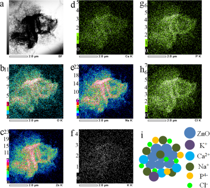
We observed that ZnO and CuO NPs can bind various constituents from biological environments to form NM bio-complexes or ion corona. The reason that we call the formed clusters as bio-complex is that the NMs are used for biological applications, though it may not involve biological molecules. Figure 1 presents the transmission electron microscopy (TEM) elemental maps of ZnO NPs dispersed in high-glucose Dulbecco's modified Eagle's medium (DMEM) without fetal bovine serum (FBS). In addition to aggregation/agglomeration of the NPs15, the elemental maps clearly illustrate the binding of ZnO NPs to components of the medium to form NP bio-complexes. We detected Ca, Na, K, P, and Cl originating from the medium (components of DMEM are listed in Supplementary Information). For comparison, we examined the effects of FBS and PBS on the formation of NP bio-complexes. High-resolution TEM images (Fig. 2a and Supplementary Fig. S1) show that in medium supplemented with 10% FBS the crystalline ZnO NPs (see Supplementary Fig. S1) are surrounded by compounds, indicating NP bio-complex formation. Furthermore, we found that these bio-complexes also formed when the ZnO NPs were dispersed in PBS, either with (Supplementary Figs. S2–3) or without FBS (Fig. 2b). We confirmed that the observed highly ordered crystalline structures are ZnO NPs because no crystalline structures were detected in the control suspensions where no ZnO NP was dispersed in medium or PBS (Supplementary Figs. S4–7, S10). These results clearly suggest that the formation of NP bio-complexes in biological environments (medium and PBS) occurs independently of serum proteins. The formation of protein coronas and the adsorption of different proteins onto NPs affect the uptake and transport of NPs in biological systems7,8,9,10,11. We believe that the biological behavior of NPs could be similarly modified by the formation of NP bio-complexes.
Figure 1. Formation of ZnO nanoparticle bio-complexes in medium without fetal bovine serum.
(a) TEM image showing where the elemental maps were obtained; (b) TEM/EDS O-K map; (c) TEM/EDS Zn-K map; (d) TEM/EDS Ca-K map; (e) TEM/EDS Na-K map; (f) TEM/EDS K-K map; (g) TEM/EDS P-K map; (h) TEM/EDS Cl-K map; (i) A simple model of ZnO bio-complexes.
Figure 2. Surrounded nanoparticles.
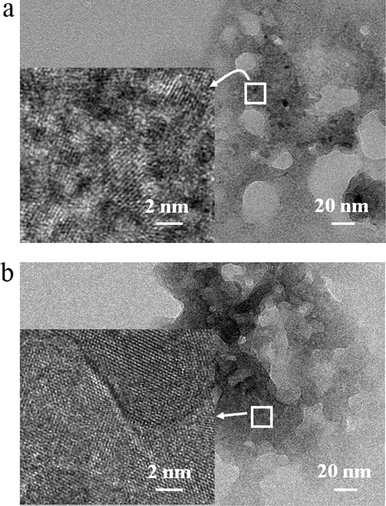
(a) TEM image of ZnO NPs dispersed in medium supplemented with fetal bovine serum; (b) TEM image of ZnO NPs dispersed in PBS without fetal bovine serum. The inset high-resolution TEM images clearly suggest that ZnO NP (dark-colored) bio-complexes were formed.
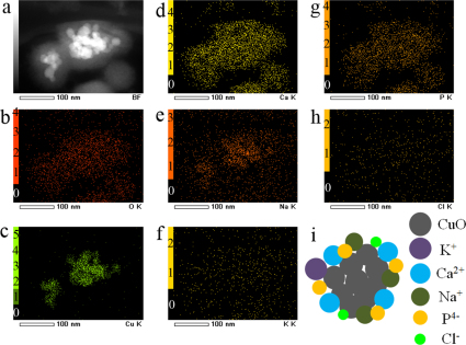
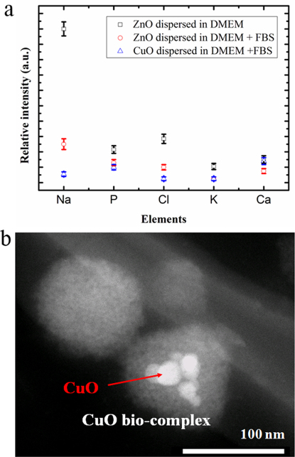
Figure 3 displays the TEM elemental maps of CuO NPs dispersed in the DMEM supplemented with 10% FBS. By comparing the relative contrast of the maps in Fig. 1 and Supplementary Fig. S1 to that in Fig. 3, we determined that the ZnO and CuO NPs exhibited different binding affinities for the various components of the medium. As plotted in Fig. 4a, in the case of the NPs dispersed in DMEM with FBS, the ZnO NPs bound more Na than Ca and more P than Cl, whereas the CuO NPs bound more Ca than Na and more P than Cl. This selective adsorption may be similar to that seen in the formation of protein coronas, which is reported to be influenced by particle chemistry9. Furthermore, FBS has effort on the binding behavior of ions as we can see from Fig. 4a that the ZnO NPs which were dispersed in DMEM with FBS bound more Cl than P, indicating biological environment may also influence the binding process. In addition, as highlighted in Fig. 4b, we observed NP-like clusters that apparently do not contain CuO or ZnO NPs; we believe they arose from components of the cell culture medium. Hence, the bio-complexes and the NP-like clusters can influence size distribution of particles in the biological systems, suggesting caution should be taken as addressing size effects of NMs on biological behaviors.
Figure 3. Formation of CuO nanoparticle bio-complexes in medium with fetal bovine serum.
(a) Dark-field TEM image showing where the elemental maps were obtained; (b) TEM/EDS O-K map; (c) TEM/EDS Zn-K map; (d) TEM/EDS Ca-K map; (e) TEM/EDS Na-K map; (f) TEM/EDS K-K map; (g) TEM/EDS P-K map; (h) TEM/EDS Cl-K map; (i) A simple model of CuO bio-complexes.
Figure 4. Selective binding of ions by ZnO and CuO NPs.
(a) Scattering plot of bound elements by ZnO and CuO NPs from the DMEM or DMEM with FBS, comparatively showing different binding affinities for the various components of the DMEM or DMEM with FBS by the NPs. The standard deviation error bars were based on the results obtained at three different sample regions; (b) Dark-field TEM image of CuO NPs suspension (25 µg/mL) in medium with FBS, showing nanoparticle-like clusters that apparently do not contain CuO NPs.
To confirm the selective binding of ions by the NPs, we used X-ray photoelectron spectroscopy (XPS) to investigate the ensemble of ZnO NPs and CuO NPs after dispersed in the DMEM or DMEM with FBS. Table 1 summarizes the atomic concentrations of the main elements bound by the NPs. In the case of the NPs ensemble dispersed in the DMEM with FBS, the orders of estimated atomic concentration of elements bound by ZnO NPs and by CuO NPs are Na1s (2.8%) > P2s (0.8%) > Ca2p (0.7%) > Cl2p (0.6%) and Na1s (2.6%) > Ca2p (1.8%) > P2s (1.0%) > Cl2p (0.5%), respectively. From Table 1, we also found that FBS affected the ion binding as compared the ZnO NPs dispersed in DMEM to that dispersed in DMEM with FBS. These XPS results are in good agreement with the TEM observation on the individual NPs.
Table 1. Atomic concentration (at%) of main elements estimated on the basis of the wide XPS spectra of ZnO and CuO NPs dispersed in DMEM or DMEM with FBS.
| N1s | O1s | Na1s | P2s | Cl2p | K2p | Ca2p | Zn2p3/2 | Cu2p3/2 | |
|---|---|---|---|---|---|---|---|---|---|
| #1 | 2.4 | 45.6 | 9.4 | 1.7 | 0.7 | 0.2 | 2.2 | 19.5 | |
| #2 | 9.8 | 28.0 | 2.8 | 0.8 | 0.5 | 0.1 | 0.7 | 5.4 | |
| #3 | 10.3 | 30.1 | 2.6 | 1.0 | 0.5 | 0.1 | 1.8 | 5.9 |
#1, #2 and #3 represent ZnO NPs dispersed in DMEM, ZnO NPs dispersed in DMEM with FBS and CuO NPs dispersed in DMEM with FBS, respectively.
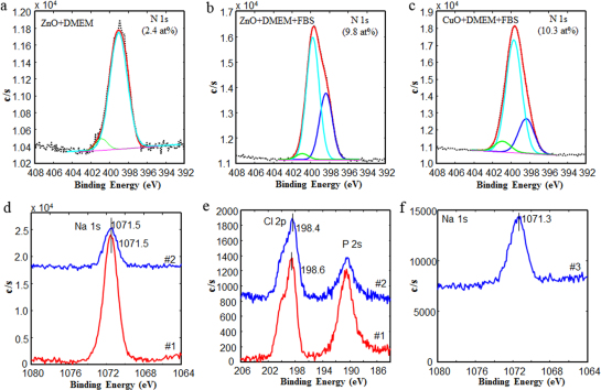
Although we did not detect obvious signal of N, a main component of protein, by TEM, we detected it by XPS due to much lower energy of the used X-ray than the electron beam used for TEM observation. The N1s atomic concentration of the ZnO NPs dispersed in DMEM with FBS is much higher than that of the ZnO NPs dispersed in DMEM. Furthermore, the binding energy of one of the fit peaks of N1s spectra shifted to 399.76 eV (Fig. 5b) or 399.70 eV (Fig. 5c) from 399.41 eV of the pristine FBS (Supplementary Fig. S15b and Table S1). The shift suggests protein molecules in the FBS may bind to the NPs, forming protein corona.
Figure 5. XPS characterization of selectively bound ions by ZnO and CuO NPs.
(a) N1s curve fit of ZnO NPs dispersed in DMEM; (b) N1s curve fit of ZnO NPs dispersed in DMEM with FBS; (c) N1s curve fit of CuO NPs dispersed in DMEM with FBS; (d) Na1s spectra of ZnO NPs dispersed in DMEM (#1) and in DMEM with FBS (#2); (e) Cl2p spectra of ZnO NPs dispersed in DMEM (#1) and in DMEM with FBS (#2); (f) Na1s spectra of CuO NPs dispersed in DMEM with FBS (#3). Compared to the binding energies of the elements of the pristine DMEM and FBS, the binding energy positions in the spectra were shifted.
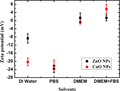
As shown in Figs. 5d–f and Supplementary Figs. S15c–d, we detected different shifts, toward a lower binding energy in contrast to a higher binding energy of the N1s (Figs. 5b–c), of the Na1s and Cl2p binding energies as they contacted with ZnO NPs in DMEM or DMEM with FBS from the pristine DMEM and FBS. The different shift may indicate various interactions between the ions and ZnO NPs in the different solution environments. The various interactions reflect the change of zeta potential of the NPs dispersed in the different solvents as shown in Fig. 6. For instance, the zeta potential of ZnO NPs changed from a negative value in de-ionized water (DI water) to a positive value as dispersed in DMEM or DMEM with FBS. The change of zeta potential of ZnO and CuO NPs is believed to be due to the binding of ions from the solvents, which is consistent with the selective binding of ions by the ZnO and CuO NPs.
Figure 6. Various zeta potentials of ZnO and CuO NPs dispersed in different solvents.
The change of zeta potential suggests ZnO and CuO NPs bound various ions in the solutions. The error bars represent standard deviation based on three independent measurements of each sample.
Discussion
We have shown that ZnO and CuO NPs formed bio-complex or ion corona by selectively absorbing components or ions from the DMEM, DMEM supplemented with FBS, and PBS. The formation of bio-complexes is not associated with serum proteins. The discovery of bio-complex formation offers new insights for interpreting the observation that the uptake of negatively charged NPs by cells is greater than that of non-functionalized NPs18,19. This phenomenon is contradictory to the well-established concept that positively charged NPs have high affinity to the negatively charged cell membrane, and the unfavorable interaction was partially attributed to the formation of NP protein corona19. For instance, it has recently been shown that negatively charged Au NPs are more toxic than positively charged and neutral Au NP counterparts, due to the interaction of the outer Au NP components with cellular components20. The interaction of the negatively charged Au NPs with cellular components could in turn influence the mitochondrial membrane potential by releasing positively charged calcium ions from the matrix into the cytosol20. Thus, previous work related to the effects of surface charge and surface modification on cell-specific responses and cytotoxicity21,22,23 may need to be re-evaluated. That is, the surface may not the one as expected from the intended surface modification. Our finding that NP bio-complexes form in biological environments could be used to help design the NP surfaces for effective drug, DNA and siRAN delivery systems and more accurate nanotoxicity studies.
We did not detect signals from key components of serum proteins, such as N, due to the high energy of the electron beam used in the TEM technique. However, we did observe the disappearance of certain matters during the TEM observation because biomolecules can be easily damaged by high energy beam, which may have been the serum proteins. Previously, Maiorano et al. made the assumption that an observed cloud surrounding Au NPs was caused by proteins, based on TEM observations without corresponding elemental maps24. Despite this assumption, protein coronas can mediate cellular responses6 and the toxic effects of NMs24,25. We believe that identification of the constituents of NP bio-complexes would significantly improve our understanding of what is selectively acquired by NMs in biological environments; how those acquired constituents influence the reactivity, transport, transformation, bioavailability, and toxicity of NMs; and how the NMs interact with living cells26,27.
Although there is implication that the binding components from cellular environment had great effects on the toxicity of Au NPs20, it is currently impossible to describe with certainty all the interactions play at the nano-bio interface in biological environments. The concept of the NP protein corona is important in understanding the dynamic surface properties of NMs in biological fluids. However, even at this moment, it is less clear how the spontaneous formation of a protein corona influences the materials' interactions with biological surfaces and receptors and contributes to biological outcomes of materials. Although it is not surprising that NPs would aggregate in salt rich solutions as this is well known from colloidal theory, the present observation of NMs bio-complexes is certainly new, and such kind of ion corona may be assumed as protein corona if there is no elemental identification24. We observed the formation of bio-complexes after naturally drying of the ZnO and CuO NPs immersed in the biological solutions. It remains unclear how dynamically biological environments influence the attaching and detaching of the components from the solution, which is under investigation. Despite challenges to probe events happened at the nano-biointerface, the basic principles of nano-biointerface are being investigated by establishing new imaging techniques, such as TEM and scanning probe microscopy, and biological approaches5. The well known colloid chemistry and physics might be considered to be helpful to understand the formation of bio-complexes, however, the behaviors in a biological environment is conceptually different from the well-known colloidal phenomena6. By contrast, the methods for protein corona investigation may be extended to explore dynamics of bio-complexes26,27.
In summary, we have discovered the formation of NM bio-complexes originating from the adsorption of various components in a biological environment onto NMs, independent of proteins. The interaction between NMs and the local environment adds complexity to the challenge of determining the biological outcome of using NMs. Thus, identification of the constituents of these bio-complexes would provide new insights into our understanding of how NMs are modified in biological environments and what cells really see. These results are expected to improve our understanding of the interactions between NMs and live cells. The results of our study are a reminder that our understanding of the nano-bio interactions is still in its infancy.
Methods
Nanoparticles
High-purity ZnO and CuO NPs were obtained from Sigma-Aldrich (Sigma-Aldrich Co., Japan). The primary dimensions and sizes of the oxides were provided in the data sheet, as determined by X-ray diffraction (XRD) and Brunauer–Emmett–Teller (BET) analysis. The average sizes of the ZnO and CuO NPs were 60 nm and 50 nm, respectively, but were not uniform, as previously observed by electron microscopy16. In contrast to Au and silica NPs, it is almost impossible to synthesize homogeneous ZnO or CuO NPs with identical shape and size. We characterized the primary size, shape, and composition of the NPs by scanning electron microscopy (SEM) equipped with energy dispersive spectroscopy (EDS) (JSM-7001F, JEOL Inc., Japan) and by X-ray photoelectron spectroscopy (XPS) (PHI Quantera SXM, ULVAC-PHI, Japan)16. Note that here we used the averaged primary size as many reports by others17. In the regard of varying size of the ZnO and CuO NPs, our observed formation of bio-complex is independent on the size of materials.
Dispersion solvents
DMEM (pH = 7.4) (Invitrogen,) with or without 10% (v/v) FBS (SAFC Bioscience Inc.,) was supplemented with 100 units/mL penicillin and 100 µg/mL streptomycin. The composition of DMEM can be found in the Supplementary Information section. Phosphate buffer saline (PBS) contains NaHPO4, Na2HPO4, and NaCl.
Nanoparticle suspension preparation
ZnO and CuO were dispersed in medium with FBS, medium without FBS, PBS, or PBS with FBS. The concentration of the NP suspensions was 25 µg/mL.
TEM characterization
The dark-field TEM images, high-resolution TEM (HRTEM) images, elemental mapping, and EDS were analyzed using a JEM-2100F high-resolution transmission electron microscope (JEOL Inc., Japan) with acceleration voltage of 200 kV. The samples for TEM observation were prepared by immersing TEM mesh into the NP suspension (25 µg/mL) for 24 h at 37°C in the dark with 5% CO2, the typical cell culture conditions. After naturally drying in air, the TEM mesh was observed.
XPS characterization
PHI Quantera SXM (ULVAC-PHI) with monochromatic Al Kα X-ray (1.5×0.1 mm, 100 W) (pass energy of 55 eV and step of 0.2 eV for narrow spectrum and pass energy of 280 eV and step of 0.5 eV for survey spectrum) was used. Binding energy corrections for the spectra were carried out by using C1s peaks at 285.0 eV. The sample for XPS investigation was prepared by immersing Au substrate into the suspension of NPs in DMEM or DMEM with FBS.
Zeta potential
The surface zeta potentials of the ZnO NPs and CuO NPs were measured by using a laser electrophoresis zeta-potential analyzer (LEZA-600, Otsuka Electronics, Japan). The NPs were dispersed in the solvents with a concentration of 25 μg/mL by sonication for ∼60 min to reduce aggregation/agglomeration. The cell unit of the equipment was washed by using the same solvent as that for dispersing NPs prior to measurement. We measured three times for each sample.
Author Contributions
M.S.X. originated the work and designed the experiments. M.S.X. performed the experiments, analyzed the data and prepared the manuscript. J.L. and N.H. performed zeta potential experiments. H.I. performed XPS measurement and data analysis. Q.S.M. carried out XRD measurement and data analysis. All discussed the results.
Supplementary Material
Supplementary information
Acknowledgments
This work was partially supported by the National Natural Science Foundation of China (Nos. 51011130028, 50990063 and 50973095), by Zhejiang Provincial Natural Science Foundation of China (Youth Talent Program: R4110030), Science and Technology Department of Zhejiang Provincial (Qianjiang Talent Program: 2011R10077), the Fundamental Research Funds for the Central Universities of China (No.: 2011QNA4030), and by the Interdisciplinary Laboratory for Nanoscale Science and Technology & Material Analysis Station, National Institute for Materials Science (NIMS), Japan. We thank Y. Nemoto and H. Gao for assistance with TEM measurements.
References
- Chatterjee D. K., Gnanasammandhan M. K. & Zhang Y. Small upconverting fluorescent nanoparticles for biomedical applications. Small 6, 2781–2795 (2010). [DOI] [PubMed] [Google Scholar]
- Kudgus R. A., Bhattacharya R. & Mukherjee P. Cancer nanotechnology: emerging role of gold nanoconjugates. Anticancer Agents Med. Chem. 11, 965–973 (2011). [DOI] [PMC free article] [PubMed] [Google Scholar]
- Farokhzad O. C. & Langer R. Impact of nanotechnology on drug delivery. ACS Nano 3, 16–20 (2009). [DOI] [PubMed] [Google Scholar]
- Chen A. M., Taratula O., Wei D. G., Yen H. I., Thomas T., Thomas T. J., Minko T. & He H. X. Labile catalytic packaging of DNA/siRNA: control of gold nanoparticles “out” of DNA/siRNA complexes. ACS Nano 4, 3679–3688 (2010). [DOI] [PMC free article] [PubMed] [Google Scholar]
- Nel A. E., Mädler L., Velegol D., Xia T., Hoek E. M. V., Somasundaran P., Klaessig F., Castranova V. & Thompson M. Understanding biophysicochemical interactions at the nano–bio interface. Nat. Mater. 8, 543–557 (2009). [DOI] [PubMed] [Google Scholar]
- Park S. & Hamad-Schifferli K. Nanoscale interfaces to biology. Curr. Opin. Chem. Biol. 14, 616–622 (2010). [DOI] [PMC free article] [PubMed] [Google Scholar]
- Keselowsky B. G., Collard D. M. & Garcia A. J. Surface chemistry modulates focal adhesion composition and signaling through changes in integrin binding. Biomaterials 25, 5947–5954 (2004). [DOI] [PubMed] [Google Scholar]
- Cedervall T., Lynch I., Lindman S., Berggard T., Thulin E., Nilsson H., Dawson K. A. & Linse S. Understanding the nanoparticle-protein corona using methods to quantify exchange rates and affinities of proteins for nanoparticles. Proc. Natl. Acad. Sci. U.S.A. 104, 2050–2055 (2007). [DOI] [PMC free article] [PubMed] [Google Scholar]
- Lunqvist M., Stigler J., Elia G., Lynch I., Cedervall T. & Dawson K. A. Nanoparticle size and surface properties determine the protein corona with possible implications for biological impact. Proc. Natl. Acad. Sci. U.S.A. 105, 14265–14270 (2008). [DOI] [PMC free article] [PubMed] [Google Scholar]
- Ehrenberg M. S., Friedman A. E., Finkelstein J. N., Oberdorster G. & McGrath J. L. The influence of protein adsorption on nanoparticle association with cultured endothelial cells. Biomaterials 30, 603–610 (2009). [DOI] [PubMed] [Google Scholar]
- Mahmoudi M., Lynch I., Ejtehadi M. R., Monopoli M. P., Bombelli F. B. & Laurent S. Protein-nanoparticle interactions: opportunities and challenges. Chem. Rev. 111, 5610–5637 (2011). [DOI] [PubMed] [Google Scholar]
- Maynard A. D., Warheit D. B. & Philbert M. A. The new toxicology of sophisticated materials: nanotoxicicology and beyond. Toxicol. Sci. 120, S109–S129 (2011). [DOI] [PMC free article] [PubMed] [Google Scholar]
- Frug H. F. & Wick P. Nanotoxicology: an interdisciplinary challenge. Angew. Chem. Int. Ed. 50, 1260–1278 (2011). [DOI] [PubMed] [Google Scholar]
- Warheit D. B. Debunking some misconceptions about nanotoxicology. Nano Lett. 10, 4777–4782 (2010). [DOI] [PubMed] [Google Scholar]
- Jiang J., Oberdörster G. & Biswas P. Characterization of size, surface charge, and agglomeration state of nanoparticle dispersions for toxicological studies. J. Nanopart. Res. 11, 77–89 (2009). [Google Scholar]
- Xu M. S., Fujita D., Kajiwara S., Minowa T., Li X. L., Takemura T., Iwai H. & Hanagata N. Contribution of physicochemical characteristics of nano-oxides to cytotoxicity. Biomaterials 31, 8022–8031 (2010). [DOI] [PubMed] [Google Scholar]
- Xia T., Kovochich M., Liong M., Madler L., Gilbert B., Shi H., Yeh J. I., Zink J. I. & Nel A. E. Comparison of the mechanism of toxicity of zinc oxide and cerium oxide nanoparticles based on dissolution and oxidative stress properties. ACS Nano 2, 2121–2134 (2008). [DOI] [PMC free article] [PubMed] [Google Scholar]
- Villanueva A., Canete M., Roca A. G., Calero M., Veintemillas-Verdaguer S., Serna C. J., Morales M. D. & Miranda R. The influence of surface functionalization on the enhanced internalization of magnetic nanoparticles in cancer cells. Nanotechnology 20, 115103 (2009). [DOI] [PubMed] [Google Scholar]
- Verma A. & Stellacci F. Effect of surface properties on nanoparticle-cell interactions. Small 6, 12–21 (2010). [DOI] [PubMed] [Google Scholar]
- Schaeublin N. M., Braydich-Stolle L. K., Schrand A. M., Miller J. M., Hutchison J., Schlager J. J. & Hussain S. M. Surface charge of gold nanoparticles mediates mechanism of toxicity. Nanoscale 3, 410–420 (2011). [DOI] [PubMed] [Google Scholar]
- Asati A., Santra S., Kaittanis C. & Perez J. M. Surface-charge-dependent cell localization and cytotoxicity of cerium oxide nanoparticles. ACS Nano 4, 5321–5331 (2010). [DOI] [PMC free article] [PubMed] [Google Scholar]
- Bhattacharjee S., de Haan L. H. J., Evers N. M., Jiang X., Marcelis A. T. M., Zuilhof H., Rietjens I. M. C. M. & Alink G. M. Role of surface charge and oxidative stress in cytotoxicity of organic monolayer-coated silicon nanoparticles towards macrophage NR8383 cells. Particle Fibre Toxicol. 7, 25 (2010). [DOI] [PMC free article] [PubMed] [Google Scholar]
- Thevenot P., Cho J., Wavhal D., Timmons R. B. & Tang L. Surface chemistry influences cancer killing effect of TiO2 nanoparticles. Nanomed. 4, 226–236 (2008). [DOI] [PMC free article] [PubMed] [Google Scholar]
- Maiorano G., Sabella S., Sorce B., Brunetti V., Malvindi M. A., Cingolani R. & Pompa P. P. Effects of cell culture media on the dynamic formation of protein−nanoparticle bio-conjugates and influence on the cellular response. ACS Nano 4, 7481–7491 (2010). [DOI] [PubMed] [Google Scholar]
- Hu W., Peng C., Lv M., Li X., Zhang Y., Chen N., Fan C. & Huang Q. Protein corona-mediated mitigation of cytotoxicity of graphene oxide. ACS Nano 5, 3693–3700 (2011). [DOI] [PubMed] [Google Scholar]
- Casals E., Pfaller T., Duschl A., Oostingh G. J. & Puntes V. Time evolution of the nanoparticle protein corona. ACS Nano 4, 3623–3632 (2010). [DOI] [PubMed] [Google Scholar]
- Walczyk D., Bombelli F. B., Monopoli M. P., Lynch I. & Dawson K. A. What the cell “sees” in bionanoscience, .J. Am. Chem. Soc. 132, 5761–5768 (2010). [DOI] [PubMed] [Google Scholar]
Associated Data
This section collects any data citations, data availability statements, or supplementary materials included in this article.
Supplementary Materials
Supplementary information


