Abstract
Introduction:
To evaluate the hepatoprotective activity of active phytochemicals, picroliv, curcumin, and ellagic acid in comparison to silymarin in the mice model of carbon tetrachloride (CCl4) induced liver toxicity. In addition, attempts were made to elucidate their possible mechanism(s) of action.
Materials and Methods:
Oxidative stress was induced in Swiss albino mice by a single injection (s.c.) of CCl4, 1 ml/kg body weight, diluted with arachis oil at a 1:1 ratio. The phytochemicals were administered once a day for 7& days (p.o.) as pretreatment at two dose levels (50 and 100 mg/kg/day).
Results:
CCl4-induced hepatotoxicity was manifested by an increase in the activities of liver enzymes (alanine transaminase, P < 0.001, aspartate transaminase, P < 0.001 and alkaline phosphatase, P < 0.001), malondialdehyde (MDA, P < 0.001)) levels and a decrease in activity of reduced glutathione (P < 0.001) and catalase in liver tissues. The histopathological examination of liver sections revealed centrizonal necrosis, fatty changes, and inflammatory reactions. The pretreatment with picroliv, curcumin, and ellagic acid normalized serum aminotransferase activities (P < 0.001), decreased levels of MDA (P < 0.001), improved the antioxidant status, and normalized the hepatic histo-architecture. The restoration of phenobarbitone-induced sleeping time also suggested the normalization of liver cytochrome P450 enzymes.
Conclusion:
This study supports the use of these active phytochemicals against toxic liver injury, which may act by preventing lipid peroxidation, augmenting the antioxidant defense system or by regenerating the hepatocytes.
Keywords: Hepatotoxicity, herbal drugs, lipid peroxidation, liver enzymes
INTRODUCTION
Liver diseases are one of the major causes of mortality and morbidity worldwide.[1] Drug-induced liver toxicity is a major cause of hepatic dysfunction.[1] An estimated 1000 drugs have been implicated in causing liver injury, and it is the most frequent reason for withdrawing approved drugs from the market. It accounts for 50% of the cases of acute liver failure. Drug-induced liver failure can mimic all forms of naturally occurring acute and chronic hepatobiliary diseases.[1,2]
Oxidative stress is considered as a mechanism in contributing to the initiation and progression of hepatic damage in a variety of liver disorders. Cell damage occurs when there is an excess of reactive species derived from oxygen and nitrogen or deficiency of antioxidants.[2,3]
Antioxidants obtained from plants represent a logical therapeutic strategy for treatment of liver diseases. There are many plant derived chemicals with potent antioxidant properties which can serve as primary compounds for development as hepatoprotective drugs.[3,4] Picroliv is a standardized iridoid glycoside mixture isolated from the roots and rhizomes of the plant Picrorrhiza kurroa. It contains at least 60% of a 1:1.5 mixture of picroside I and kutkoside; the remainder (40%) is a mixture of iridoid as well as cucurbitacin glycosides. Picroliv was reported to possess hepatoprotective activities by their anti-lipid peroxidative and free radical scavenging properties.[5] Curcumin is the major biologically active phenolic compound from Curcuma longa with strong antioxidant, anti-inflammatory, and hepatoprotective activities.[3] Ellagic acid is a polyphenolic compound found in grapes, strawberries, black currants, and raspberries, which have potent antioxidant property. Ellagic acid has been reported to reduce the production of hydroxyproline and fibrous connective tissue formation indicating its antifibrotic activity.[6] Ellagic acid also slowed down the conversion of hepatic stellate cells (HSC) into their activated forms, which produce extracellular matrix and result in liver fibrosis.[6] Although individual reports are available on the hepatoprotective activities of picroliv, curcumin, and ellagic acid, their relative efficacy is unknown. This study will enable us to select the most effective lead compound for further development as hepatoprotective drugs. Silymarin, a flavonolignan obtained from Silybum marianum, the most researched hepatoprotective agent, is used as the comparator drug.[4]
Oxidative stress caused by the highly reactive metabolite produces depletion of glutathione, and through nuclear factor-kappa B (NF-κB) induced proinflammatory cytokines and chemokines, cyclooxygenase-2 (COX-2) and inducible nitric oxide synthase (iNOS). Inflammatory liver injury is caused by tumor necrosis factor alpha (TNF-α), interleukin-12 (IL-12), monocyte chemotactic protein-1 (MCP-1), and macrophage inflammatory protein-2 (MIP-2). TNF-α in turn leads to apoptosis and cell death.[7] It is reported that ellagic acid can inhibit NADPH oxidase-induced overproduction of superoxide, suppress the release of nitric oxide by down-regulating iNOS, enhance cellular antioxidant defences, and attenuate oxidized LDL-induced lipoxygenase-1 up-regulation and endothelial nitric oxide synthase down-regulation.[8] Therefore, it is speculated that phytochemicals such as curcumin, ellagic acid, and picroliv can act at one or several sites of these cascade of events.[9]
In this study, we have attempted to evaluate the hepatoprotective activity of picroliv, curcumin, and ellagic acid in comparison to silymarin, in carbon tetrachloride (CCl4) induced acute liver toxicity in mice. We also aimed to study whether the antioxidant properties of these phytochemicals have a role in their hepatoprotective actions.
MATERIALS AND METHODS
Drugs and chemicals
All the chemicals and reagents used for this study were of analytical grade. Silymarin was purchased from Microlabs (Pondicherry, India), curcumin from Sigma (St. Louis, MO, USA), ellagic acid from Fluka (USA), and picroliv were kindly gifted by Dr. S. Singh, CDRI, Lucknow, India. CCl4 was obtained from Merck Ltd (Mumbai, India).
Animals
Swiss albino mice (20–30 g) of either sex, bred in Central Animal House, JIPMER, Pondicherry, were used for the study. The animals were allowed standard food pellets (Hindustan Lever Ltd., Mumbai) and water ad libitum and were maintained in standard laboratory conditions (12 h:12 h dark and light cycle and 25±2 °C temperature). The study was approved by ‘The Institute Animal Ethics Committee’, JIPMER, Pondicherry and was done according to the ethical guidelines laid down by Committee for the Purpose of Control and Supervision of Experiment on Animals.
Treatment groups
The animals were divided into 30 groups (n = 6/group) with 10 groups for biochemical (estimation of serum levels of liver enzymes, namely alanine transaminase (ALT), aspartate transaminase (AST), and alkaline phosphatase (ALP)) and histopathological parameters, 10 groups for pharmacological parameters (phenobarbitone-induced sleeping time) and another 10 for oxidative stress parameters (malondialdehyde (MDA), reduced glutathione (GSH), and catalase).
Group 1: Animals received distilled water for 7 days and served as normal control.
Group 2: The animals in this group received distilled water as in the previous group and given CCl4, 1 ml/kg body weight diluted with arachis oil at 1: 1 ratio, s.c., once on day 8.
Groups 3–6: Four phytochemicals, picroliv, curcumin, ellagic acid, and silymarin were administered once daily for 7 days p.o. (50 mg/kg body weight/day) followed by a single s.c. dose of CCl4 (1 ml/kg body weight) on day 8.
Groups 7–10: A double dose pretreatment (p.o. 100 mg/kg body weight/day) with the phytochemicals were administered like the previous groups (3–6) followed by a single s.c. dose of CCl4 (1 ml/kg body weight) on day 8.
Groups 11–20: As stated earlier, 10 groups of animals were used for phenobarbitone-induced sleeping time.
Groups 21–30: Another 10 groups of animals were used for the study of oxidative stress parameters as stated earlier.
The dose of the hepatoprotective drugs were chosen according to the previous studies and the responses shown by these drugs in our study.[10–12] It was observed that the protection was improved further by increasing the dose to twice (100 mg/kg). Therefore, we chose the 50 and 100 mg/ kg BW dose of phytochemicals for our study. We used the same dose for all the four phytochemicals, so as to make the comparison easy.
Estimation of biochemical and histopathological parameters
After 24 h of CCl4 administration, animals were anesthetized using ether and 1 ml of blood was collected by cardiac puncture. The blood was allowed to clot and centrifuged (Remi, Mumbai) at 350 g for 10 min. The serum was separated and used for assay of ALT, AST, and ALP by standard methods using enzyme assay kits (Span Diagnostics Limited, India) adapted to Microlab 200 semi-auto analyzer (E. Merck, Germany).[13,14] The animals were killed by cervical dislocation and livers excised, washed in phosphate buffer and dried using a tissue paper. The liver was weighed by using electronic balance (Sartorius, Germany) and transferred to a 10% formalin fixative solution for 48 h. The liver tissues were processed for paraffin embedding and sections of 5 μm thickness were taken in a microtome. After staining with hematoxylin and eosin, slides were examined under a microscope (Olympus, Japan) at 100 × magnification for histopathological changes.
Phenobarbitone-induced sleeping time
After 24 h of CCl4 administration, phenobarbitone was administered at a dose of 40 mg/kg body weight i.p., and sleeping time was recorded in minutes from onset of sleep to its natural arousal, i.e. loss of righting reflex to its recovery.[15] We modified the method by using phenobarbitone instead of pentobarbitone.
Estimation of MDA, GSH, and catalase in liver tissue
After 24 h of CCl4 injection, the liver tissues were removed and immediately transferred into cold phosphate buffer, blotted free of blood and tissue fluids and then weighed on an electronic balance. The tissues were chopped into small pieces with scissors and homogenized in ice-cold phosphate buffer (pH 8) at a concentration of 15% (weight by volume). They were then centrifuged in cooling centrifuge (Hettich Zentrifugen, Germany) at 960 g for 5 min. The supernatant was separated and further centrifuged at 7840 g for 45 min at 4°C. The final clear supernatant was used for evaluation of MDA, GSH, and catalase by standard methods.[16–18]
Estimation of proteins
Proteins in the liver tissue homogenate were estimated by the method of Lowry.[19]
Statistical analysis
Results were expressed as mean ± SEM. The data were analyzed by one way analysis of variance followed by the Student–Newman–Keuls test using Graphpad Instat version 3.06. A P value of less than 0.05 was considered as statistically significant.
RESULTS
The hepatic injury induced by CCl4 (1 ml/kg, s.c.) resulted in an increase in serum ALT, AST, and ALP levels as compared to the normal control group [Figures 1 and 2]. The pretreatment with picroliv, curcumin, and ellagic acid (50 and 100 mg/kg) significantly reduced serum transaminase levels as compared to the CCl4-treated group. This observation was comparable to that of the standard drug, silymarin.
Figure 1.
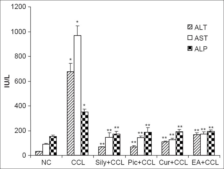
The effect of pretreatment with picroliv, curcumin, and ellagic acid (50 mg/kg/day for 7 days) on serum levels of liver enzymes of mice in carbon tetrachloride (CCl4) induced hepatotoxicity. The values are expressed as mean± SEM (n=6 mice/group). *P<0.001 compared to the normal control (NC) group, **P<0.001 compared to the CCl4 group, by one-way ANOVA followed by Student–Newman–Keuls test as the post hoc test. ALT: Alanine transaminase; AST: Aspartate transaminase; ALP: Alkaline phosphatase; CCL: Carbon tetrachloride; Sily: Silymarin; Pic: Picroliv; Cur: Curcumin; EA: Ellagic acid
Figure 2.
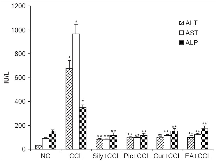
The effect of pretreatment with picroliv, curcumin, and ellagic acid (100 mg/kg/day for 7 days) on serum levels of liver enzymes of mice in carbon tetrachloride (CCl4) induced hepatotoxicity. The values are expressed as mean± SEM (n = 6 mice/group). *P<0.001 compared to the normal control (NC) group, **P<0.001 compared to the CCl4 group, by one-way ANOVA followed by the Student–Newman–Keuls test as post hoc test. ALT: Alanine transaminase; AST: Aspartate transaminase; ALP: Alkaline phosphatase; CCL: Carbon tetrachloride; Sily: Silymarin; Pic: Picroliv; Cur: Curcumin; EA: Ellagic acid
The administration of phenobarbitone in CCl4-treated animals caused an enhancement in the mean duration of sleeping time as compared to that of normal control. Pretreatment with hepatoprotective phytochemicals (50 mg/kg) reduced and restored the phenobarbitone-induced sleeping time in CCl4-induced hepatotoxicity except in the curcumin pretreatment group [Table 1]. When a dose was increased to 100 mg/kg, all the active phytochemicals significantly reduced and restored the prolonged sleeping time. The dose-dependent effect of curcumin was also observed at higher doses.
Table 1.
The effect of pretreatment with picroliv, curcumin, and ellagic acid on phenobarbitoneinduced sleeping time and the liver weight of mice in carbon tetrachloride (CCl4) induced hepatotoxicity

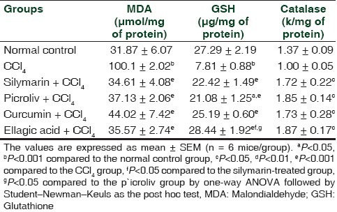
CCl4-induced hepatotoxicity resulted in oxidative stress and lipid peroxidation. This was reflected by an increase in the MDA levels from that of normal control [Tables 2 and 3]. The pretreatment with the phytochemicals (50 and 100 mg/kg) effectively restored the elevated levels of MDA, which was comparable to silymarin. The GSH and catalase concentrations were significantly reduced with the administration of CCl4 [Tables 2 and 3]. A significant increase in GSH levels was observed with 50 and 100 mg/kg dose of picroliv, curcumin, ellagic acid, and silymarin pretreatment. The catalase activity was improved only at 100 mg/kg/day dose of the phytochemicals.
Table 2.
The effect of pretreatment with picroliv, curcumin, and ellagic acid at 50mg/kg/day for 7 days on oxidative stress parameters of mice in carbon tetrachloride (CCl4) induced hepatotoxicity
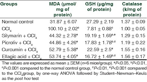
Table 3.
The effect of pretreatment with picroliv, curcumin, and ellagic acid at 100 mg/kg/day for 7 days on oxidative stress parameters of mice in carbon tetrachloride (CCl4) induced hepatotoxicity
With CCl4 treatment, there was no significant difference in the mean (±SEM) liver weight in mice with that of normal control [Table 1]. The pretreatment with phytochemicals (50 mg/kg) resulted in an increase in the mean liver weights of the silymarin, curcumin, and ellagic acid treated groups as compared to normal control as well as that of CCl4 groups. A similar increase in liver weight was observed at the 100 mg/ kg dose level only with picroliv and ellagic acid. However, at both dose levels studied, ellagic acid produced the maximum gain in liver weight.
The normal control group animals showed the typical architecture of liver tissue with a central vein (CV) and chords of hepatocytes radiating [Figure 3a]. CCl4 treatment produced extensive necrosis of hepatocytes which was more pronounced in the centrizonal (zone 3) area. The fatty changes were of macrovesicular type which was evident in centrizonal and portal areas with inflammatory reactions [Figure 3b]. Pretreatment with low dose of phytochemicals (50 mg/kg) showed partial hepatic protection with reduction in the extent of hepatic necrotic areas, fatty infiltration, and mild portal inflammation [Figures 3c–f]. When administered with higher doses (100 mg/kg), picroliv, curcumin, and ellagic acid completely protected the liver as evidenced by restoration of a normal histoarchitecture of the liver similar to the silymarin pretreatment group [Figures 3g–j].
Figure 3.
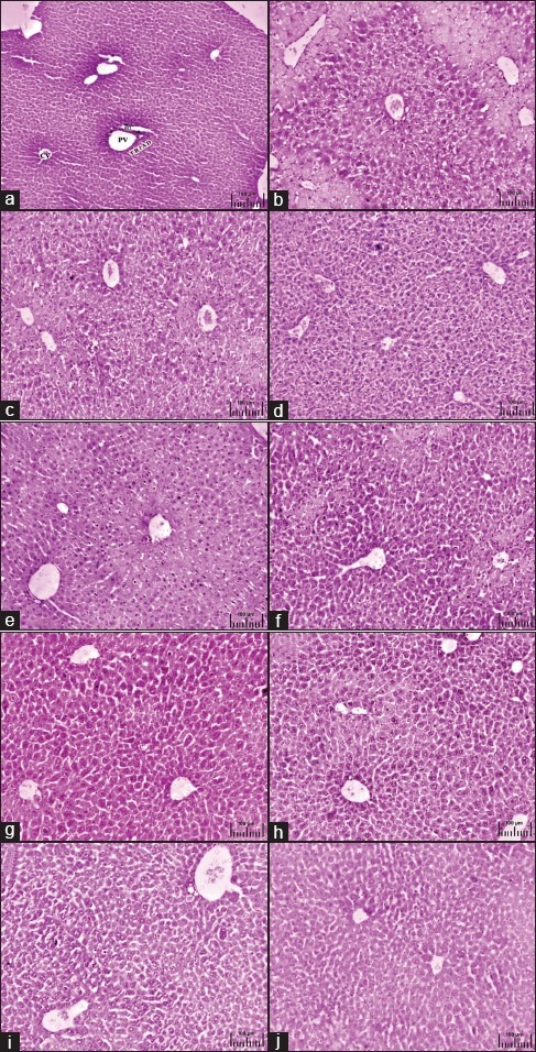
Effect of pretreatment with different active phytoconstituents on light micrographic changes of liver after carbon tetrachloride induced hepatotoxicity. (a) The liver section of normal control mouse showing CV with radiating hepatocytes and portal triad (H and E, ×40). (b) Carbon tetrachloride-treated mouse liver section showing centrizonal necrotic areas and fatty degeneration (H and E, ×100). Pretreatment with 50 mg/kg/day of (c) picroliv, (d) curcumin, (e) ellagic acid, and (f) silymarin in carbon-tetrachloride-induced hepatotoxicity showing partial protection of liver with amelioration of necrosis with mild fatty changes (H and E, ×100). Pretreatment with 100 mg/kg of (g) picroliv, (h) curcumin, (i) ellagic acid, and (j) silymarin in carbon tetrachloride induced hepatotoxicity showing normalization of hepatic architecture without any necrotic changes (H and E, ×100)
DISCUSSION
In this study, hepatoprotective and antioxidant activities of picroliv, curcumin, and ellagic acid were compared with silymarin in the mice model of CCl4 induced liver toxicity. To the best of our knowledge, this is the first study to directly compare four known antioxidants in the setting of acute severe liver injury-induced by CCl4. In the dose ranges studied by us, the hepatoprotection offered by the phytochemicals was profound; however, the precise mechanisms of action of these drugs are still unclear.
CCl4 is a well known hepatotoxin which is widely used to induce toxic liver injury and to study the cellular mechanisms behind oxidative damage in laboratory animals.[20] CCl4 needs metabolic activation by mixed function oxidases to produce the hepatotoxicity. It is activated by cytochrome P 450 2E1 (CYP2E1), CYP2B1 or CYP2B2 and possibly by CYP3A to form the trichloromethyl radical (CCl3●). This toxic free radical reacts with various biologically important substances such as amino acids, nucleotides, and fatty acids as well as proteins, nucleic acids, and lipids. The impairment of lipid metabolism can result in steatosis. Formation of DNA-CCl3● adducts is considered as the initial step in hepatic cancer. The radical can also react with oxygen to form CCl3OO●, a highly reactive species. CCl3OO● initiates a chain reaction of lipid peroxidation which attacks and destroys poly-unsaturated fatty acid, in particular those associated with phospholipids. This affects the permeability of mitochondrial endoplasmic reticulum and plasma membrane resulting in the loss of Ca2+ sequestration and homeostasis. This can heavily contribute to the cellular damage.[20,21]
The oxidative injury caused by CCl4 disrupted the hepatocellular plasma membrane and the enzymes normally present in the cytosol were released into the blood stream.[22] ALP is excreted normally in bile. The liver injury due to toxins could result in defective excretion of bile by hepatocytes which were reflected in their increase in levels in serum.[22] In the CCl4 group animals, pretreatment with picroliv, curcumin, and ellagic acid restored serum transaminase values to normal levels, showing the hepatoprotective activity. The protective effects may be the result of stabilization of plasma membrane thereby preserving the structural integrity of cells as well as the repair of hepatic tissue damage caused by CCl4.[23]
The CCl4 induced hepatic injury decreased the activity of cytochrome P450 enzymes and thereby the metabolic functional activity of the hepatocytes. This caused delay in the barbiturate metabolism and slowed down the excretion of phenobarbitone, thereby prolonging the sleeping time. Pretreatment with hepatoprotective phytochemicals restored the phenobarbitone-induced sleeping time in a dose-related fashion. This is an indicator of normalization of cytochrome P450 and related hepatic mixed function oxidase enzymes system.[24] Moreover, curcumin was reported to protect against CCl4-induced hepatic cytochrome P450 enzyme inactivation via its antioxidant properties.[25] The results were similar to the groups which received silymarin which has been reported to inhibit human microsomal cytochrome P450 activities.[26]
The increased levels of MDA in liver tissue homogenate of mice treated with CCl4 reflected lipid peroxidation and damage to plasma membrane, as a consequence of oxidative stress. Picroliv, curcumin, and ellagic acid restored these values toward normal indicating their anti-lipid peroxidative properties. Picroliv acted as an oxygen-free radical scavenger that limits lipid peroxidation involved in membrane damage elicited by hepatotoxins.[27] Curcumin by scavenging or neutralizing free radicals, interacting with oxidative cascade, quenching oxygen, inhibiting oxidative enzymes like cytochrome P450, and by chelating metal ions, maintained cell membrane integrity and their functions. Further, curcumin may stabilize the cell membrane and significantly reduce the extent of lipid peroxidation in the liver, lung, and kidney.[27]
The tripeptide thiol, GSH, is the most important of the sulfur-containing nonenzymatic antioxidant molecules. GSH can also conjugate with free radicals directly, earmarking them for renal excretion, which is especially important for dealing with the products of hepatic cytochrome P450 enzyme activity. The sulfhydryl (–SH) portion of the GSH can be used to reduce a variety of free radicals in a reaction catalyzed by the antioxidant enzyme, glutathione peroxidase.[28] Catalase, an antioxidant enzyme that protects cell from oxidative stress of hydrogen peroxide, is induced on the generation of free radicals in cells. Catalase acts as a preventative antioxidant and plays an important role in protection against the deleterious effects of lipid peroxidation.[28,29]
The reduction in concentrations of GSH and catalase observed in this study may be related to the excessive production of free radicals generated by the metabolism of CCl4. The antioxidant enzymes are inactivated or exhausted by lipid peroxides and/or reactive oxygen species which result in decreased activities of these enzymes in CCl4 toxicity. An increase in GSH levels observed with 50 mg/kg dose of silymarin pretreatment was in agreement with the previous study.[30] There was normalization of both GSH and catalase levels with picroliv, curcumin, and ellagic acid at 100 mg/kg dose. Picroliv has been reported to prevent the depletion of reduced glutathione which is needed for the glutathione-S-transferase for detoxification reaction and rise in levels of lipid peroxides in the liver.[27] Curcumin can lower lipid peroxidation by maintaining the activation of antioxidant enzymes like superoxide dismutase, catalase, and glutathione peroxidase at higher levels. Curcumin and ellagic acid are capable of scavenging oxygen free radicals such as superoxide anions and hydroxyl radicals which are important in the initiation of lipid peroxidation.[3,11] Ellagic acid has the capacity to regulate intimate intracellular mechanisms by direct interaction with double stranded DNA.[31] It can also act on drug metabolizing enzymes and prevent the formation of toxic metabolites.[32] Thus, the increased levels of GSH and catalase with the treatment of phytochemicals may be involved in the protective mechanism against liver toxicity produced by CCl4.
The structural activity of the phytochemicals revealed that the antioxidant potential of picroliv could be related to electrophilic free radical scavenging nature of its iridoid glycosides, whereas curcumin contains the phenolic group which is important for its antioxidant activity.[7,33] It has been reported that two lactone groups of ellagic acid can act as a hydrogen bond donor or acceptor, which might be involved in the free radical scavenging action and decreased free radicals mediated lipid peroxidation.[33]
The pretreatment with the active phytochemicals resulted in an increase in liver weight as compared to normal control and CCl4-treated groups. This may be due to the promotion of the assembly of ribosomes on endoplasmic reticulum to facilitate protein synthesis and regenerative activity.[34] This effect was more with the ellagic acid pretreatment at both the dose levels (50 and 100 mg/kg).
Histopathological profile of the liver of CCl4-treated mice showed zone 3 necrosis and fatty degeneration with infiltration of inflammatory mediators. The animals treated with active phytochemicals (50 mg/kg) showed a significant improvement of CCl4-induced liver injury evident from the presence of normal hepatic cords, the absence of necrosis, and a lesser degree of fatty infiltration. At the dose of 100 mg/kg, the liver architecture was near normal with only mild fatty changes. These observations were well correlated with the biochemical findings and gives clear evidence that there was not only improvement in the liver functions with the treatment of phytochemicals, but also in the hepatic architecture.
CONCLUSIONS
In this study, the phytochemicals picroliv, curcumin, and ellagic acid showed hepatoprotective activities comparable to silymarin in CCl4-induced hepatotoxicity in mice. The protective action was improved further by doubling the dose of the phytochemicals. Apart from the anti-lipidperoxidative and antioxidant actions, these active phytochemicals might have played a role in restoring the cytochrome P450 enzyme system or promoted the liver regenerative activity. In future, the derivatives of these phytochemicals or their combinations may show efficacy in various experimental toxic models. They may be developed as future drugs for use in human liver diseases with antioxidant, antifibrotic, immunomodulatory, antiviral, and regenerative properties.
ACKNOWLEDGMENTS
The first author acknowledges the institutional grant-in-aid for this research work. Thanks are also due to Dr. Steven A Dkhar, Professor, Department of Pharmacology, JIPMER, Pondicherry for the constructive comments on the manuscript. There is no conflict of interest.
Footnotes
Source of Support: Nil
Conflict of Interest: None declared.
REFERENCES
- 1.Abbound G, Kaplowitz N. Drug induced liver injury. Drug Saf. 2007;30:277–94. doi: 10.2165/00002018-200730040-00001. [DOI] [PubMed] [Google Scholar]
- 2.Medina J, Moreno-Otero R. Pathophysiological basis for antioxidant therapy in chronic liver disease. Drugs. 2005;65:2445–61. doi: 10.2165/00003495-200565170-00003. [DOI] [PubMed] [Google Scholar]
- 3.Girish C, Pradhan SC. Drug development for liver diseases; focus on picroliv, ellagic acid and curcumin. Fundam Clin Pharmacol. 2008;22:623–32. doi: 10.1111/j.1472-8206.2008.00618.x. [DOI] [PubMed] [Google Scholar]
- 4.Pradhan SC, Girish C. Hepatoprotective herbal drug, silymarin from experimental pharmacology to clinical medicine. Indian J Med Res. 2006;124:491–504. [PubMed] [Google Scholar]
- 5.Chander R, Kapoor NK, Dhawan BN. Picroliv, picroside-1 and kutkoside from Picrorhiza kurroa are scavengers of superoxide anions. Biochem Pharmacol. 1992;44:180–3. doi: 10.1016/0006-2952(92)90054-m. [DOI] [PubMed] [Google Scholar]
- 6.Buniatian GH. Stages of activation of hepatic stellate cells: Effects of ellagic acid, an inhibitor of liver fibrosis, on their differentiation in culture. Cell Prolif. 2003;36:307–19. doi: 10.1046/j.1365-2184.2003.00287.x. [DOI] [PMC free article] [PubMed] [Google Scholar]
- 7.Manna SK, Mukhopadhyay A, Van NT, Aggarwal BB. Silymarin suppresses TNF-induced activation of NF-kappa B, c-Jun N-terminal kinase, and apoptosis. J Immunol. 1999;163:6800–9. [PubMed] [Google Scholar]
- 8.Lee WJ, Ou HC, Hsu WC, Chou MM, Tseng JJ, Hsu SL, et al. Ellagic acid inhibits oxidized LDL-mediated LOX-1 expression, ROS generation, and inflammation in human endothelial cells. J Vasc Surg. 2010;52:1290–300. doi: 10.1016/j.jvs.2010.04.085. [DOI] [PubMed] [Google Scholar]
- 9.Anand P, Kunnumakkara AB, Harikumar KB, Ahn KS, Badmaev V, Aggarwal BB. Modification of cysteine residue in p65 subunit of nuclear factor- kappa B (NF-kappaB) by picroliv suppresses NF-kappa B-regulated gene products and potentiates apoptosis. Cancer Res. 2008;68:8861–70. doi: 10.1158/0008-5472.CAN-08-1902. [DOI] [PMC free article] [PubMed] [Google Scholar] [Retracted]
- 10.Devipriya N, Sudheer AR, Menon VP. Dose-response effect of ellagic acid on circulatory antioxidants and lipids during alcohol-induced toxicity in experimental rats. Fundam Clin Pharmacol. 2007;21:621–30. doi: 10.1111/j.1472-8206.2007.00551.x. [DOI] [PubMed] [Google Scholar]
- 11.Kaur G, Tirkey N, Bharrhan S, Chanana V, Rishi PK, Chopra K. Inhibition of oxidative stress and cytokine activity by curcumin in amelioration of endotoxin-induced experimental hepatoxicity in rodents. Clin Exp Immunol. 2006;145:313–21. doi: 10.1111/j.1365-2249.2006.03108.x. [DOI] [PMC free article] [PubMed] [Google Scholar]
- 12.Girish C, Koner BC, Jayanthi S, Rao KR, Rajesh B, Pradhan SC. Hepatoprotective activity of picroliv, curcumin and ellagic acid compared to silymarin on paracetamol induced liver toxicity in mice. Fundam Clin Pharmacol. 2009;23:735–45. doi: 10.1111/j.1472-8206.2009.00722.x. [DOI] [PubMed] [Google Scholar]
- 13.Kind PR, King EJ. Estimation of plasma phosphatase by determination of hydrolysed phenol with amino-antipyrine. J Clin Pathol. 1954;7:322–6. doi: 10.1136/jcp.7.4.322. [DOI] [PMC free article] [PubMed] [Google Scholar]
- 14.Reitman S, Frankel S. A colorimetric method for the determination of serum glutamic oxaloacetic acid and glutamic pyruvic transaminases. Am J Clin Pathol. 1957;28:56–63. doi: 10.1093/ajcp/28.1.56. [DOI] [PubMed] [Google Scholar]
- 15.Handa SS, Sharma A. Hepatoprotective activity of andrographolide from Andographis paniculta against carbon tetrachloride. Indian J Med Res. 1990;92:276–83. [PubMed] [Google Scholar]
- 16.Utley HG, Bernheim F, Hochstein P. Effect of sulfhydryl reagents on peroxidation in microsomes. Arch Biochem Biophys. 1967;118:29–32. [Google Scholar]
- 17.Smith IK, Vierheller TL, Thorne CA. Assay of glutathione reductase in crude tissue homogenates using 5,5’-dithiobis (2-nitrobenzoic acid) Anal Biochem. 1988;175:408–13. doi: 10.1016/0003-2697(88)90564-7. [DOI] [PubMed] [Google Scholar]
- 18.Aebi H. Catalase in vitro. In: Fleischer S, Fleischer B, editors. Methods in enzymology. Vol. 105. San Diego: Academic Press; 1984. pp. 121–6. [DOI] [PubMed] [Google Scholar]
- 19.Lowry OH, Rosebrough NJ, Farr AL, Randall RJ. Protein measurement with the folin phenol reagent. J Biol Chem. 1951;193:265–75. [PubMed] [Google Scholar]
- 20.Weber LW, Boll M, Stampfl A. Hepatotoxicity and mechanism of action of haloalkanes: Carbon tetrachloride as a toxicological model. Crit Rev Toxicol. 2003;33:105–36. doi: 10.1080/713611034. [DOI] [PubMed] [Google Scholar]
- 21.Rang HP, Dale MM, Ritter JM, Flower RJ. Pharmacology. 6th ed. Edinburgh: Churchill Livingstone; 2007. [Google Scholar]
- 22.Rajesh MG, Latha MS. Preliminary evaluation of the antihepatotoxic activity of Kamilari, a polyherbal formulation. J Ethnopharmacol. 2004;91:99–104. doi: 10.1016/j.jep.2003.12.011. [DOI] [PubMed] [Google Scholar]
- 23.Pari L, Murugan P. Protective role of tetrahydrocurcumin against erythromycin estolate-induced hepatotoxicity. Pharmacol Res. 2004;49:481–6. doi: 10.1016/j.phrs.2003.11.005. [DOI] [PubMed] [Google Scholar]
- 24.Girish C, Koner BC, Jayanthi S, Rao KR, Rajesh B, Pradhan SC. Hepatoprotective activity of six polyherbal formulations in paracetamol induced liver toxicity in mice. Indian J Med Res. 2009;129:105–14. [PubMed] [Google Scholar]
- 25.Sugiyama T, Nagata J, Yamagishi A, Endoh K, Saito M, Yamada K, et al. Selective protection of curcumin against carbon tetrachloride-induced inactivation of hepatic cytochrome P450 isozymes in rats. Life Sci. 2006;78:2188–93. doi: 10.1016/j.lfs.2005.09.025. [DOI] [PubMed] [Google Scholar]
- 26.Zuber R, Modrianský M, Dvorák Z, Rohovský P, Ulrichová J, Simánek V, et al. Effect of silybin and its congeners on human liver microsomal cytochrome P450 activities. Phytother Res. 2002;16:632–8. doi: 10.1002/ptr.1000. [DOI] [PubMed] [Google Scholar]
- 27.Rastogi R, Srivastava AK, Rastogi AK. Long term effect of aflatoxin B (1) on lipid peroxidation in rat liver and kidney: Effect of picroliv and silymarin. Phytother Res. 2001;15:307–10. doi: 10.1002/ptr.722. [DOI] [PubMed] [Google Scholar]
- 28.Webb C, Twedt D. Oxidative stress and liver disease. Vet Clin North Am Small Anim Pract. 2008;38:125–35. doi: 10.1016/j.cvsm.2007.10.001. [DOI] [PubMed] [Google Scholar]
- 29.Ookhtens M, Kaplowitz N. Role of the liver in interorgan homeostasis of glutathione and cyst(e)ine. Semin Liver Dis. 1998;18:313–29. doi: 10.1055/s-2007-1007167. [DOI] [PubMed] [Google Scholar]
- 30.Campos R, Garrido A, Guerra R, Valenzuela A. Silybin dihemisuccinate protects against glutathione depletion and lipid peroxidation induced by acetaminophen on rat liver. Planta Med. 1989;55:417–9. doi: 10.1055/s-2006-962055. [DOI] [PubMed] [Google Scholar]
- 31.Thulstrup PW, Thormann T, Spanget-Larsen J, Bisgaard HC. Interaction between ellagic acid and calf thymus DNA studied with flow linear dichroism UV-VIS spectroscopy. Biochem Biophys Res Commun. 1999;265:416–21. doi: 10.1006/bbrc.1999.1694. [DOI] [PubMed] [Google Scholar]
- 32.Hayeshi R, Mutingwende I, Mavengere W, Masiyanise V, Mukanganyama S. The inhibition of human glutathione S-transferases activity by plant polyphenolic compounds ellagic acid and curcumin. Food Chem Toxicol. 2007;45:286–95. doi: 10.1016/j.fct.2006.07.027. [DOI] [PubMed] [Google Scholar]
- 33.Venkatesan P, Rao MN. Structural activity relationships for the inhibition of lipid peroxidation and the scavenging of free radicals by synthetic symmetrical curcumin analogues. J Pharm Pharmacol. 2000;52:1123–8. doi: 10.1211/0022357001774886. [DOI] [PubMed] [Google Scholar]
- 34.Girish C, Koner BC, Jayanthi S, Rao KR, Rajesh B, Pradhan SC. Hepatoprotective activity of six polyherbal formulations in carbon tetrachloride induced liver toxicity in mice. Indian J Experimental Biol. 2009;47:257–63. [PubMed] [Google Scholar]


