Abstract
In this present era, when a significant number of patients seeking orthodontic treatment are adults, importance of multidisciplinary treatment approach cannot be overemphasized. Higher susceptibility of plaque accumulation in patients undergoing orthodontic treatment makes involvement of periodontist almost unavoidable. Also, orthodontic treatment frequently results in undesirable periodontal changes which require immediate attention. More recently, orthodontics has been used as an adjunct to periodontics to increase connective tissue support and alveolar bone height. The purpose of this article is to review the adverse effects of orthodontic treatment on the periodontal tissues and to discuss the mutually beneficial relationship shared between the two specialties.
Keywords: Orthodontics, periodontium, recession
INTRODUCTION
The term synergy refers to two or more distinct influences or agents acting together to create an effect greater than that predicted by knowing only the separate effects of the individual agents. This definition is applicable to the classic relationship between orthodontic and periodontics specialties in treating patients. Understanding the biologic basis of periodontal surgical procedures, recent advancements in tissue engineering and research development can yield more productive clinical endpoints than ever before. Making the most of what these two specialties offer each other begins with the identification of periodontal problems that could become more complicated during orthodontic therapy and, conversely, those that could benefit from orthodontic therapy.[1]
Kingsley[2] stated in the late 19th century that age is hardly a limiting factor as far as tooth movement is concerned. But since then, for a long time, orthodontists limited their services to children and adolescents. However in the 1960s and 70s, prevention of oral diseases like caries and periodontal breakdown became the primary reason for seeking orthodontic treatment. Since then orthodontic treatment has been available to adults as well.
Reasons for adults seeking orthodontic treatment were enlisted by Perregaard.[3] According to him, although 50% of adults seeking orthodontic treatment report with the chief complaint of untreated malocclusion, a significant percentage of patients (12%) seek orthodontic treatment to prevent occurrence or progression of periodontal diseases. Better compliance offered by adult patients compensate for the slower tissue response.
Orthodontic patients can be classified into three categories: (1) Patients with good oral health; (2) Patients with periodontal disease and/or loss of permanent teeth; and (3) Patients with severe skeletal discrepancies.[4] A multidisciplinary approach involving an orthodontist and a periodontist is required to treat patients belonging to the second category. While treating such patients, both specialists should be involved in treatment planning, and the treatment progress should be evaluated and shared.
Mechanisms of tissue damage
Unfestooned orthodontic bands are particularly suspects as possibly complicating factors jeopardizing interproximal periodontal support, and at the present time “special periodontally friendly bands” are being designed in research and design laboratories. These challenging effects of band impingement may directly compromise local resistance related to subgingival pathogens in susceptible patients and result in damage to both interproximal gingival tissues and alveolar crestal bone in a manner similar to that produced by faulty crown margins. Periodontal support might also be damaged during tooth intrusion where patients have active periodontitis or gingival infection significant enough to convert to periodontal disease. In these kinds of susceptible patients a screening examination for the interleukin (IL) family of inflammatory mediators may be wise. The details of genetic screening involve studying the genetic potential of exaggerated immunologic reactions of host response to bacterial challenge such as those that recruit IL-1β.
The etiology of periodontal problems may not simply rely on exaggerated host immunologic reactions. Mattingly and coworkers[5] and others[6–8] reflect the view that long-term fixed appliances can contribute to unfortunate but predictable qualitative alterations in the subgingival bacterial biofilms that become progressively periodontopathic with time.
On a practical level it seems that an absence of bleeding on probing is a better forecasting parameter of health than bleeding on probing is a predictor of progressive disease. In other words, an absence of bleeding on probing, despite the pocket depth can justifiably be used as a test of “healthy gums.” On the other hand, while bleeding on probing is certainly an indication of infection of the gingivae, it is one of many risk factors associated with progressive bone loss due to periodontitis. However, the test is not spontaneous bleeding or even bleeding on brushing and flossing. That elicits only superficial disease, one that contributes significantly to caries and marginal decalcification. The best test is “bleeding on probing” elicited by stroking the sulci with a flexible plastic periodontal probe at a comfortable range of force between 10 and 20 g . Those orthodontic patients who present with persistent bleeding on such probing should be notified that they are “at risk” and that prudence dictates a more intensive regimen of periodontal therapy than those who present with little or no bleeding on probing. Since bleeding swollen gingiva is ubiquitous in the orthodontic population, universal caution should be employed and supportive periodontal care recommended routinely as an integral part of orthodontic therapy. Studies have pointed out the importance of a full-mouth examination, six sites per tooth, for a comprehensive description of periodontal status in orthodontic patients.
The conclusion that seems most logical is that some periodontal damage may occur, particularly in those patients who exhibit poor oral hygiene during fixed appliance therapy, but the contribution of orthodontic care is generally minor, occasionally severe enough to justify periodontal therapy and prevalent enough to indicate concomitant supportive periodontal therapy as a routine preventive tactic during fixed appliance therapy. It is advisable that professional scaling and root planning, where indicated, be performed by a periodontist.[9,10]
Mucogingival changes during orthodontic treatment
It has been widely believed that appropriately applied orthodontic forces do not damage the periodontium. However, insufficient width of attached gingival is widely believed to be a predisposing factor for recession. Lang and Loe[10] concluded from their study that 2 mm of keratinized gingiva is adequate to maintain gingival health.
It is believed that alveolar bone dehiscence is a predisposing factor for the development of gingival recession.[11,12] So as long as a tooth is housed within the alveolar bone, orthodontic tooth movement (OTM) will not result in recession. Batenhorst[13] concluded from his experiment on monkeys that facial tipping, extrusion, and bodily movements of incisors results in apical shift of labial gingival margin and loss of attachment. However a study done on humans[14] failed to prove the same.
Steiner[11] suggested that tension in the marginal tissue created by the orthodontic forces could be an important factor in causing gingival recession. This means, the thickness of the gingival tissue at the pressure side and not its apico-coronal width, is an indicator of possible recession. An experimental study was done on monkeys to confirm this hypothesis.[12] Following extensive bodily movement of incisors in a labial direction, most teeth showed clinically some apical displacement of the gingival margin as well as loss of probing attachment, but no loss of connective tissue attachment when evaluated histologically.
Periodontal response to different types of orthodontic forces
Greenbaum[15] studied the effects of slow and rapid maxillary expansion on the periodontium. They concluded that patients subjected to rapid maxillary expansion showed significantly lesser bone relative to the cemento-enamel junction when compared to patients treated with slow expansion and the control group. However, they did not find any significant difference in probing depth and width of attached gingival between the groups.
Siew Han Chay[16] has shown that gingival margin can be moved incisally by as much as 9 mm using orthodontic extrusion. Erkan[17] observed that gingival margin and mucogingival junction moved in the same direction along with teeth by 79 and 62%, respectively, when mandibular incisor was intruded orthodontically.
Extrusion of mandibular incisor produces gingival margin and the mucogingival junction movement in the same direction as the extruded teeth by 80 and 52.5%, respectively.[16] This also results in reduction of the sulcus depth without significant reduction in the width of attached gingival. Also, no attachment loss was observed.[17]
A longitudinal study conducted by Alstad[18] on patients undergoing orthodontic treatment did not report any significant loss of attachment. They concluded that if a professional preventive program is pursued throughout the course of orthodontic treatment, loss of attachment can be limited to less than 0.1 mm per surface.
Prevention of periodontal breakdown during orthodontic treatment
Orthodontic bands, brackets and wires not only test the patient's ability to maintain good oral hygiene, but also compromises the self-cleansing property of the dentition.[19] Orthodontic attachments have the potential to cause plaque accumulation and increase the pathogenicity of the microbes.[20] This tendency is often dealt with by thorough professional prophylaxis. Repeating the oral hygiene instructions on each visit and rubber cup prophylaxis are effective measures to prevent plaque accumulation and gingival enlargement.[21] Costa et al.,[22] compared the efficacy of manual, electric and ultrasonic toothbrushes in patients undergoing fixed orthodontic therapy. They concluded that plaque scores on the buccal surfaces of teeth were lowered in patients using ultrasonic toothbrush. Also, S. mutans count reduction was seen in patients using ultrasonic and electric toothbrushes. According to Hannah,[23] oral hygiene can be improved in orthodontic patients by using a sanguinaria-containing toothpaste along with a sanguinaria-containing oral rinse.
Orthodontics as an adjunct to periodontal therapy
Orthodontics can serve as an adjunct to periodontal treatment procedures to improve oral health in a number of situations. Pathological tooth migration is one of the few evident signs of periodontitis that affects dentofacial esthetics. This phenomenon is more commonly seen in the anterior dentition due to lack of stable occlusal and sagittal contacts with the opposing teeth.[24] Achieving an esthetically acceptable result in such cases may require various OTMs like intrusion, rotation, and uprighting. This can also help control periodontal breakdown and restore good oral function.[25]
Tulloch[26] is of the opinion that fixed appliance therapy is more preferable if OTM is desired in a patient suffering from periodontitis. Fixed appliance allows easy splinting of teeth to achieve stable anchorage. He also highlights the importance of reducing the force magnitude and applying counteracting moments to reduce the stress on periodontal ligament fibres. Lijian[27] has enlisted the various precautions to be taken when attempting tooth movement in height-reduced periodontium, which includes achieving stable anchorage and long-term periodontal maintenance care.
Deepa[28] reported the use of orthodontic soft aligners in repositioning a periodontally involved tooth. Light and intermittent forces generated by the soft aligner allow regeneration of tissue during tooth movement. Along with periodontal procedures, orthodontically assisted occlusal improvement may be required in treatment of patients with severely attrited lower anterior teeth.[29]
Patient's compliance, motivation, and oral hygiene maintenance will help determine the best time to start adjunctive orthodontic treatment. It is suggested that tooth movement can be undertaken 6 months after completion of active periodontal treatment if there is sufficient evidence of complete resolution of inflammation.[26] Sanders[30] has recommended a three-step comprehensive protocol to be followed before, during, and after adjunctive orthodontic therapy.
In patients diagnosed with vertical bony defects, adjunctive orthodontic procedures can help improve the condition. Shoichiro[31] reported improvement in alveolar bone defects, gingival esthetics, and the crown-root ratio in patients with one- or two-wall isolated vertical infrabony defects with a combination of tooth extrusion and periodontal treatment. Orthodontic intrusion has also been shown to improve periodontal condition.[32] However, elimination of pockets was undertaken prior to intrusion in order to prevent apical displacement of plaque.[33]
Periodontics as an adjunct to orthodontic treatment
On many occasions, a stable and esthetically acceptable outcome cannot be achieved with orthodontics without adjunctive periodontal procedures. For instance, a high labial frenum attachment is considered to be a causative factor of midline diastema. Frenectomy is recommended in such cases as the fibres are thought to prevent the mesial migration of the central incisors. However, the timing of periodontal intervention has been a topic of much debate.
According to Vanarsdall,[34] surgical removal of a maxillary labial frenum should be delayed until after orthodontic treatment unless the tissue prevents space closure or becomes painful and traumatized.
Forced eruption of a labially or palatally impacted tooth is now a common orthodontic treatment procedure. Careful exposure of the impacted tooth while preserving keratinized tissue requires the expertise of a periodontist. Preservation of keratinized tissue is important to prevent loss of attachment. The preferred surgical procedure is primarily an apically or laterally positioned pedicle graft.[35]
Retention of orthodontically achieved tooth rotation is a problem that has always plagued the orthodontist. Circumferential supracrestal fiberotomy (CSF) is a procedure that is frequently used to enhance post-treatment stability.[36] Edwards[37] concluded from his long-term prospective study that CSF is more successful in preventing relapse in the maxillary arch. According to him, CSF does not affect the periodontium adversely.
Mucogingival surgeries may be needed during the course of orthodontic treatment to maintain sufficient width of attached gingival.[35] Also, crown lengthening procedures can facilitate easy placement of orthodontic attachments on teeth with short clinical crowns. This procedure can also be used for smile designing.[38] Alveolar ridge augmentation and placements of dental implants[39] are the other adjunctive periodontal treatment procedures undertaken to facilitate achievement of orthodontic treatment goals.
There is an ever increasing concern for dentofacial esthetics in adult population. The primary motivating factor for seeking orthodontic treatment is dental appearance. Pathologic migration of anterior teeth is a common cause of esthetic concern among adults. The disruption of equilibrium in tooth position may be caused by several etiologic factors. These include periodontal attachment loss, pressure from inflamed tissues, occlusal factors, oral habits such as tongue thrusting and bruxism, loss of teeth without replacement, gingival enlargement and iatrogenic factors. However, according to the literature, destruction of tooth supporting structures is the most relevant factor. The periodontal disease and its sequela such as diastema, pathological migration, labial tipping or missing teeth often lead to functional and esthetic problems either alone or with restorative problems. Advanced periodontal disease is characterized by severe attachment loss, reduced alveolar bone support, tooth mobility and gingival recession. Orthodontic treatment is initiated only after periodontal disease is brought under control. This communication highlights good treatment outcome achieved in a patient with impaired dentofacial aesthetics and advanced periodontal disease.[1]
Lt. Col. M. Panwar et al.[40] in 2010 presented a case report on combined periodontal and orthodontic treatment of pathologic migration of anterior teeth. Comprehensive orthodontics was initiated with pre-adjusted edgewise appliances using very light force, which resulted in optimal biological response. Since there was trauma from lower anterior teeth, anterior bite plane allowed posterior eruption of teeth, which resulted in the opening of the bite. The periodontal health improved the moment trauma was relieved. Periodontal treatment and the patient's co-operation in oral hygiene were also continued as supportive therapy.[40]
Michael et al. in 2009 provided the treatment options for the significant dental midline diastema. After the required prosthetic intervention, periodontal tissues were altered by gingivoplasty and crown lengthening and provided optimal result with favorable esthetic, functional, and biologic consequences.[41]
Interdisciplinary approach to improve smile esthetics
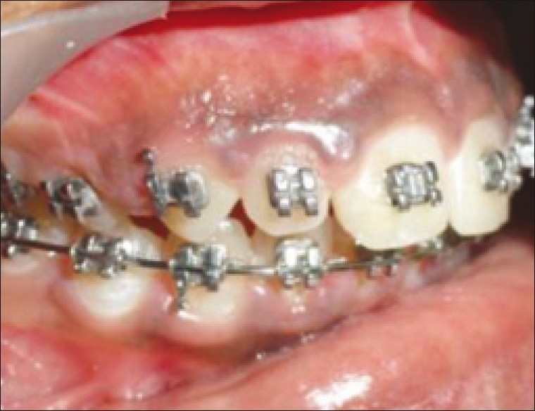
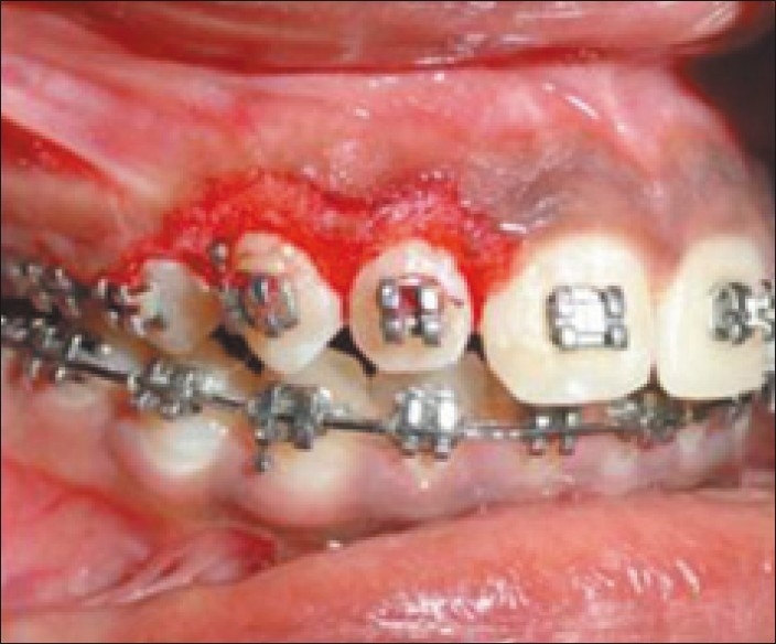
Periodontal intervention, along with OTM helps achieve esthetically acceptable results in adult patients suffering from periodontitis. This association has great potential considering the ever increasing focus on gingival appearance in smile designing. Gummy smile can be a result of vertical maxillary excess, in which case, orthognathic surgery should be the preferred treatment plan. However, gummy smile can be a result of delayed apical migration of gingival [Figure 1]. Gingivectomy can be considered as a treatment option in the latter case [Figure 2].[38]
Figure 1.

Delayed apical migration of gingival (gingival enlargement)
Figure 2.

Gingivectomy procedure done
Missing interdental papilla can be an esthetic concern in patients undergoing orthodontic treatment. OTM alone or enameloplasty may not solve the problem in many such cases. A competent periodontist can solve the problem using a papilla creation procedure as described by Zetu.[41] Similarly, Crown lengthening procedure combined with orthodontic extrusion and incisal edge leveling can help correct gingival margin discrepancy.[38]
Biocompatible orthodontic materials
It is a well known fact that orthodontic appliances provide a good environment for oral microbes to thrive and cause diseases like dental caries or even periodontitis.[24] It is thus natural for clinicians to try out more biocompatible materials and reduce the chances for microbial colonization. Chun et al.[42] suggested surface modification of orthodontic wires with photocatalytic TiO2 as a method to prevent the development of dental plaque during orthodontic treatment. From their experiment, they concluded that bacterial mass bound to the TiO2-coated orthodontic wires during treatment was significantly lower than that of the uncoated wires. They also demonstrated the bactericidal effect of TiO2-coated orthodontic wires on S. mutans and P. gingivalis.[43]
Since the last two decades or so, glass ionomer cements (GICs) are the most commonly used material for band cementation. This material exhibits a continuous release and uptake of fluoride, which has certain antibacterial activities. However, their antibacterial effect is limited to a relatively narrow spectrum of bacteria. Recent studies have shown that the addition of chlorhexidine (CHD) to resin composites and glass ionomer cements for cementation significantly improves the antibacterial effect. GICs with the addition of 18% CHD showed significant inhibition of bacteria in comparison with the control groups without compromising on the mechanical properties.[44]
Accelerated osteogenic orthodontics and periodontal implications
In an attempt to reduce the treatment duration, procedures like accelerated osteogenic orthodontics (AOOs) are being popularized. Kim et al.[45] reported rapid tooth movement when temporary anchorage devices were used in combination with AOO. However, this procedure involves decortications and subsequent placement of graft material. Hence, continuous periodontal monitoring is required when this technique is employed. With the increasing popularity of these invasive procedures, partnership with a periodontist will become indispensible for orthodontists in the near future.
CONCLUSION
Patient education, motivation, enhanced oral hygiene maintenance and regular periodontal care are essential during orthodontic treatment. Certain adjunctive periodontal procedures may help an orthodontist achieve more stable and esthetically acceptable results. Close co-operation between the periodontist and orthodontist can ensure excellent results with long-term stability.
Footnotes
Source of Support: Nil,
Conflict of Interest: None declared.
REFERENCES
- 1.Palomo L, Palomo Jm, Bissada NF. Semin Orthod. 2008;14:229–45. [Google Scholar]
- 2.Kingsley NW. New York: Appleton; 1880. [Last cited on 2011 Sep 03]. Treatise on oral deformities as a branch of mechanical surgery. Available from: http://www.bibliopolis.com/main/books/author/Kingsley,%20Norman.html . [Google Scholar]
- 3.Perrigaard J, Blixencrone-Moller T. Why do adults seek orthodontic treatment? In: Proceedings of the 64th congress of the European orthodontic society. London: European orthodontic Society; 1988. Jul, p. 61A. [Google Scholar]
- 4.Moyers RE, Dryland-Vig KW, Fonsece RJ. Adult treatment. In: Moyers RE, editor. Handbook of orthodontics. 4th ed. Chicago: Year Book medical Publishers Inc; 1998. pp. 472–510. [Google Scholar]
- 5.Mattingly JA, Sauer GJ, Yancey JM, Arnold RR. Enhancement of streptococcus mutans colonization by direct bonded orthodontic appliances. J Dent Res. 1983;62:1209–11. doi: 10.1177/00220345830620120601. [DOI] [PubMed] [Google Scholar]
- 6.Paolantonio M, Festa F, di Placido G, D’Attilio M, Catamo G, Piccolomini R. Site-specific subgingival colonization by actinobacillus actinomycetemcomitans in orthodontic patients. Am J Orthod Dentofacial Orthop. 1999;115:423–8. doi: 10.1016/s0889-5406(99)70263-5. [DOI] [PubMed] [Google Scholar]
- 7.Sallum EJ, Nover DF, Klein MI, Gonçalves RB, Machion L, Wilson Sallum A, et al. Clinical and microbiologic changes after removal of orthodontic appliances. Am J Orthod Dentofacial Orthop. 2004;126:363–6. doi: 10.1016/j.ajodo.2004.04.017. [DOI] [PubMed] [Google Scholar]
- 8.Perinetti G, Paolantonio M, Serra E, D’Archivio D, D’Ercole S, Festa F, et al. Longitudinal monitoring of subgingival colonization by actinobacillus actinomycetemcomitans, and crevicular alkaline phosphatise and aspartate aminotransferase activities around orthodontically treated teeth. J Clin Periodontol. 2004;31:60–7. doi: 10.1111/j.0303-6979.2004.00450.x. [DOI] [PubMed] [Google Scholar]
- 9.Jin L. Periodontic—orthodontic interactions-rationale, sequence and clinical implications. Hong Kong Dent J. 2007;4:60–4. [Google Scholar]
- 10.Lang NP, Loe H. The relationship between the width of keratinized gingiva and gingival health. J Periodontol. 1972;43:623–7. doi: 10.1902/jop.1972.43.10.623. [DOI] [PubMed] [Google Scholar]
- 11.Steiner GG, Pearson JK, Ainamo J. Changes of the marginal periodontium as a result of labial tooth movement in monkeys. J Periodontol. 1981;52:314–20. doi: 10.1902/jop.1981.52.6.314. [DOI] [PubMed] [Google Scholar]
- 12.Wennstrom JL, Lindhe J, Sinclair F, Thilander B. Some periodontal tissue reactions to orthodontic tooth movement in monkeys. J Clin Periodontol. 1987;14:121–9. doi: 10.1111/j.1600-051x.1987.tb00954.x. [DOI] [PubMed] [Google Scholar]
- 13.Batenhorst KF, Bowers GM, Williams JE. Tissue changes resulting from facial tipping and extrusion of incisors in monkeys. J Periodontol. 1974;45:660–8. doi: 10.1902/jop.1974.45.9.660. [DOI] [PubMed] [Google Scholar]
- 14.Rateitschak KH, Herzog-Specht F, Hotz R. Reaktion und Regene-ration des Parodonts auf BehandlungmitfestsitzendenApparaten und abnehmbaren Flatten. Fortschritte der Kieferorthopadie. 1968;29:415–35. doi: 10.1007/BF02165844. [DOI] [PubMed] [Google Scholar]
- 15.Greenbaum KF, Zachrisson BU. The effect of palatal expansion therapy on the periodontal supporting tissues. Am J Orthod. 1982;81:12–21. doi: 10.1016/0002-9416(82)90283-4. [DOI] [PubMed] [Google Scholar]
- 16.Chay SH, Rabie AB. Repositioning of the gingival margin by extrusion. Am J Orthod. 2002;122:95–102. doi: 10.1067/mod.2002.122397. [DOI] [PubMed] [Google Scholar]
- 17.Erkan M, Pikdoken L, Usumez S. Gingival response to mandibular incisor intrusion. Am J Orthod. 2007;132:143e9–13. doi: 10.1016/j.ajodo.2006.10.015. [DOI] [PubMed] [Google Scholar]
- 18.Alstad S, Zachrisson BU. Longitudinal study of petiodontal condition associated with orthodontic treatment in adolescents. Am J orthod. 1979;76:277–86. doi: 10.1016/0002-9416(79)90024-1. [DOI] [PubMed] [Google Scholar]
- 19.Bloom RH, Brown LR. A study of the effects of orthodontic appliances on the oral microbial flora. Oral Surg Oral Med Oral Pathol. 1964;17:758–67. doi: 10.1016/0030-4220(64)90373-1. [DOI] [PubMed] [Google Scholar]
- 20.Balenseifen J, Madonia J. Study of dental plaque in orthodontic patients. J Dent Res. 1970;49:320–4. doi: 10.1177/00220345700490022101. [DOI] [PubMed] [Google Scholar]
- 21.Huber SJ, Vernino AR, Nanda RS. Professional prophylaxis and its effect on the periodontium of full-banded orthodontic patients. Am J Orthod Dentofacial Orthop. 1987;91:321–7. doi: 10.1016/0889-5406(87)90174-0. [DOI] [PubMed] [Google Scholar]
- 22.Costa MR, Silva VC, Miqui MN, Sakima T, Spolidorio DM, Cirell JA. Efficacy of ultrasonic, electric and manual toothbrushes in patients with fixed orthodontic appliances. Angle Orthod. 2007;77:361–6. doi: 10.2319/0003-3219(2007)077[0361:EOUEAM]2.0.CO;2. [DOI] [PubMed] [Google Scholar]
- 23.Hannah J, Johnson JD, Kuftinec MM. Long-term clinical evaluation of toothpaste and oral rinse containing sanguinaria extract in controlling plaque, gingival inflammation, and sulcular bleeding during orthodontic treatment. Am J Orthod Dentofacial Orthop. 1989;96:199–207. doi: 10.1016/0889-5406(89)90456-3. [DOI] [PubMed] [Google Scholar]
- 24.Melsen B, Agerbaek N, Markenstam G. Intrusion of incisors in adult patients with marginal bone loss. Am J Orthod Dentofacial Orthop. 1989;96:232–41. doi: 10.1016/0889-5406(89)90460-5. [DOI] [PubMed] [Google Scholar]
- 25.Zachrisson BU. Orthodontics and periodontics. In: Lindhe J, Karring T, Lang NP, editors. Clinical periodontology and implant dentistry. 4th ed. Oxford: Blackwell Munksgaard; 2003. pp. 744–80. [Google Scholar]
- 26.Tulloch JF. Contemporary orthodontics. In: Proffit WR, Fields HW Jr, editors. Contemporary orthodontics. Louis: Mosby; 2000. pp. 616–43. [Google Scholar]
- 27.Lijian Jin. Periodontic-orthodontic interactions—rationale, sequence and clinical implications. Hong Kong Dental Journal. 2007;4:60–4. [Google Scholar]
- 28.Deepa D, Mehta DS, Puri VK, Shetty S. Combined periodontic-orthodontic-endodontic interdisciplinary approach in the treatment of periodontally compromised tooth. J Indian Soc Periodontol. 2010;14:139–43. doi: 10.4103/0972-124X.70837. [DOI] [PMC free article] [PubMed] [Google Scholar]
- 29.Padmanabhan S, Reddy VL. Inter-disciplinary management of a patient with severely attrited teeth. J Indian Soc Periodontol. 2010;14:190–4. doi: 10.4103/0972-124X.75916. [DOI] [PMC free article] [PubMed] [Google Scholar]
- 30.Sanders NL. Evidence-based care in orthodontics and periodontics: A review of the literature. J Am Dent Assoc. 1999;130:521–7. doi: 10.14219/jada.archive.1999.0246. [DOI] [PubMed] [Google Scholar]
- 31.Iino S, Taira K, Machigashira M, Miyawaki S. Isolated vertical infrabony defects treated by orthodontic tooth extrusion. Angle Orthod. 2008;78:728–36. doi: 10.2319/0003-3219(2008)078[0728:IVIDTB]2.0.CO;2. [DOI] [PubMed] [Google Scholar]
- 32.Sam K, Rabie AB, King NM. Orthodontic intrusion of periodontally involved teeth. J Clin Orthod. 2001;35:325–30. [PubMed] [Google Scholar]
- 33.Melsen B. Tissue reaction following application of extrusive and intrusive forces to teeth in adult monkeys. Am J Orthod. 1986;89:469–75. doi: 10.1016/0002-9416(86)90002-3. [DOI] [PubMed] [Google Scholar]
- 34.Vanarsdall RE. Periodontal/orthodontic interrelationships. In: Graber TM, Vanarsdall RE, editors. Orthodontics- current principles and technique. 3rd ed. Louis: Mosby; 2000. pp. 801–38. [Google Scholar]
- 35.Vanarsdall RL, Corn H. Soft-tissue management of labially positioned unerupted teeth. Am J Orthod. 1977;72:53–64. doi: 10.1016/0002-9416(77)90124-5. [DOI] [PubMed] [Google Scholar]
- 36.Edwards JG. A surgical procedure to eliminate rotational relapse. Am J Orthod. 1970;57:35–46. doi: 10.1016/0002-9416(70)90203-4. [DOI] [PubMed] [Google Scholar]
- 37.Edwards JG. A long-term prospective evaluation of the circumferential supracrestalfiberotomy in alleviating orthodontic relapse. Am J Orthod Dentofacial Orthop. 1988;93:380–7. doi: 10.1016/0889-5406(88)90096-0. [DOI] [PubMed] [Google Scholar]
- 38.Kokich VG. Esthetics: The orthodontic-periodontic restorative connection. Semin Orthod. 1996;2:21–30. doi: 10.1016/s1073-8746(96)80036-3. [DOI] [PubMed] [Google Scholar]
- 39.Huang LH, Shotwell JL, Wang HL. Dental implants for orthodonticanchorage. Am J Orthod Dentofacial Orthop. 2005;127:713–22. doi: 10.1016/j.ajodo.2004.02.019. [DOI] [PubMed] [Google Scholar]
- 40.Panwar M, Jayan B. Combined periodontal and orthodontic treatment of pathologic migration of anterior teeth. MJAFI. 2010;66:67–9. doi: 10.1016/S0377-1237(10)80100-5. [DOI] [PMC free article] [PubMed] [Google Scholar]
- 41.Michael R. Treatment options for the significant dental midline diastema inside dentistry May 2009.http:/www. 2009 May; Available from: http://www.dentalaegis.com/id/2009/05/clinical-treatment-options-treatmentoptionsfor-the-significant-dental-midline-diastema . [Google Scholar]
- 42.Zetu L, Wang HL. Management of inter-dental/inter-implant papilla. J Clin Periodontol. 2005;32:831–9. doi: 10.1111/j.1600-051X.2005.00748.x. [DOI] [PubMed] [Google Scholar]
- 43.Chun MJ, Shim E, Kho EH, Park KJ, Jung J, Kim JM, et al. Surface modification of orthodontic wires with photocatalytic titanium oxide for its antiadherent and antibacterial properties. Angle Orthod. 2007;77:483–8. doi: 10.2319/0003-3219(2007)077[0483:SMOOWW]2.0.CO;2. [DOI] [PubMed] [Google Scholar]
- 44.Farret MM, de Lima EM, Mota EG, Oshima HM, Barth V, de Oliveira SD. Can we add chlorhexidine into glass ionomer cements for band cementation.? Angle Orthod. 2011;81:496–502. doi: 10.2319/090310-518.1. [DOI] [PMC free article] [PubMed] [Google Scholar]
- 45.Kim SH, Kook YA, Jeong DM, Lee W, Chung KR, Nelson G. Clinical application of accelerated osteogenic orthodontics and partially osseointegrated mini-implants for minor tooth movement. Am J Orthod Dentofacial Orthop. 2009;136:431–9. doi: 10.1016/j.ajodo.2007.08.025. [DOI] [PubMed] [Google Scholar]


